
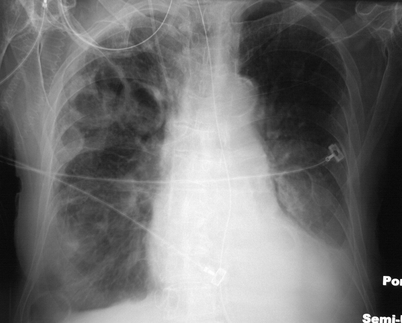
76year old female presents with dyspnea sepsis and bacteremia
CXR
Thick Walled Cystic lesions
Ashley Davidoff TheCommonVein.net 33005
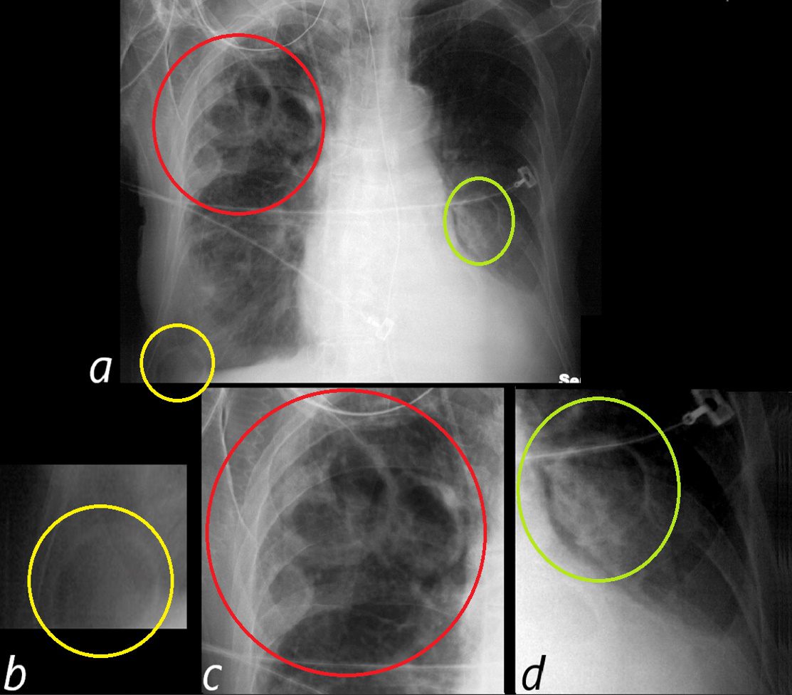
Frontal CXR reveals thick walled cystic changes clustered in the right apex (red ring a and c) and similar single thick walled cystic spaces in the right costophrenic angle yellow ring, magnified in a and b) and alongside the left heart border (green ring in a and d). There is silhouetting of the left hemidiaphragm. Echo showed tricuspid valve vegetations. Diagnosis is consistent with cavitating septic emboli
Ashley Davidoff TheCommonVein.net 33005cL
CT
Thick Walled Cystic Lesions
Bacteremia – Septic Emboli
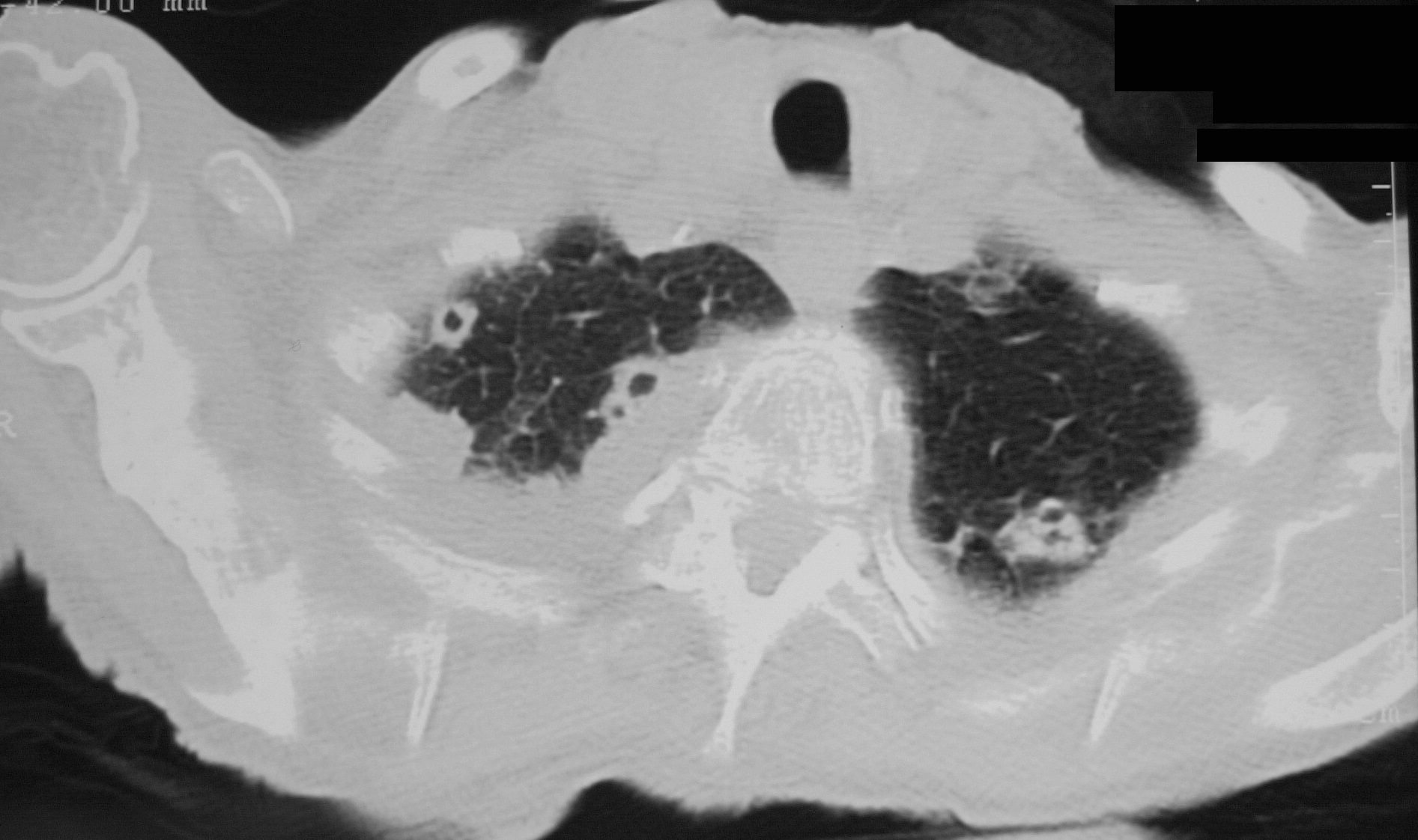
Axial CT reveals multiple small thick walled cystic changes clustered in the apices bilaterally, and thickening of the interlobular septa There is a large right pleural effusion. Echo showed tricuspid valve vegetations. Diagnosis is consistent with cavitating septic emboli
Ashley Davidoff TheCommonVein.net 33011
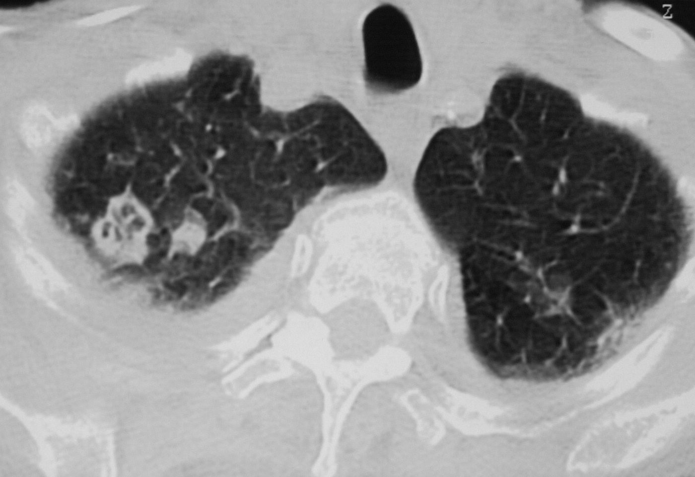
Axial CT reveals a thick walled complex cystic nodule alongside a solid nodule clustered in the right apex, and thickening of the interlobular septa There is a large right pleural effusion. Echo showed tricuspid valve vegetations. Diagnosis is consistent with cavitating septic emboli
Ashley Davidoff TheCommonVein.net 33010
Wedge Shaped Cystic Complex with
Feeding Bronchovascular Bundle
Cavitating Hampton’s Hump
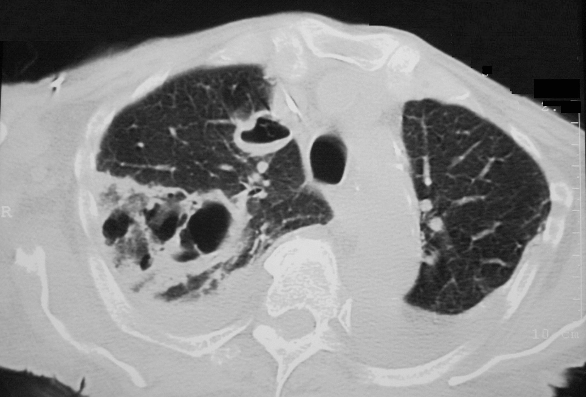
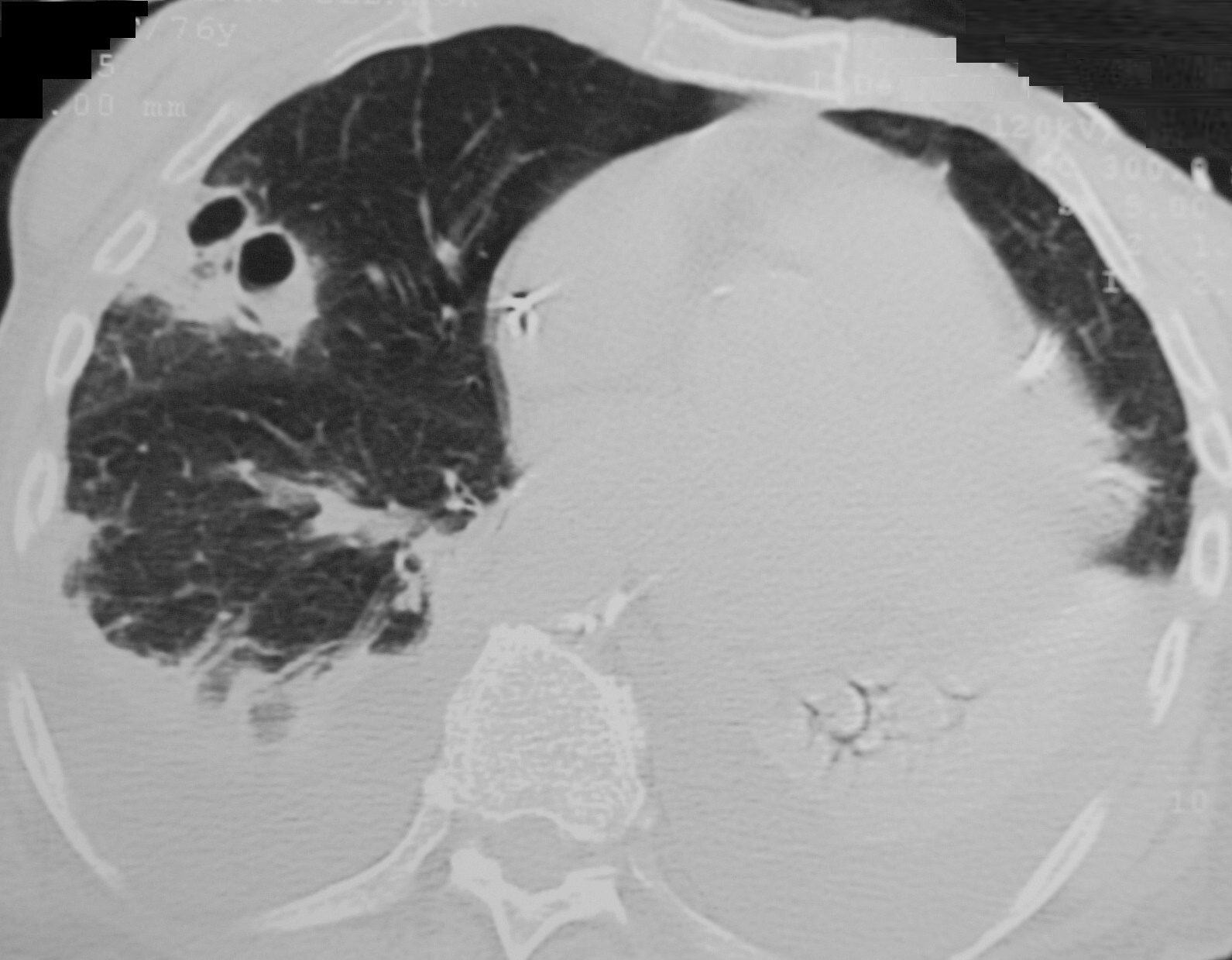
Axial CT reveals a large wedge shaped thick walled complex multicystic lesion associated with a feeding bronchovascular bundle (feeding vessel sign) in the right apex consistent with a cavitating infarction (cavitating Hampton’s hump). In addition there is a second smaller unilocular thick-walled cyst with a small air fluid level suggesting infection. There are pleural effusions. Echo showed tricuspid valve vegetations. Diagnosis is consistent with cavitating septic emboli
Ashley Davidoff TheCommonVein.net 33012
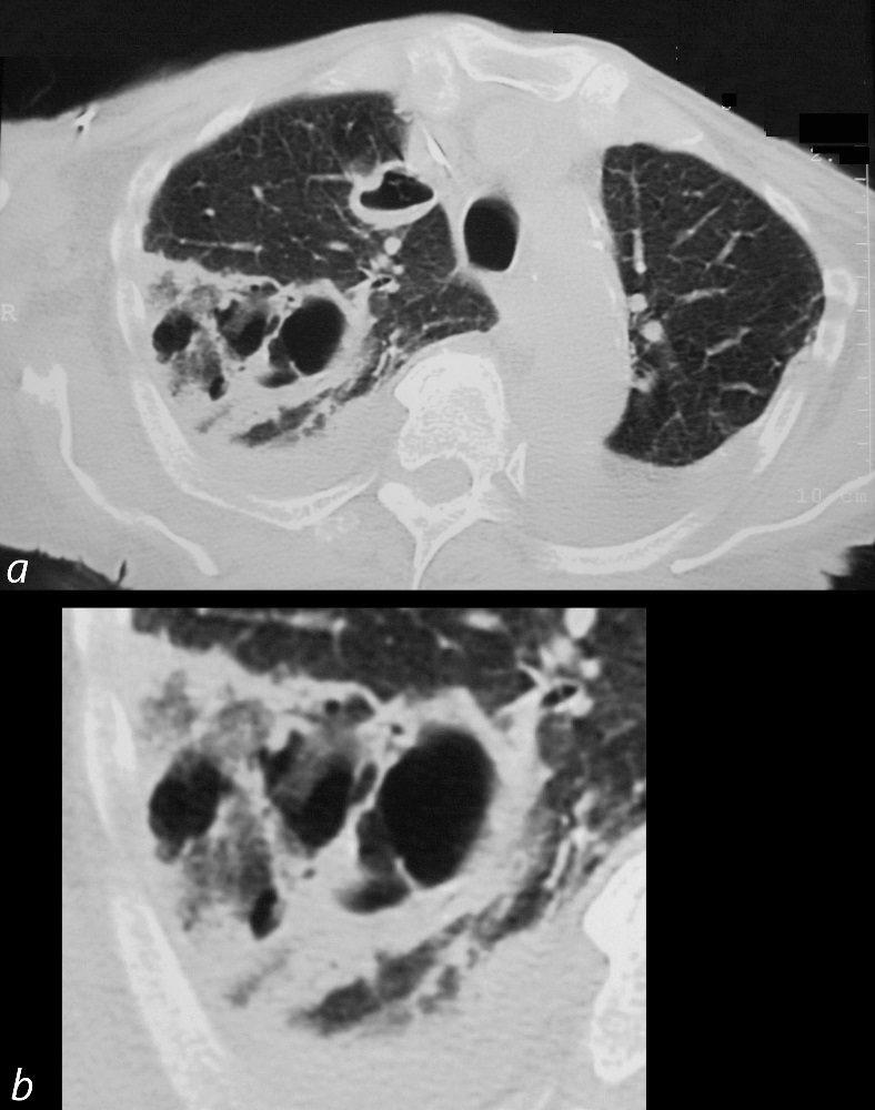
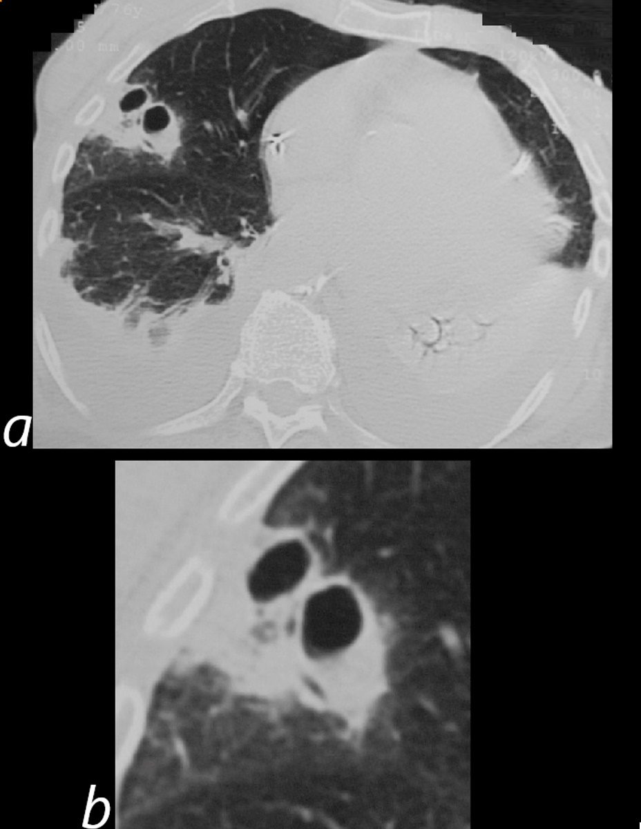
Axial CT reveals a large wedge shaped thick walled complex multicystic lesion associated with a feeding bronchovascular bundle (feeding vessel sign) in the right apex consistent with a cavitating infarction (cavitating Hampton’s hump). In addition there is a second smaller unilocular thick-walled cyst with a small air fluid level suggesting infection. There are pleural effusions. Echo showed tricuspid valve vegetations. Diagnosis is consistent with cavitating septic emboli with pulmonary infarction.
Ashley Davidoff TheCommonVein.net 33012
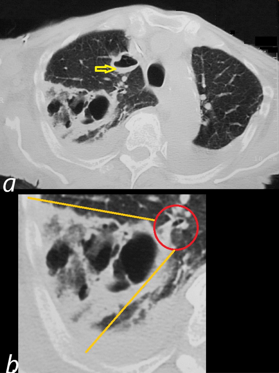
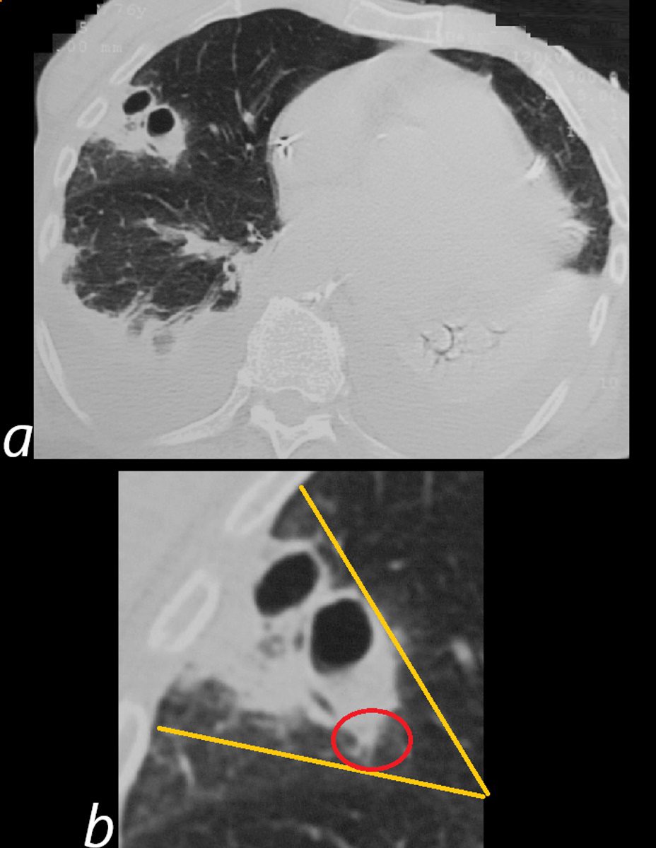
Axial CT reveals a large wedge shaped thick walled complex multicystic lesion ( bordered by orange lines in b) associated with a feeding bronchovascular bundle (red ring b -feeding vessel sign) in the right apex consistent with a cavitating infarction (cavitating Hampton’s hump). In addition there is a second smaller unilocular thick-walled cyst with a small air fluid level (yellow arrow, a) suggesting additional purulence in this clinical context. There are bilateral pleural effusions. Echo showed tricuspid valve vegetations. Diagnosis is consistent with cavitating septic emboli with pulmonary infarction.
Ashley Davidoff TheCommonVein.net 33012
Additional Cavitating Nodules and a
Small Nodule with a Halo
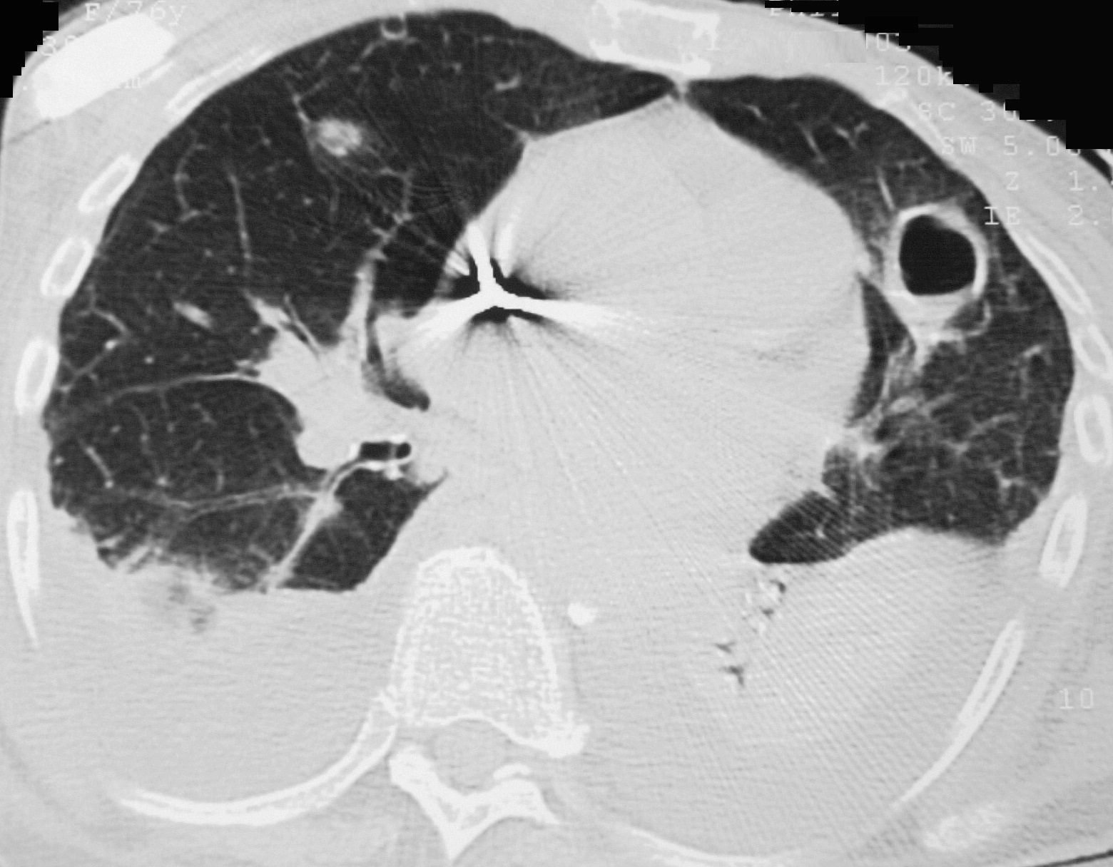
Axial CT reveals a thick walled complex cystic nodule alongside in the lingula with a small air fluid level and a smaller right upper lobe nodule with a ground glass halo. There are large bilateral pleural effusions associated with compressive atelectasis. Echo showed tricuspid valve vegetations. Diagnosis is consistent with cavitating septic emboli
Ashley Davidoff TheCommonVein.net 33014
Axial CT reveals a thick walled wedge shaped bilocular cystic lesion in a region of subsegmental consolidation associated with a feeding bronchovascular bundle (feeding vessel sign). There are large bilateral pleural effusions associated with compressive atelectasis. Echo showed tricuspid valve vegetations. Diagnosis is consistent with cavitating septic emboli
Ashley Davidoff TheCommonVein.net 33015
Axial CT reveals a thick walled wedge shaped bilocular cystic lesion in a region of subsegmental consolidation associated with a feeding bronchovascular bundle (feeding vessel sign) magnified in image b. There are large bilateral pleural effusions associated with compressive atelectasis. Echo showed tricuspid valve vegetations. Diagnosis is consistent with cavitating septic emboli
Ashley Davidoff TheCommonVein.net 33015c
Axial CT reveals a thick walled wedge shaped (orange triangle in b) bilocular cystic lesion in a region of subsegmental consolidation associated with a feeding bronchovascular bundle (red ring b – feeding vessel sign). There are large bilateral pleural effusions associated with compressive atelectasis. Echo showed tricuspid valve vegetations. Diagnosis is consistent with cavitating septic emboli
Ashley Davidoff TheCommonVein.net 33015cL