Lung Azygoesophageal Recess
- What is it?
- The azygoesophageal recess is a vertical interface formed between the medial right lower lobe of the lung and adjacent structures, including the azygos vein and esophagus.
- It is created by the apposition of the lung parenchyma with the mediastinal structures.
- Parts:
- Medial right lower lobe lung interface.
- Adjacent azygos vein.
- Esophagus.
- Posterior mediastinum.
- Size and Shape:
- Typically, a vertically oriented and concave structure.
- The degree of concavity can vary with the size of the azygos vein and mediastinal pathology.
- Position:
- Located on the right side of the posterior mediastinum.
- Extends vertically from the level of the azygos vein arch superiorly to the diaphragm inferiorly.
- Embryology:
- Forms during lung development as the right lung expands to meet the mediastinal structures, including the developing azygos vein and esophagus.
- Applied Anatomy:
- Imaging Application:
- The azygoesophageal recess is an important radiological landmark on chest imaging.
- Changes in its contour can indicate pathology, such as:
- Right lower lobe atelectasis or consolidation.
- Mediastinal lymphadenopathy.
- Esophageal masses or dilation.
- Azygos vein enlargement.
- Posterior mediastinal masses or abnormalities.
- Imaging Application:
-
- CXR:
- On chest radiographs, the azygoesophageal recess appears as a thin, smooth, and concave line on the right side of the mediastinum.
- Distortion, obliteration, or convexity of this line suggests underlying pathology, such as:
- Right lower lobe consolidation or atelectasis.
- Mediastinal lymphadenopathy.
- Posterior mediastinal mass or esophageal abnormalities.
- CT scan:
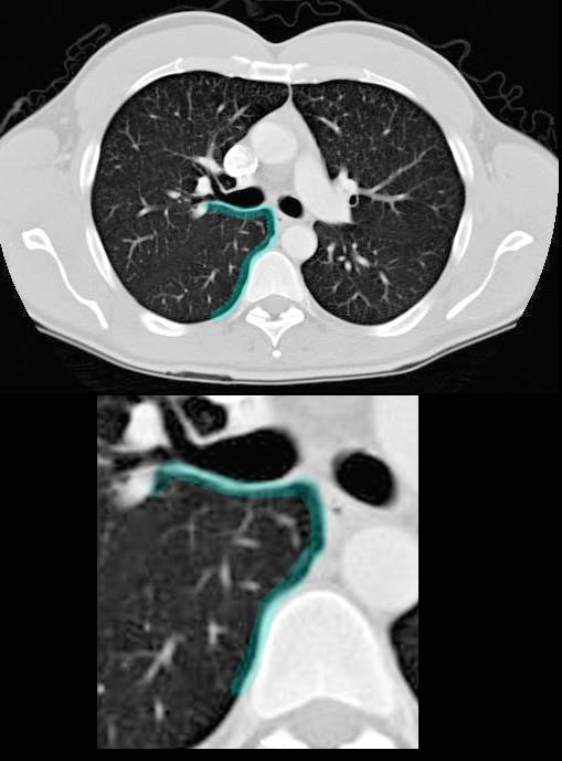
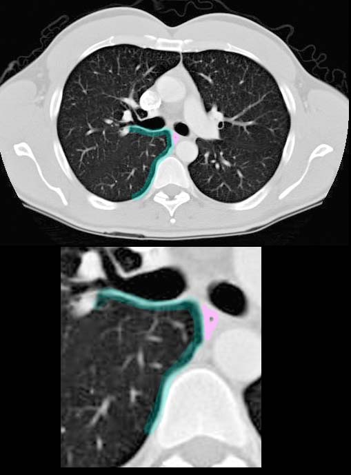
- On CT imaging, the azygoesophageal recess is seen as a sharp interface between the medial right lower lobe and adjacent mediastinal structures, specifically the azygos vein and esophagus.
- Normal appearance: a thin, well-defined curvilinear interface with no masses or distortions.
- Abnormal findings:
- Bulging or obliteration due to lymphadenopathy, posterior mediastinal masses, or esophageal pathology.
- Displacement caused by right lower lobe collapse or consolidation.
- Thickening of the recess associated with inflammatory or neoplastic conditions.
- Key Points and Pearls:
- The azygoesophageal recess should normally appear as a smooth, concave interface on imaging.
- Deviation, obliteration, or bulging of the recess can signal significant mediastinal or pulmonary pathology.
- Careful evaluation of the recess on chest radiographs and CT is critical for detecting subtle mediastinal abnormalities.
- When distorted, the recess can provide clues to the underlying condition, such as lymphadenopathy, esophageal disease, or vascular abnormalities.

CT scan at the level of the carina shows the azygo-esophageal recess (outlined in teal)
Ashley Davidoff MD
TheCommonVein.net
38803c

CT scan at the level of the carina shows the azygo-esophageal recess (outlined in teal) abutting the esophagus (outlined in pink)
Ashley Davidoff MD
TheCommonVein.net
38803cL
