

Definition
Colon carcinoma is a malignant disease and is characterized by insidious but progressive nature and late clinical presentation.
It is the third most common cancer and cause of cancer deaths in the United States according to the American Cancer Society (American Cancer Society, 2014).
The disease has multifactorial causes with the most common cause having no single known specific etiological factor. The strongest causative link relates to patients with familial adenomatous polyposis. One hundred percent of patients with this entity will develop carcinoma of the colon and a large percentage of patients with longstanding ulcerative colitis will develop carcinoma as well. It is therefore imperative that these patients undergo colectomies before the risk of carcinoma becomes a reality.
As a malignant epithelial tumor in a capacious organ, the clues to its presence are few in the early stages of the disease. The earliest clinical clue in the unsuspected patient is the presence of blood in the stool (hemoccult test) or the presence of anemia.
The neoplastic proliferation usually starts out as an adenomatous polyp and progresses over many years resulting in a clinical or pathological presentation.
Complications include systemic and local metastatic disease, obstruction and perforation.
The diagnosis is suspected clinically in middle-aged or elderly patients who are anemic or who have guaiac positive stools and it is confirmed by colonoscopy with biopsy.
If detected early, colorectal cancer is curable by surgery.
Imaging includes the use of endoscopy, barium enema and CT colonography.
Treatment options depend on the staging of the disease but include surgery, systemic and locoregional chemotherapy. Adjuvant chemotherapy can prolong survival in disease that has reached the lymph nodes. Radiotherapy is used in cases of rectal cancer to reduce local recurrence.
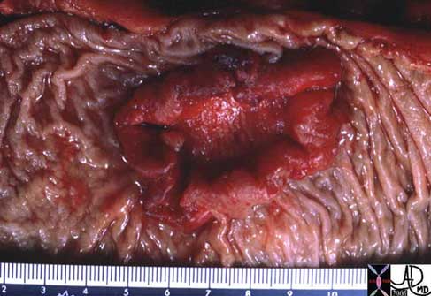
This specimen has obvious malignant characteristics macroscopically, characterized by heaped edges and central ulceration.
Courtesy of: Gutkin, M.D.
Principles of Structure
Colon carcinoma starts in the cell and if left unimpeded it will burrow through the walls of the bowel. When discovered, staging and treatment will depend on the histological staging of the tumor which in turn depends on the extent of the histological depth of invasion. It is therefore important to understand the histological makeup of the wall of the bowel.
Like many other tubes in the body its wall has four layers. The inner layer that lines the lumen is called the mucosa which rests on its basement membrane. The mucosa consists of a single layer of rectangular cells called the epithelium. Because of the rectangular shape of the cells they are called columnar cells and so the layer is called a simple, columnar epithelium. The second layer is the submucosa which contains blood vessels, lymphatics, nerves and loose connective tissue. The third layer is the muscular layer called the muscularis and the fourth layer is called the serosa or adventitia which acts as a protective outer ?skin? for the colon. This layer is called a serosa when it is surrounded by peritoneum (transverse colon and sigmoid colon) and called an adventitia when it is retroperitoneal (ascending colon, descending colon and part of the rectum).
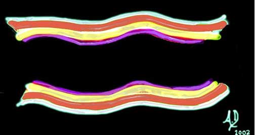
This diagram illustrates the 4 basic layers of the colon. The inner pink layer is the mucosa, the yellow layer beneath the mucosa is called the submucosa, while the red layer is the muscular layer (muscularis) and the 4th layer is called the serosa or adventitia.
Courtesy Ashley Davidoff, M.D.
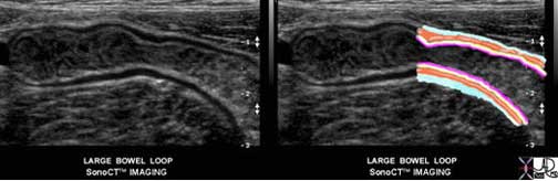
This ultrasound image of the colon demonstrates the 4 layers of the large bowel quite beautifully. The luminal contents are heterogeneous and are bordered by an echogenic line that is the combination of the mucosa and submucosa. The lucent line we see is the muscular layer while the outer echogenic line represents the serosa or adventitia
Courtesy Philips Medical Systems

The mucosa consists of a simple columnar epithelium which means it is a single layer of epithelial cells that are each shaped like a column – similar to a tall rectangular block of apartments.
Courtesy Ashley Davidoff, M.D.
The submucosa contains the vital lifelines including the terminal branches of the arteries, the capillary network, the smallest and earliest venous branches and the lymphatics. There are nerve endings in the submucosa as well as the muscular layer. Circulating white cells that perform the vital local defense functions police the submucosa for potentially harmful organisms and substances. The muscularis contains spindle-shaped smooth muscle cells while the serosa and adventitia consist of relatively strong connective tissue, and in the case of the serosa, a layer of epithelial cells derived from the peritoneal lining.

The lifelines together with the small rounded lymphoid cells are illustrated in submucosa. A capillary network between arteries and veins forms creating the end of one part of the circulation and the beginning of the next. The spindle shaped smooth muscle cells are illustrated in the red muscular layer and the outer layer in white is made of connective tissue and in the case of the serosa a layer of flat epithelial cells derived from the peritoneum.
Courtesy Ashley Davidoff, M.D.
This overlay of the transverse colon reveals the three outstanding morphological features of the colon including the bright yellow appendices epiploicae, one of the three longitudinal muscles called the taenia coli in maroon and the sacculations called haustra caused by pleating of the colon by the taenia.
Courtesy Ashley Davidoff, M.D.
Histology
The wall of the colon requires a very substantial vascular supply and effective nerve and lymphatic system in order to coordinate its function with other physiological events occurring within the gastrointestinal system specifically and within the body at large. The neural, lymphatic, and vascular channels enter through supporting mesentery and penetrate the wall (just like the cable systems, water systems, electrical and sewerage enter and leave our homes). These penetrations produce weakening of the wall and remain the potential site for diverticuli to develop. The vessels terminate in the submucosa.

Courtesy Ashley Davidoff MD
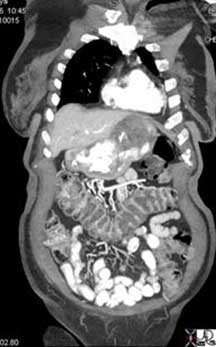
The coronal reformat of an abdominal CT shows branches of the middle colic artery in the transverse mesocolon just about to penetrate the wall of the transverse colon. There is a hint in the middle of the section of transverse colon of smaller vessels running along the mucosal folds.
Courtesy Ashley Davidoff, M.D.
The mucosa and wall of the bowel are not isolated structures. John Donne, a clergyman and poet from the Renaissance period, stated that ?? No man is an island ?? In the same way no organ is an island. Each is connected and dependent on the other organs and the colon and its substructures are no different. It is not surprising therefore that disease of the colon, for example, is associated with disease in other parts of the body. The association of ulcerative colitis and sclerosing cholangitis is a prime example. The psyche and the bowel are also intimately related. Acute anxiety is notorious for inducing diarrhea.
The proximal large bowel is presented with the sludge-like byproducts of small bowel digestion consisting of mostly water and undigested complex carbohydrates. Colonic mucosa absorbs most of the water and together with churning action and bacterial digestion, the nature of the sludgy, semi-liquid stool is transformed into more solid and compacted stool. This function is not affected by malignancy.
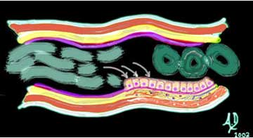
In the above image the fluid filled chyme represented in the left part of image (light green), is transformed into the gas filled compacted solid stool (dark green) seen on the right side of image.
Courtesy Ashley Davidoff, M.D.
Applied Histology
There are both absorptive cells (enterocytes) as well as a significant proportion of goblet cells which secrete mucus. Malignant cell populations may arise from either of these populations. If the malignancy is mucus secreting it may manifest radiologically with excessive mucus in the tumor and or the metastases. The imaging characteristics of mucus (fluid) are distinctly different from purely solid tumors. In addition, dystrophic calcification occurs more commonly in mucus secreting tumors and is a helpful means of characterizing the disease.
Parts of the Colon
The colon is made up proximally of the cecum which is the widest portion which links the large bowel to the small bowel at the ileocecal junction. It contains the appendix which is a tubular structure about 3-5 mm in diameter and between 3 and 10cm long. Thereafter, comes the ascending colon which is situated in the retroperitoneal position, followed by the transverse colon. The transverse colon hangs like a washing line within the peritoneal cavity between the two flexures – hepatic flexure on the right and the splenic flexure on the left. The descending colon is also in a retroperitoneal position while the sigmoid subtended on the sigmoid mesocolon becomes intraperitoneal. The rectum is bound down by the peritoneum as it exits at the anus.
Each component of the colon has an inner endothelium, a submucosa, a middle muscular layer called the muscularis and an outer layer called a serosa or adventitia. Minor but characteristic components include the taenia coli and appendices epiploica.

These two diagrams give an approximate sense of the parts of the colon with no consistent divisions marking the transitions from one part into the next. The cecum (green) is well demarcated by the entrance of the ileum, with the ascending colon (light blue) transitioning into the hepatic flexure (royal blue) just before the turn below the liver. The transverse colon (pink) is of variable length and then makes a turn at the splenic flexure (yellow) just under the spleen. The descending colon (orange) extends along the left side of the abdomen after which it escapes from the retroperitoneum and becomes the sigmoid colon (gold). The proximal part of the rectum (brown) becomes bound by peritoneum and transitions into the anal canal a few centimeters shy of the anus which is the last part of the colon.
Courtesy Ashley Davidoff, M.D.
Blood Supply
The superior mesenteric artery (SMA), inferior mesenteric artery (IMA), and internal iliac arteries are the vessels that supply the colon. The SMA is the artery of the midgut while the IMA is the artery of the hindgut. The distal and terminal portion of the hindgut is supplied by the rectal branches of the internal iliac arteries.
Blood Supply

The diagram shows the branches of the SMA including the ileocolic (1), right colic (2) and the middle colic (3). These vessels connect through the marginal artery (4) which runs along the inner or mesenteric border of the colon (4) and gives rise to the vasa recta that run over the colon before branching into the wall of the colon to finally enter the submucosa. The IMA (6) gives rise to the left colic artery (7), three sigmoid branches (8) and the superior rectal artery [also known as the superior hemorrhoidal artery (9)]. The internal iliac artery (10) supplies the distal part of the rectum and gives rise to the middle rectal artery (11), and inferior rectal (12) arteries. These are paired arteries arising from the left and right iliac and they are connected through collaterals with the superior rectal artery.
Courtesy of: Ashley Davidoff, M.D.
The circulation of the gastrointestinal tract is a fascination with areas of obvious deficiencies and areas of excess. With respect to the colon, the areas of deficiencies are in the region of the splenic flexure, since it is as stated above, the watershed region where the terminal circulations of the middle colic and left colic arteries meet. As a terminal circulation, it implies that if there is a deficiency of circulation such as might occur in the patient who is hypotensive, then this area is last in the food line. On the other hand, the rectum has an abundant blood supply and it would be rare and almost unheard of, for the rectum to undergo infarction. The other interesting part of the rectal circulation is its connection with the systemic circulation, so that rectal circulation does play an important role in the patient with peripheral vascular disease who has a significant common iliac stenosis.
These facts are most important to the colorectal surgeon performing colonic resection for cancer, since surgery involves segmental resection of the colon together with the arteries, veins and lymphatics. Knowledge of the vascular anatomy is essential.
Venous Drainage
The final common pathway of colonic venous drainage is dominantly via the portal system but also via the systemic venous system using the internal iliac veins to gain access to the IVC. The veins in general follow the course of the arteries and are similarly named. Thus, there is a superior mesenteric vein (SMV), inferior mesenteric vein (IMV) and rectal or hemorrhoidal veins. The SMV usually drains with the splenic vein into the portal vein and in fact their confluence gives rise to the portal vein. The IMV usually drains into the splenic vein. The middle rectal vein and inferior rectal vein drain into the internal iliac veins and then into the IVC as stated above.
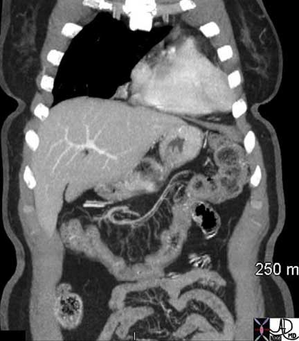
This coronally reconstructed CT of the abdomen shows middle colic venules and arterioles in the transverse mesocolon becoming confluent to form the right and left branches of the middle colic vessels.
Courtesy Ashley Davidoff, M.D.
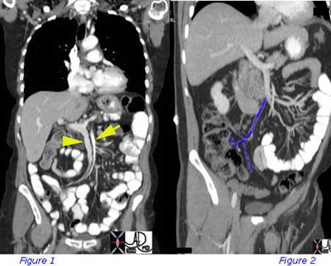
Figure 1 is a coronal CT scan that shows the normal appearance of the superior mesenteric artery and superior mesenteric vein. The artery is to the left (yellow arrow), smaller in diameter and in this instance slightly brighter. The SMV is to the right (yellow arrowhead), larger, and becomes confluent with the splenic vein to form the portal vein which courses to the liver. Figure 2 shows jejunal veins to the left of the patient’s body and ileocolic vein (blue) draining the proximal ascending colon. The SMV, splenic vein and portal vein are again demonstrated.
Courtesy of: Ashley Davidoff, M.D.
Hematogenous spread of tumor via the mesenteric circulation will be transported into the portal system and will be filtered in the liver. Hematogenous metastases from the inferior hemorrhoidal vein will drain into the systemic venous system and will be filtered by the lung. Thus, it is incumbent on physicians following patients with low rectal carcinomas to follow progress with both an abdominal and chest CT when metastases are suspected.
Lymphatic Drainage
The lymph nodes are named mostly according to the vessels along which they travel. Thus, the names include the ileocolic, right colic, middle colic, left colic, sigmoid, superior rectal, middle rectal, and inferior rectal lymph nodes. These in turn drain to the appropriate group of nodes situated at the origin of the main mesenteric vessels including the SMA, IMA and iliac nodal stations. The nodes that are along the mesenteric border where the marginal artery is located are called the paracolic nodes. In addition there are two special groups of nodes around the cecum. One group is called prececal and the second is called retrocecal ? both named according to their position with respect to the cecum.
The solitary lymphatic nodules of submucosal lymphoid tissue in the large intestine are most abundant in the cecum and appendix, but are also found throughout the rest of the colon.
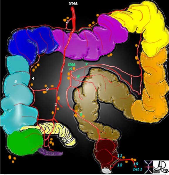
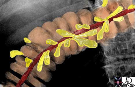
The lymph nodes are named according to the blood vessels with which they travel. Thus there is the ileocolic group (1) right colic (2), middle colic (3) left colic (7), sigmoid (8), superior rectal (9), middle rectal (11), and inferior rectal (12). These nodes drain into the regional halfway houses for the nodes including the SMA, IMA and iliac groups. The paracolic groups are located along the marginal artery (4) and the prececal and retrocecal are seen in relation to the cecum.
Courtesy Ashley Davidoff, M.D.
This elderly man has lymphoma and the enlarged lymph nodes are clustered around the SMV (maroon) and SMA (red) like a bunch of grapes. Smaller nodes are also seen in the small bowel mesentery and along the left colic and left paracolic region.
Courtesy Ashley Davidoff, M.D.
Staging of colon cancer is based on the degree of penetration of the tumor through the layers of the bowel wall, involvement of the regional lymph nodes, and finally on distant metastases. Once the lymph nodes are involved, the tumor is at least stage III in the TNM classification and stage C in the Duke classification. Therapy is based on the staging of the tumor, often best evaluated after surgery is performed and the lymph nodes are examined microscopically.
Principles of Disease
The mucosa is a metabolically active structure with rapid turnover. Intermittently, new cells with either spontaneous mutation or mutations as a result of an environmental pathogen or carcinogen proliferate. Malignant growth is characterized by unrelenting expansion of the malignant cell population without regard for the organism. When the malignant growth arises from an epithelium, it develops into a carcinoma, and when it arises from stromal tissue it becomes a sarcoma. Since the mucosa of the colon has glandular elements, the carcinoma arising from the colon is called an adenocarcinoma. The colonic mucosa also secretes mucin and so histopathologically the entity may present as a mucinous adenocarcinoma.
The colon is a capacious tube and the tumor is usually far advanced before the transport function becomes impeded. Thus, obstruction is a late manifestation. Because of the occult nature of the disease, diagnostic endeavors are focused on screening for early detection to enable the chance for prevention and cure.
Principles of Cancer in General
The normal process of cell growth and cell death is orderly. There is a time to live and a time to die. The process of timed cell death is called apoptosis literally meaning “dropping off” from the Greek apo = from and ptosis = falling, and is best equated to a similar event in botany of a petal falling off a flower. A series of biochemical events over time results in structural change in the nucleus, cytoplasm and cell membrane, with eventual “falling off” of the cell from the population at large. In health the cell is replaced. Between 50-70 billion cells die each day in the human adult.
In cancer, the orderly process of new cell growth and cell death is altered. There is uncontrolled cell division and growth and reduction in cell death. An imbalance occurs with a shift that favors the anti-apoptotic mechanisms over the pro-apoptotic mechanisms and cells live past their age and do not die.
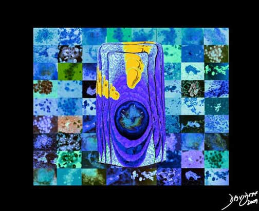
The image represents the life of a single set of columnar cells showing a progression of generations as the cell lives, dies and is regenerated. The orange secretions of the cell are seen in the background of the pink cytoplasm and the purple nucleus. The nucleus of the newest generation and cell is seen as a clock that has become distorted causing time to become disordered. This is the forerunner of a malignant process.
Courtesy of: Ashley Davidoff, M.D.
Intermittently new cells with either spontaneous mutation or mutations as a result of an environmental pathogen or carcinogen proliferate. Malignant growth is characterized by unrelenting expansion of the malignant cell population without regard for the organism. When the malignant growth arises from an epithelium it develops into a carcinoma and when it arises from stromal tissue it becomes a sarcoma. Since the mucosa of the colon has glandular elements the carcinoma arising from the colon is called an adenocarcinoma. The colonic mucosa also secretes mucin and so histopathologically the entity may present as a mucinous adenocarcinoma.
Pathogenesis
Adenocarcinomas of the colon arise from preexisting adenomatous polyps that develop in the normal colonic mucosa. There is a well recognized adenoma-carcinoma sequence. The most common site for tumor development is the mucosa. The mucosa is a highly active structure and cell turnover is rapid, with the reproduction of new cells and death of old cells occurring about every four days.
The single aberrant cell that develops at the initiation of the disease process, multiplies and forms its own community of cells that enlarges with time – and in its infancy is seen as a benign, adenomatous polyp. Most colonic polyps, however, (90%) are not adenomatous but are hyperplastic (size < 5 mm) meaning that they are still under the control of normal regulatory mechanisms of the body. Hyperplastic polyps are benign and, in most instances, are not considered premalignant. On the other hand, 10% are adenomatous polyps and are premalignant.
The endoscopist cannot distinguish between a hyperplastic polyp and an adenomatous polyp and therefore, the endoscopist removes all polyps.
When polyps are less than 1 cm the possibility that they are malignant is less than 5%. As they grow into the 1-2 cm range the chances of malignancy are in the 10% range. Once they grow beyond 2 cm the chances of malignancy increase to 50%. There are three types of adenomatous polyps: tubular, tubulovillous, and villous. Tubular adenomas are the most common while villous adenoma has the greatest potential to become malignant.
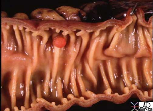
This 5 mm polyp is a common finding on colonic evaluation and at this size is a benign abnormality. Left intact it can grow into a monster. Hence it is usually removed by the endoscopist.
Courtesy Ashley Davidoff, M.D.

This series of images from a virtual colonoscopy shows an 8 mm polyp seen on the surface rendering images (salmon colored images, cross marks the polyp) referenced to the sigmoid colon (bottom left) and with conventional CT imaging (bottom middle). At this size the polyp is almost certainly benign and can be removed through the colonoscope safely.
Courtesy Scott Tsai, M.D.
An adenomatous polyp, if it is destined to become malignant, usually takes 10-15 years to evolve. It will slowly invade the space of the lumen, but more importantly the space of the wall of the bowel, first into the submucosa and subsequently into the muscularis, serosa, adventitia lymphatics and finally into the systemic circulation.

The gross pathology specimen shows an inflamed, angry looking mass in the colon that has central necrotic area surrounded by heaped tissue.
Courtesy of: Barbara Banner, M.D.
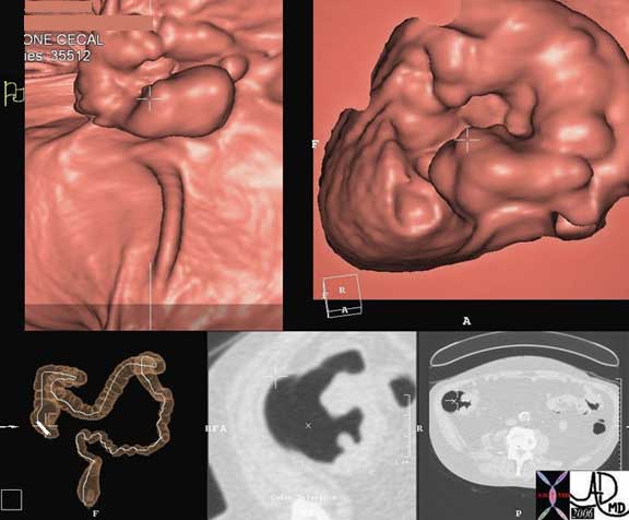
This series of images from a virtual colonoscopy shows a 5 cm ulcerated mass (cross marks the mass) seen on the surface rendering images (salmon colored images) referenced to the cecum (bottom left) and with conventional CT imaging (bottom middle). At this size and shape the mass is almost certainly malignant and cannot be removed through the colonoscope safely. Note how similar the morphology of this tumor is to the pathology of the specimen above (different patient).
Courtesy Scott Tsai, M.D.
As the disease progresses there is expansion of the tumor in the submucosa so that a circumferential lesion evolves. There may be extension into the deep layers of the wall, into lymphatics and portal venous system and eventually into the liver. As the tumor grows locally it spreads circumferentially and eventually totally surrounds the bowel in a “napkin-ring” like manner as depicted in the image below. Radiologically, the “napkin-ring” translates into an apple core lesion when viewed radiologically and is best appreciated with a double contrast barium enema.
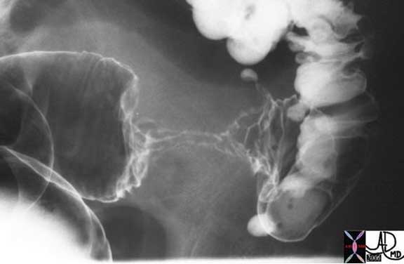
The double contrast barium enema shows an apple core lesion caused by circumferential growth of a malignant tumor of the colon in ?napkin-ring? fashion. The lumen is compromised. Note how the distal colon which is receiving air and barium via the enema tip is relatively dilated when compared to the descending colon which is relatively decompressed suggesting an obstructive process. Note also the presence of contrast filled diverticula at the rectosigmoid junction. The two entities of carcinoma and diverticulosis often coexist since diverticulosis is very common in Westernized nations.
Courtesy Ashley Davidoff, M.D.
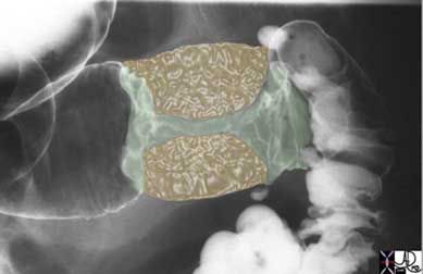
The overlay image shows the yellow mass of tumor that has surrounded the lumen in napkin ring fashion essentially strangling the lumen. Note how the distal colon which is receiving air and barium via the enema tip is relatively dilated when compared to the descending colon which is relatively decompressed suggesting an obstructive process. Note also the presence of contrast filled diverticula at the rectosigmoid junction. The two entities of carcinoma and diverticulosis often coexist since diverticulosis is very common in Westernized nations.
Courtesy Ashley Davidoff, M.D.
Cause and Predisposing Factors
Colon cancer is a disease that is its highest in developed countries such as the United States and Japan, and lowest in developing countries in Africa and Asia. It is interesting to note that in the US it has its highest incidence in African American men.
It is thought or appears to be closely linked to Western diet where high fat content and relatively low fiber content are considered major factors in the cause of the disease. In patients who develop colon cancer, it is thought that diet and other factors alter the genetics of a cell or a group of cells and an adenomatous polyp results. Most polyps will not evolve into cancer. An entity called multiple aberrant mutations seems to be the responsible process that will cause about 10% of adenomatous polyps to evolve into cancer. This process disinhibits normal regenerative processes and enables the mucosa to advance from an adenomatous polyp into a carcinoma over 10-15 years. Evolving work on the genetic aberrancies is allowing advances in both the diagnosis and treatment of the disease. Identifying the genetic mutation of a cell in the stool may prove to be an important screening technique. Another example is the DCC gene, which causes the neoplastic cells to adhere to each other. The absence of this gene in the mutated gene would decrease cell adherence and theoretically enable malignant cells to metastasize easily. Thus, testing for the DCC gene may shed light on the virulence and natural history of colon carcinoma in a particular patient and thus, aid in prognostication.
Other known associated diseases include a prior history of colon carcinoma (metachronous = multiple separate occurrences – 4-5% of colon cancers), synchronous lesions (two or more at the same time = 1%) and inflammatory bowel diseases (IBD). Ulcerative colitis is a well established associated disease, while the association with Crohn’s colitis is less common.
Hereditary factors that predispose to colon cancer are seen in conditions such as familial polyposis and Gardener’s syndrome, where there is a very strong association. Other conditions such as prior history of breast or endometrial carcinoma and retinitis pigmentosa also raise the awareness of associated colonic carcinoma.
Cause and Predisposing Factors: Familial Adenomatous Polyposis
Familial adenomatous polyposis (FAP) is the most common adenomatous polyposis syndrome and it is characterized by the presence of innumerable neoplastic tubular adenomatous polyps of the colon. The cause of the disease relates to an autosomal dominant inherited disorder. In early life (about 16 years) the genetic defect evolves into and results in a colonic mucosa that is carpeted with hundreds to thousands of adenomatous polyps. In 100% of patients the syndrome is complicated by the development of cancer at the age of 30 ? 40 years and hence, all patients should be treated with colectomy in early years of the disease (teenage to early 20’s).
Associated diseases include desmoid tumors (4-30%) and other rarer associations including medulloblastoma, thyroid cancer, adrenal cancer, gastric cancer and pancreatic cancer.
The diagnosis is established in patients who have a positive family history and by colonoscopy demonstrate more than 100 polyps. Clinically, it is usually asymptomatic and only presents once the patient has developed cancer. Symptoms include abdominal pain, diarrhea, mucus diarrhea, and bloody diarrhea.
Colonoscopy is the imaging and diagnostic study of choice since the diagnosis can be established and biopsy of suspicious lesions can be accomplished.
Colectomy with mucosal proctectomy and ileoanal pouch pull-through is the therapeutic procedure of choice.
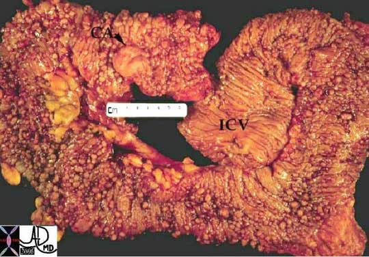
The normal mucosa in this patient has been replaced by an inumerable carpet of polyps extending throughout the colon. A malignancy has evolved (“ca” = carcinoma) at the distal end of the colon. IVC = ileocecal valve.
Courtesy of: Ashley Davidoff, M.D.
Ulcerative Colitis and Cancer
Ulcerative colitis is an idiopathic inflammatory process of the mucosa of the colon and rectum and is characterized by its involvement of the rectum, and retrograde progressive involvement of sigmoid then descending, transverse and ascending colon. It is in essence a mucosal disease. The cause of the disease is unknown but the result is mucosal inflammation and ulceration. The risk of cancer increases with the chronicity and extent of the disease. About 5% of patients with UC develop colon cancer. As chronicity increases and extent of involvement increases the incidence of colon cancer also increases. Carcinoma is usually preceded by a dysplastic phase. Patients who have had a pancolitis for 8 years should receive a screening colonoscopy and biopsy, while those whose disease is restricted to the left colon should have screening colonoscopy and biopsy after about 12 years from the onset of disease. The biopsy is taken every 10 cms at a random location. All patients with IBD should have repeat colonoscopy and biopsy every 2-3 years. These recommendations have been endorsed by the American Cancer Society, the American College of Gastroenterology, the American Society of Colon and Rectal Surgeons, and the Crohn?s & Colitis Foundation of America.
Statistics
Adenocarcinomas of the colon are the most common cancers seen in the GI tract in western society. One in 20 Americans will develop colon cancer and 136,830 new diagnoses will be made each year and about 50,000 patients will die of the disease per year (American Cancer Society, 2014). While it ranks third among both men and women in mortality in western society, it is really second to lung cancer in women and men.
Men and women face a lifetime risk of nearly 6% for the development of invasive colorectal cancer. The disease is most common after the age of 70, but in the patient at high risk such as those with familial syndromes, colonic carcinoma could present in the late twenties or 30’s but overall, 90% of new cases and 93% of deaths occur in people 50 and older (American Cancer Society, 2014).
Pathology
Most tumors are adenocarcinomas. The most common site is the sigmoid colon (20%) followed by rectum (15%), transverse colon (12%), descending colon (10%), ascending colon (8%) and cecum (8%). As the population ages, there is a tendency for the right-sided colonic carcinomas to become more common. In ulcerative colitis, since the longest standing inflammation is left-sided it is not surprising that left-sided colon carcinoma is more common.
This specimen has obvious malignant characteristics macroscopically, characterized by heaped edges and central ulceration.
Courtesy of: Gutkin, M.D.
Potential Complications
The most common site for metastases is the liver (25%), mesenteric and retroperitoneal nodes (15%), and kidney due to obstruction (10%). Large bowel obstruction, perforation and intussusception are potential complications of the disease.
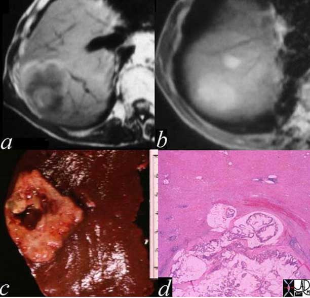
The MRI of the liver shows a space occupying abnormality at the dome of the liver with central hyperintensity on T1-weighted image, (a) as well as diffuse hyperintensity on T2-weighting (b) representing a hemorrhagic mucin secreting adenocarcinoma seen on the gross specimen (c) and on the histological; section (d).
Courtesy of: Ashley Davidoff, M.D.

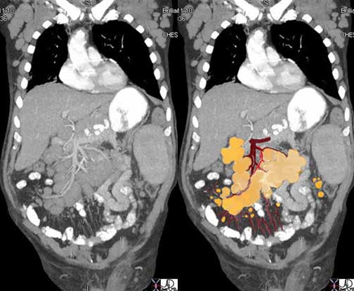
The abdominal CT is from a middle aged female with right sided discomfort. The image reveals multiple space occupying lesions (light green) in the liver. The patient was found to have a primary colonic carcinoma. The capsule of the liver is involved by tumor extending to the surface of the liver and deforming the surface (dark green). The disease is also close to the diaphragm (maroon). The involvement of the liver capsule and possibly the right hemidiaphragm is likely the cause of the patient?s pain. There is also a small amount of ascites present (yellow) either as a response to involvement of the liver capsule or the early development of malignant ascites.
Courtesy of: Ashley Davidoff, M.D.

The positron emission tomography (PET) scan in this elderly female with known carcinoma of the sigmoid colon shows metastatic disease to the liver, (dark green), lungs (light green) and mediastinum (light green with arrows) There is normal increased activity in the heart (red) and excretion of the FDG into minimally dilated collecting systems (yellow).
Image courtesy of: Ashley Davidoff, M.D.
Lymph Node Metastases
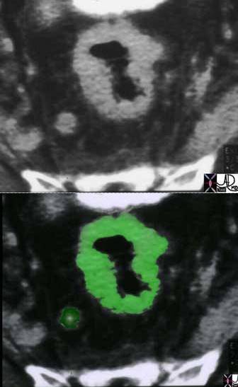
The CT scan of the rectosigmoid region shows diffuse thickening of the rectum (light green) with an abnormal lymph node in the perirectal fat (dark green) in this patient with carcinoma.
Courtesy of: Ashley Davidoff, M.D.
Complications: Bowel Obstruction

X-ray of the abdomen shows dilated colon in a patient presenting with rectal carcinoma and bowel obstruction
Courtesy Ashley Davidoff MD

Image a, is a CT scan of the abdomen and shows dilated upstream colon in a patient presenting with ascending colonic carcinoma and bowel obstruction
In image b, the dilated upstream colon contains retained fluid content (orange) and the carcinomatous mass is overlaid in green
Courtesy Ashley Davidoff MD
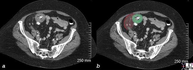
Image a, is a CT scan of the lower abdomen and shows circumferential thickening of the cecum (overlaid in green in b) stranding of the paracolic fat. with thickened peritoneum, and 2 nodules in the fat, likely representing metastatic disease.
Images courtesy of: Ashley Davidoff, M.D.
Diagnosis
In general, colon carcinoma is symptomatically occult and patients in general are asymptomatic in the early stages but may present with anemia and hemoccult positivity. As the anemia progresses the patients may present with weakness and weight loss and occasionally with abdominal pain.
The diagnosis of colon carcinoma should be on the mind of caregivers in all patients starting at about the age of forty where unexplained hemoccult positive test or occult anemia should be viewed with high suspicion. Such a patient should undergo colonoscopy as the diagnostic study of choice. All patients should be screened with colonoscopy after the age of 50 years and thereafter, every 10 years.
In addition to the colonoscopy, annual fecal occult blood testing (FOBT) and periodic flexible sigmoidoscopy should be done. Alternatively, barium enema should be used every 3-5 years (Helm).
Despite the guidelines, a large proportion of the US population is not being screened because of insufficient numbers of trained colonoscopists to meet the need, fear of the procedure, or ignorance of the guidelines. CT colonography is an excellent alternative for screening when colonoscopy is not possible. Sensitivity for small polyps is high and is similar to that of optical colonoscopy.
Lab Tests
In the patient with suspected colon carcinoma, blood tests should include cell count and hematocrit for the evaluation of anemia. The serum carcinoembryonic antigen (CEA) is a tumor marker most often used for the evaluation of gastrointestinal tract malignancies. In about 60% of patients with colon carcinoma it is elevated, and it is therefore a worthwhile test to obtain. It is also reasonable to evaluate liver function tests in the patient with suspected colon carcinoma to evaluate for the presence of liver metastases, though a negative result does not exclude the diagnosis.
Imaging
Optical colonoscopy is the study of choice for the patient with suspected carcinoma since it offers the opportunity to biopsy suspicious lesions and to remove small polyps. When this method fails or is not possible, then CT colonography has now become the second line diagnostic study of choice.
Once the diagnosis is established, a CT scan of the abdomen is used for staging the disease preoperatively. The local disease and extension into pericolic fat and lymph nodes can sometimes be established. Liver involvement is also best evaluated at this time with, and if necessary, suspicious lesions can be biopsied or further characterized with MRI.
It is also reasonable to perform a chest x-ray preoperatively to aid in staging, though sensitivity for small lesions is low.
In this instance, a small nodule that is about 5mm is spotted on the fillet view (top); its position in the hepatic flexure is confirmed on the scout view; the axial view is not as clear, but in the split or cuboid view it can be rotated and confirmed to be a small polyp. The radiologist can use either the fillet view or the fly through view as the primary navigation tool.
Courtesy of: Ashley Davidoff, M.D.
Treatment and Management: Surgery
In general, almost all patients should have the tumor removed regardless of staging because of the risk of obstruction. A right, transverse, or left hemicolectomy encompassing the lymph drainage area is standard surgical care. At the time, additional staging procedures are critical in the management. Regional lymph nodes should be removed for pathological evaluation; nodular lesions in the peritoneal cavity should also be evaluated, and the liver should be examined with ultrasound and suspicious lesions biopsied as well.
Thereafter, it is important to stage the disease when all the data is gathered to assess the post operative management.
Treatment and Management: Staging
Two classifications have been of use: the TNM (primary) tumor, (regional lymph) node, (remote) metastasis staging and the Dukes classification.
TNM Staging System for Colon Cancer
|
Stage |
Primary Tumor (T) |
Regional Lymph Node (N) |
Remote Metastasis (M) |
| Stage 0 | Carcinoma in situ | N0 | M0 |
| Stage I | Tumor may invade submucosa (T1) or muscularis (T2). | N0 | M0 |
| Stage II | Tumor invades muscularis (T3) or perirectal tissues (T4). | N0 | M0 |
| Stage IIIA | T1-4 | N1 | M0 |
| Stage IIIB | T1-4 | N2 | M0 |
| Stage IV | T1-4 | N2 | M1 |
*N0: No regional lymph node metastasis.
N1: Metastasis in 1 to 3 regional lymph nodes.
N2: Metastasis in 4 or more regional lymph nodes.
Dukes Classification
|
Stage |
Characteristics |
| Dukes stage A | Carcinoma in situ limited to mucosa or submucosa (T1, N0, M0) |
| Dukes stage B | Cancer that extends into the muscularis (B1), into or through the serosa (B2) |
| Dukes stage C | Cancer that extends to regional lymph nodes (T1-4, N1, M0) |
| Dukes stage D | Modified classification; cancer that has metastasized to distant sites (T1-4, N1-3, M1) |
Stage 1 and 2 cancers are treated with surgery alone and have a very high cure rate. Stage 1 disease has a 90 to 95 percent five-year survival and early stage 2 disease (B-1) has an 85 percent five-year survival. Once the tumor invades the muscularis layer (advanced stage 2 or B-2, T3N0M0) treatment protocols are more controversial as to whether post operative chemotherapy offers advantage. The overall five-year survival with surgery alone in this group is 70 percent.
Stage 3 disease (node positive) benefits from postoperative chemotherapy. The overall five-year survival in this stage ranges from 25 percent to 60 percent.
Stage 4 metastatic colon cancer occurs in approximately 30% of all patients diagnosed with the disease. If metastatic disease is not identified within 5 years of diagnosis it would be quite unusual for it to develop at all. Patients with isolated liver metastases may benefit from resection, and if surgery is contraindicated then alternative techniques such as cryoablation, radiofrequency ablation or stereotactic radiation. Systemic chemotherapy is an option for patients who cannot undergo the above options and combination chemotherapy may cause regression in 20-25% of patients. The average survival for patients with metastatic colon cancer is between 12 and 18 months and 5 year survival is approximately 5%.
Conclusion
Colon carcinoma is a common disease and its pathogenesis from an adenomatous polyp to a malignancy is between 10-15 years. The colon offers a unique window for the endoscopist or radiologist by exposing its entire mucosa to the examining scope, and thus small early lesions can be visualized, biopsied and treated. Hence, if all patients and their caregivers would adhere to screening protocols for both the common and uncommon forms, early detection and hence treatment and cure could be accomplished. The challenge is to create a system where screening techniques are made available to all.