Co Author – Dr. Ilan Yavitz, M.D.
Learning Objectives
-
-
- Discuss the causes of the headache
- Describe the applied anatomy
- Explain the physiology and pathophysiology
- Discuss the characteristic clinical presentations of headache
- Recognize the usual diagnostic algorithm and procedures that are performed
- Explain the general approach to the management
-
Definition
Headache is defined as pain in the head that is located above the eyes or the ears, behind the head (occipital), or in the back of the upper neck.
There are many causes for headache but among the most common causes are migraine and tension headaches. The vast majorities of headaches are benign and self-limiting and result in spontaneous resolution. Treatment is symptomatic and aided with the use of appropriate analgesics. The most feared headache is the life-threatening event of a ruptured berry aneurysm.
It is estimated that three out of four Americans had a headache at least once during the past year, and approximately forty-five million Americans suffer from chronic headaches, accounting for 80 million doctors’ office visits and more than 400 million dollars spent on over-the-counter pain relievers each year.
Principles
The brain in itself is not sensitive to pain because it lacks pain-sensitive nerve fibers, but several areas of the head can hurt, including the muscles of the neck and head, the meninges and the blood vessels, which do have pain perception, as well as several nerves which extend over the scalp. Inflammation, traction, compression, malignant infiltration, and other disturbances of pain-sensitive structures lead to headache (1).
Applied Anatomy
Only certain cranial structures are sensitive to pain but there are well over 100 causes of pain and this relates to the number of anatomical sites that are the source of pain.
The structures that may be the source of headache are divided into two sites:
Extracranial sources
Intracranial sources
Extracranial Sources of Headache
Skin
Scalp muscle
Skull
Arteries – both carotid and vertebral arteries and their branches
Paranasal sinuses
Orbits and eyes
Mouth, teeth, and pharynx
Ears
Cervical spine and ligaments
Cervical muscles

This CT shows a reconstruction of the face with attention to the air-filled structures. Of particular interest to headache are the paranasal sinuses. The maxillary sinuses are overlaid in orange, while the ethmoid sinus and the more posterior sphenoid sinuses cannot be separated in this anteroposterior view (green). Lastly, the frontal sinuses are seen overlaid in dark orange just above the orbits. Other sources of pain demonstrated in this image include the eyes and orbits, the ears, mouth, teeth and pharynx.
Courtesy of: Ashley Davidoff, M.D.
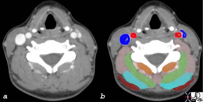
This cross sectional CT scan of the neck shows the cervical muscles which may be the source of pain. In general, the major muscles of the neck include the trapezius and the paraspinal muscles. The carotid artery (bright red) and the jugular vein (royal blue), the cervical spine (white) and the pharynx (black) may all be the source of headache. In addition, the skin of the back of the scalp could be the source.
Courtesy of: Ashley Davidoff, M.D.
Applied Anatomy: Intracranial Structures
The intracranial structures that may be sensitive to pain include:
Periosteum
Cranial nerves
Meninges
Meningeal arteries and dural sinuses
Proximal intracranial arteries
Sphenoid sinus
Thalamic nuclei
Brainstem pain – modulating centers
Intracranial Structures Associated with Headache
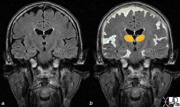
A MRI of the brain and its coverings are shown in the coronal projection above. The thalamic nuclei are overlaid in orange. The thalamic nuclei are known to be associated with some headaches. The green overlay represents the cerebrospinal fluid (CSF) in the subarachnoid space. The dura is the thickest of the meninges, and it lies superficial to the subarachnoid space, while the periosteum lies superficial to the dura. All of these structures have been associated with headaches.
Courtesy of: Ashley Davidoff, M.D.
Applied Anatomy: Vascular Structures
Several vascular structures are also sensitive to pain, like the intracranial venous sinuses and their large tributaries; the middle meningeal and superficial temporal arteries. Parts of the dura at the base of the brain and the arteries within the dura and pia-arachnoid, may also be a source of pain (6).
Pain sensation from the above mentioned structures is transmitted by the fifth (trigeminal) cranial nerve to trigeminal nuclei in the brainstem, which are largely responsible for processing pain from the face and head (7). Other nerves that can also carry painful stimuli are the facial nerve, which transmits impulses from the naso-orbital region, and the ninth and tenth cranial nerves and the first three cervical nerves, which innervate the inferior surface of the tentorium and all of the posterior fossa.
In general, pain from supratentorial structures is referred to the anterior two-thirds of the head (trigeminal nerve), and pain from infratentorial structures is referred to the back of the head and neck (upper cervical nerves)(6). The seventh, ninth, and tenth cranial nerves refer pain to the ear and naso-orbital region.

This venous phase of an angiogram in the anteroposterior projection is normal. Venous evaluation is not performed in the workup of headaches but the intracranial veins are responsible for some headaches. The exact pathogenesis remains controversial and is a source of ongoing research.
Courtesy of: Ashley Davidoff, M.D.
Applied Physiology and Pathophysiology
Several theories have been developed to try to explain the mechanisms involved in the genesis of each type of headache, but there is no general consensus and, despite being such a common condition, its pathophysiology remains poorly understood.
It used to be thought that cluster headaches and migraines were the result of an abnormal vascular reactivity, with aura of migraine being produced by vasoconstriction, and the headache being a consequence of vasodilatation. It was also believed that tension-type headaches were a consequence of increased muscle contraction in the neck and head producing vasoconstriction and ischemia. Both of these theories have now been considered insufficient to explain all the complexities involved in the genesis of the headaches.
Migraine is currently thought to have a multifactorial etiology. There is indeed a certain instability in the regulation of the vascular tone which may lead to vasodilation, and this dilatation could play a role in the throbbing head pain of the migraine, but there are also several other processes involved in the pathophysiology of migraines. There is an imbalance between excitation and inhibition of the neurons at various levels of the nervous system, each with its own consequences. At the trigeminovascular level, there seems to be an excessive release of substance P (substance known to cause pain) and other inflammatory mediators. Another important phenomenon involved is the cortical spreading depression (CSD), a self propagating wave of neuronal activation that seems to be responsible for causing the aura of migraine, and which also results in further activation of the trigeminal system, producing the release of more inflammatory mediators (3). Other mechanisms involved are beyond the scope of this work.
In the case of cluster headaches, vasodilatation appears to be secondary to neuronal dysfunction and it may be responsible for the pain and autonomic features of cluster headaches. In this case, as in migraines, dysregulation of the trigeminovascular system is likely involved in causing this vasodilatation. The autonomic symptoms of cluster headaches, such as lacrimation and pupillary constriction, appear to be due to an abnormal activation of parasympathetic and sympathetic fibers in the central nervous system. The periodicity of cluster headaches might be related to changes in the hypothalamus region of the brain, where the ?biologic clock? is located.
In the case of tension-type headaches (TTH) current theories also attribute the pain, at least in part, to abnormal vascular reactions and an increased sensitivity to painful vasodilatation compared to people that do not suffer from headaches. In this sense, TTH would be on the milder end of a “headache continuum” that has migraines on the more severe end (3).
Causes and Predisposing Factors
There are two types of headaches:
primary headaches and
secondary headaches.
Primary headaches are not associated with (caused by) other diseases.
Secondary headaches are caused by some associated disease, ranging from minor conditions to serious and life-threatening ones.
Many controversies exist in the literature regarding the nomenclature and classification of headache. The International Headache Society (IHS) developed and published a classification and diagnostic criteria which lists over 150 different types and sub-types of headaches (2). We will simplify the classification and focus on the most common types.
PRIMARY HEADACHES
The vast majority of all primary headaches fall under three categories:
-
-
- Migraine
- Tension-type
- Cluster headache
-
Causes and Predisposing Factors: Migraine Headaches
Migraine headaches are the second most common type of primary headache, affecting 18% of women and 6% of men and affecting 27.9 million people in the USA (Lipton 2001) Migraine is responsible for more than 112 million bedridden days per year, and costs American employers $13 billion per year because of missed work and decreased productivity (Hu 1999).
It is more prevalent in women than in men, and it can be divided in two major subtypes: with or without aura.
Migraine without aura is the most common subtype of migraine; it has a higher average attack frequency, and is usually more disabling than migraine with aura. It manifests in recurrent headache attacks lasting 4-72 hours. The headache is usually localized to one side of the head (it is commonly bilateral in young children), has a pulsating quality, moderate or severe intensity, and photophobia and phonophobia are common accompanying symptoms. The headache is often worsened by routine physical activity, sneezing, rapid head motion, or straining.
Migraine with aura is characterized by attacks of reversible focal neurological symptoms (aura) preceding the onset of a headache with the same features of migraine without aura.
The aura typically develops gradually over 5-20 minutes and lasts for less than one hour. It usually occurs before the onset of migraine headache, but it can also occur during or even after the headache. Auras may involve visual disturbances, sensory symptoms, speech disturbances and motor weakness.
Visual disturbances are the most common type of aura, accounting for the majority of the neurologic symptoms associated with migraine. The most characteristic visual aura of migraine is a ?scintillating scotoma? (a scotoma is an area of loss of vision), beginning as a hazy spot from the center of a visual hemifield followed by shimmering light of different patterns expanding peripherally to involve a greater part of the hemifield with scotoma.
Numbness and tingling of the lips, lower face, and fingers of one hand is the second most common type of aura. Some patients have several types of aura symptoms that vary with attacks.
Both subtypes of migraine headaches (with or without aura) may be preceded by premonitory symptoms that occur hours to two days before a migraine attack. They include various combinations of fatigue, difficulty in concentrating, neck stiffness, sensitivity to light or sound, nausea, blurred vision, yawning and pallor.
Causes and Predisposing Factors: Tension-type headaches (TTH)
Tension-type headaches (TTH) are the most common type of primary headache; as many as 80% of adults have, had or will have tension headaches and they are more prevalent among women.
They are further subdivided according to their frequency in:
-
-
- Infrequent episodic TTH (occurring once a month or less)
- Frequent episodic TTH (occurring on more than one but less than fifteen days per month)
- Chronic TTH (daily or very frequent episodes, an average of 15 or more days per month)
-
This kind of headache may feel like pressure or tightness all around the head, mild to moderate in intensity, has a tendency to wax and wane, and it often radiates to the neck muscles at the base of the skull. The pain is bilateral, non-pulsating, usually not aggravated by routine physical activity, and is devoid of typical migrainous features (e.g., nausea, vomiting, phonophobia, photophobia, and aura).
Some individuals may have overlapping features of both migraine and tension type headache. Those individuals are hard to satisfactorily label with a single diagnosis, but generally should be treated as tension type headache sufferers. The clinical features that appear to be most predictive of migraine compared with tension type headache include nausea, photophobia, phonophobia, and exacerbation by physical activity.
Cluster Headaches
Cluster headache are relatively uncommon, affecting less than one percent of the population. In contrast with migraines and TTH, an estimated 85% of cluster headache sufferers are men. Age of onset is usually 20-40 years, although headaches may begin in childhood.
They are characterized by attacks of severe, strictly unilateral pain, beginning quickly without warning and reaching maximal intensity within a few minutes. The pain is usually deep, excruciating, continuous, and explosive in quality, usually beginning in or around the eye or temple.
Attacks typically last from 15 minutes to 3 hours, occur from once every other day to 8 times a day, and are associated with one or more of the following, all of which are ipsilateral: lacrimation and redness of the eye, nasal congestion, rhinorrhea, sweating, pallor, and Horner’s syndrome (eyelid drop and constricted pupil).
Attacks usually occur in series (cluster periods) lasting for weeks or months separated by remission periods usually lasting months or years. This type of presentation, called episodic, is the most common one, but 10-15% of patients have chronic cluster headache, with attacks occurring for more than 1 year without remission or with remissions lasting less than one month.
Over 50 percent of sufferers report that alcohol is a potent precipitant of cluster headaches during a cluster bout; this sensitivity to alcohol ceases when the cluster ends (histamine and nitroglycerine are other well-known triggers). Nitroglycerin, histamine, some foods and smoking can also be precipitants of cluster headaches.
Most patients with cluster headache are restless and may pace when an attack is in progress, in contrast to migraine sufferers, who tend to rest in a dark, quiet room.
Secondary Headaches
Secondary headaches have diverse causes, ranging from serious and life-threatening conditions such as brain tumors, strokes, meningitis, and subarachnoid hemorrhages to less serious but common conditions such as withdrawal from caffeine and discontinuation of analgesics.
In contrast with the primary headaches, for most secondary headaches the characteristics of the headache itself are poorly described in the scientific literature. Even for those where it is well described, there are usually few diagnostically important features. Therefore, the correct diagnosis of secondary headaches depends on identifying the presence of a disorder known to cause headache, and a close temporal relation between the occurrence of the headache and that other disorder (or other evidence of a causal relationship). Secondary headaches are greatly reduced or resolve within three months (or less) after resolution of the causative disorder.
There are many different types of secondary headaches; we will focus on the most common ones.
Secondary Headaches : Post-traumatic Headache
Post-traumatic headache can follow direct impact to the head, or sudden acceleration and/or deceleration (whiplash injury). It can occur in different patterns, and may closely resemble primary headache disorders, most frequently tension-type headache (1).
It can be further subdivided in acute and chronic. They both occur within seven days after the head trauma, but the chronic persists for more than three months after the initial event and it?s often part of the ?post-traumatic syndrome? which includes a variety of symptoms such as equilibrium disturbance, poor concentration, decreased work ability, irritability, depressive mood, and sleep disturbances.
Post-traumatic headache can also vary in intensity, and in the most severe cases can be accompanied by loss of consciousness, mental status changes, and/or post-traumatic amnesia.
When neuroimaging techniques (e.g., CT of the head) are done after trauma, several structural abnormalities may be identified, like cerebral hematoma, subarachnoid hemorrhage, subdural hematoma, diffuse axonal lesion and skull fractures, for example.
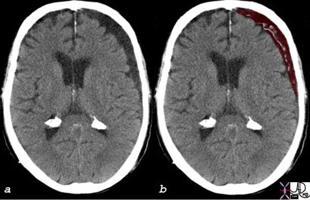
The CT scan of the head reveals a crescentic low density region in the subdural space of the left frontoparietal region and is overlaid in maroon in image b. This area represents a chronic subdural hematoma. In the acute phase it is of higher density than surrounding brain and as time progresses it becomes less dense. It is the source of headache.
Courtesy of: Ashley Davidoff, M.D.
Secondary Headaches : Medication Overuse Headache (MOH)
For the medication overuse headache, prevalence is about one percent of the population, higher in women than in men, and it may occur in patients with tension-type headache, migraine, or cluster headache. It is the consequence of an interaction between a therapeutic agent used excessively (regular overuse for more than three months) and a susceptible patient. The best example is overuse of symptomatic headache drugs causing headache in the headache-prone patient. The most common cause of chronic migraine-like headache is overuse of symptomatic migraine drugs and/or analgesics. Chronic tension-type headache is less often associated with medication overuse but it can also become chronic headache through overuse of analgesics.
This type of headache usually resolves or reverts to its previous pattern within two months after discontinuation of overused medication.
Secondary Headaches : Sinus Headache
A sinus headache is a frontal headache accompanied by pain in one or more regions of the face, developing simultaneously with an episode of acute rhinosinusitis (which may include purulence in the nasal cavity, nasal obstruction, hyposmia/anosmia and/or fever). Headache and/or facial pain resolve within seven days after remission or successful treatment of the sinusitis. Although sinus headache is commonly diagnosed by physicians and self-diagnosed by patients, many patients presenting with ?sinus headache? turn out to have migraine without aura, with headache either accompanied by prominent autonomic symptoms in the nose (rhinorrhea, nasal congestion) or triggered by nasal changes (but they don?t have purulent nasal discharge or any of the other features characteristic of acute rhinosinusitis).
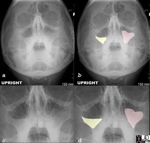
These plain film images of the sinuses show an air-fluid level in the right maxillary sinus (yellow = fluid) and opacification of the left maxillary sinus (pink). An air-fluid level represents acute sinusitis and is a common cause for headache. Total opacification may represent acute or chronic sinusitis. Images c and d are magnified views of images a and b.
Courtesy of: Ashley Davidoff, M.D.

These images reflect normal air-filled sinuses and are presented in contrast to the abnormal fluid-filled sinuses (black) in the patient described below who presented with severe headache.
Courtesy of: Ashley Davidoff, M.D.

The CT scan is from a 28-year-old male who presented with headaches. The CT scan shows easily identified pansinusitis. Image (b) (reflects an overlay of image (a) and shows light yellow total opacification of the frontal sinuses, light orange opacification of the ethmoid sinuses, and dark orange opacification of the sphenoid sinuses. Image (d) is an overlay of image (c) and shows almost total opacification of the maxillary sinuses (darkest orange) as well as the ethmoids (light orange).
Images courtesy of: Ashley Davidoff, M.D.
Secondary Headaches : Temporomandibular Dysfunction
Temporomandibular joint dysfunction syndrome (TMJ) is characterized by musculoskeletal pain with dysfunction of the masticatory system. Many patients with TMJ complain of headache and in some cases headache is its only manifestation, without the patient being aware of a TMJ.
The headache associated with TMJ is usually localized to one ear or preauricular area, and it can radiate to the jaw, temple, or neck. The pain is deep, dull, continuous, and usually worse in the morning. It typically is associated with a limitation of jaw motion and deviation of the jaw upon opening. Physical examination may reveal tenderness of the muscles of mastication and, less commonly, clicking of the joint.
Secondary Headaches : Giant Cell Arteritis
Giant cell arteritis (GCA), also known as temporal arteritis, is a chronic vasculitis of unknown etiology that commonly presents with a headache. In the US the reported incidence of GCA is approximately 15-20 cases per 100,000 people aged 50 years or older, and women are 2-4 times more likely to have GCA than men. Age is the most important risk factor for GCA, as the disease occurs mostly in patients older than 50 years, with incidence increasing with age and peaking in the eighth decade.
Headache was the most common symptom, experienced by 72% of patients at some time, and was the initial symptom in 33% (5). The head pain tends to be located over the temporal areas but can be frontal or occipital in location and the severity can range from mild to severe. Many patients report scalp tenderness on combing their hair, and tender temporal or occipital arteries are found in approximately one-third of patients.
Visual symptoms, including blurriness, double vision or loss of vision (partial or complete), are present in about a third of the patients and in as much as half of them the symptoms can become permanent. The onset of blindness from involvement of the ophthalmic artery is one of the most serious complications of GCA and it may occur without warning; about 50% are unilateral and 50% are bilateral. Newly recognized GCA should be considered a true neuro-ophthalmic emergency, as prompt treatment with steroids can prevent blindness (4).
The definitive diagnosis is usually obtained through a biopsy of the superficial temporal artery, but therapy should not be withheld pending the performance or results of the biopsy in patients with acute visual loss and high clinical suspicion for GCA.

The lateral projection angiogram of the intracerebral circulation shows spasm (white arrows) of the anterior cerebral artery. Spasm can be caused by vasculitis such as occurs in systemic lupus erythematosus (SLE). Vasculitis is a cause of headache and temporal arteritis is the most common cause.
Courtesy of: Ashley Davidoff, M.D.
Secondary Headaches : Brain Tumors
Approximately 50 percent of patients with brain tumors, either primary or metastatic, will suffer from headaches and in many of them headache is the worst symptom they have. The headaches are most frequently similar to tension type and it?s usually bifrontal but worse on the same side of the tumor.
Tumors can produce headache by two different mechanisms by:
compromising or exerting direct pressure on a pain-sensitive structure (e.g. the trigeminal nerve)
obstructing the CSF circulation causing elevated intracranial pressure
When the CSF circulation becomes impaired, headache often becomes generalized and worse in the occipitonuchal area. It?s typically described as being worse in the morning (on awakening), aggravated by coughing and straining, and often associated with nausea and vomiting (5).
Brain tumors can also present with focal neurological signs (visual, motor, sensory), altered mental status or seizures.
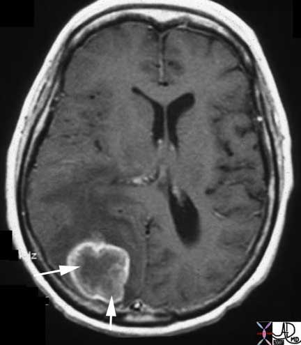
The contrast-enhanced T1-weighted MRI image above shows an enhancing mass on the surface of the posterior parietal lobe in close proximity to the meninges. It likely represents a metastasis from a known primary endometrial carcinoma and the source of headaches.
Courtesy of: Ashley Davidoff, M.D.

This 37-year-old female presented with headaches. On the T1-weighted axial MRI images before (upper images) and after contrast, (lower images) an enhancing mass (green) was pathologically shown to be lymphoma. The meninges are sensitive to pain.
Courtesy of: Ashley Davidoff, M.D.

The coronal MRI shows a gadolinium enhanced T1-weighted image (a, b) with black CSF in the dilated ventricles and a T2-weighted image with bright CSF (c, d). A large mass in the third ventricle (overlaid in green) dominates the study and is complicated by severe hydrocephalus. The mass represented a choroid plexus papilloma of the third ventricle. In patients with brain tumor one of the causes of headache is hydrocephalus. This is an extreme case of hydrocephalus.
Courtesy of: Ashley Davidoff, M.D.
Secondary Headaches : Elevated Intracranial Pressure
Elevated intracranial pressure (ICP) can occur in different settings including the presence of a brain tumor (as we mentioned above), trauma, impaired CSF circulation, impaired venous outflow from the brain, and hepatic encephalopathy.
Elevated ICP headache is characteristically worsened by straining or coughing and is usually accompanied by vomiting. Physical exam may reveal depressed global consciousness, focal neurological signs, papilledema, and in severe cases, a triad of bradycardia, respiratory depression, and hypertension known as Cushing’s triad (ominous sign).
Idiopathic intracranial hypertension (i.e., pseudotumor cerebri) is a rare condition that can occur in young, overweight women, or sometimes in patients taking certain medications like lithium, oral contraceptives, or large doses of vitamin A. It is an elevation of the intracranial pressure of unknown mechanism, usually presenting with a pulsatile headache, pulsatile tinnitus, associated with nausea or vomiting, and sometimes retroocular pain worsened by eye movement. Papilledema is usually found on funduscopic examination and the patients may have visual blurring which may become permanent if prompt therapy is not instituted.
Another rare cause of elevated ICP is cerebral venous thrombosis (CVT). The thrombosis can be localized to cerebral veins, interfering with the venous outflow of the brain, or to the dural sinuses, resulting in decreased CSF absorption. Headache is the most frequent presentation of CVT, and it?s usually of gradual onset, but can also be of explosive characteristics (known as thunderclap headache), similar to the subarachnoid hemorrhage (SAH) pain.
Secondary Headaches : Meningitis
Meningitis is an inflammation of the leptomeninges and underlying subarachnoid cerebrospinal fluid (CSF), and its incidence is 2-3 cases per 100,000 people. Meningitis and meningoencephalitis both have headache as a major symptom. Pain is often severe, retro-orbital and worsened by moving the eyes.
Headache is a key symptom of meningeal syndrome or meningism consisting usually of headache, neck stiffness and photophobia. Other classic symptoms and signs of meningitis are fever and chills, vomiting, seizures (30-40% in children, 20-30% in adults), focal neurologic symptoms, and altered sensorium (confusion may be sole presenting complaint, especially in elderly).
Meningitis has a high morbidity and mortality and it?s crucial to recognize meningitis on initial presentation and to begin antibiotic treatment as soon as possible (within the first 30 minutes in the Emergency Department or ED), for any delay in instituting treatment may lead to permanent neurological sequelae or death.
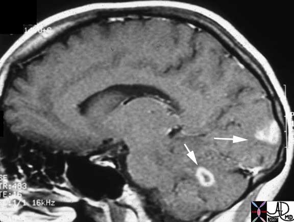
This T1-weighted MRI study with gadolinium shows two enhancing lesions. The more superior lesion is in the occipital lobe and the inferior lesion is in the cerebellum. They represent a cerebral infection with toxoplasmosis. Cerebral toxoplasmosis usually presents with persistent headache, sometimes associated with focal signs, change in mental status or seizures.
Courtesy of: Ashley Davidoff, M.D.
Secondary Headaches : Headache Associated with Refractive Errors
Eye strain: Patients frequently attribute headaches to eye strain, and the International Headache Society (IHS) recognizes headaches associated with refractive errors (HARE). Headaches are only rarely due to refractive error alone, but correcting vision may improve headache symptoms in some of these patients.
HARE presents as a recurrent mild headache, frontal and in the eyes themselves, which develops in close temporal relation to the refractive error, is absent on awakening and aggravated by prolonged visual tasks at the distance or angle where vision is impaired.
Headache and eye pain resolve within 7 days, and do not recur, after full correction of the refractive error (2).
Secondary Headaches : Arterial Hypertension
There is a common belief, particularly among patients, that hypertension can cause headaches. While this is true in the case of hypertensive emergencies, it is probably not true for typical migraine or tension headaches. Mild (140-159/90-99 mm Hg) or moderate (160-179/100-109 mm Hg) chronic arterial hypertension does not appear to cause headache. Whether moderate hypertension predisposes to headache at all remains controversial, but there is little evidence that it does. Ambulatory blood pressure monitoring in patients with mild and moderate hypertension has shown no convincing relationship between blood pressure fluctuations over a 24-hour period and presence or absence of headache (2).
Secondary Headaches : Subarachnoid Hemorrhage
Subarachnoid hemorrhage (SAH) is most commonly caused by a ruptured intracranial aneurysm, which produces a severe headache of sudden onset that patients often characterize as “the worst headache of their life.” Stiff neck is frequently found on physical examination and a decreased level of consciousness may also be noticed. Up to 60% of the patients may experience a “sentinel headache” hours to days prior to the SAH. The sentinel headache typically presents as a severe headache lasting for only a few minutes without associated neurologic abnormalities and it is caused by a minor leak from the aneurysm prior to its rupture.
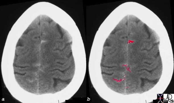
In the axial images of the brain using CT, there are subtle accumulations of high density material outlined in red in image b, representing acute subarachnoid hemorrhage. In the patient who presents with a severe acute headache, this can be a life-threatening rupture of a cerebral berry aneurysm.
Courtesy of: Ashley Davidoff, M.D.

The CT scan on the right (b, d) is from a 52-year-old male who presented to the ER with severe acute headache associated with neurological deficit. When the images are compared with a normal patient at approximately the same level, the hyperdense acute blood (pale white areas with arrowheads) can be readily seen in the sulci, (b) and around the midbrain (d ? midbrain is shaped like Mickey Mouse) with subtle accumulation in the fovea of the cerebellum. A ruptured berry aneurysm at the tip of the basilar artery was suspected.
Courtesy of: Ashley Davidoff, M.D.
Secondary Headaches : Ruptured Berry Aneurysm
The “worst headache of my life” sends shivers down the caregiver?s back, since if the headache is truly caused by a subarachnoid bleed then the patient has a 50-50 chance of coming out alive or without significant deficit.
The aneurysm is characterized by focal dilatation or sacculation of one or more of the intracranial arteries of the brain. The cause is multifactorial including congenital weakness of the wall, and acquired factors such as hypertension and turbulence at the branch points.
The focal outpouching may be complicated by rupture.
Structurally berry aneurysms, are more common in the anterior circulation, are multicentric in 20-30% of patients, and commonly occur at branch points.
Functionally, only the giant aneurysms (greater than 2.5cms) will affect function by compressing on other structures.
The risk of rupture is the most feared complication of berry aneurysms and it is the most common cause of spontaneous subarachnoid bleeding. The aneurysm may also rupture into the brain substance, into the ventricles, or into the subdural space. About 60% of patients will have a catastrophic outcome either succumbing or having a permanent significant deficit and the remaining 40% will have a relatively favorable outcome.
Clinically, the smaller aneurysms are asymptomatic. When they rupture the presentation is dramatic with the acute onset of a headache which classically is described as the most severe headache ever experienced by the patient.
The diagnosis is documented by finding acute bleed most commonly in the subarachnoid space, characterized by hyperdense blood in the sulci on a non-contrast CT. The bleed may occur or extend into the cerebral substance, or into the ventricles or subdural space. An angiogram is required to accurately localize the aneurysm and plan therapy.
Treatment options include transcatheter embolization or surgical clipping at the base of the aneurysm.
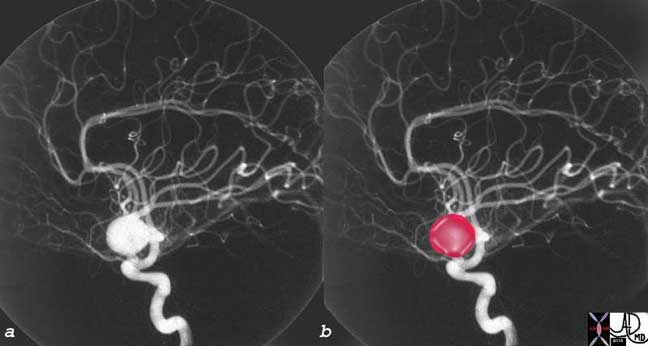
The large, rounded, contrast-filled structure overlaid in red in b, represents a berry aneurysm of the anterior communicating artery noted on this angiogram of the left internal carotid artery.
Courtesy of: Ashley Davidoff, M.D.
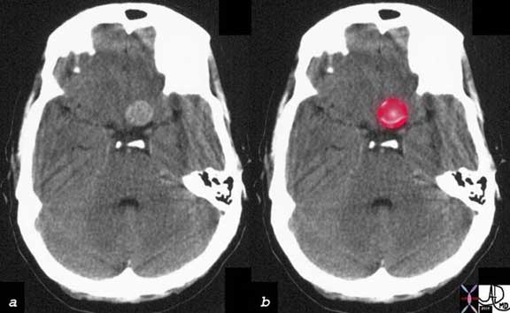
The large, rounded, dense structure overlaid in red in b, represents a berry aneurysm of the anterior communicating artery noted on this CT and correlating with the angiogram above.
Courtesy of: Ashley Davidoff, M.D.
Clinical Presentation of Headaches
A careful history and physical examination remain the most important part of the assessment of the headache patient. A thorough history is the single most important factor in establishing a headache diagnosis and determining the future work-up and treatment plan.
History and Precipitating Factors
Migraines may be precipitated by bright light, menstruation, weather changes, caffeine withdrawal, sleeping longer or less than usual and ingested substances such as alcohol or certain foods. If bending, lifting, coughing, or Valsalva?s maneuver produces a headache, an intracranial lesion must be considered; however, most exertional and cough headaches are benign. Alcohol is often a potent precipitant of cluster headaches.
Duration
Migraine pain usually lasts 6 to 36 hours, whereas cluster headache characteristically lasts 45-120 minutes. Tension-type headaches commonly build up over hours and may last days to years.
Onset
Cluster headaches often awaken patients from a sound sleep and have a tendency to occur at the same time each day in a given person. Migraines can occur at any time during the day or night but usually begins in the morning. A headache of recent onset that disturbs sleep or is worse on waking may be caused by increased intracranial pressure. TTH are typically present during much of the day and are often more severe late in the day.
Headache of instantaneous onset suggest an intracranial hemorrhage, usually in the subarachnoid space.
Character
Migraine often has a pulsating quality. Cluster headache is characteristically severe, boring, steady, and it has a certain frequency and periodicity of occurrence which helps to identify it. TTH are usually described as a feeling of fullness, tightness, or pressure, or as being like a cap, band, or vice.
Situation
Migraine is most often unilateral, commonly in the frontotemporal region, but it may be generalized. Cluster headaches are virtually always unilateral during an attack and are typically centered in, behind, or about the eye. A typical TTH is generalized, although it may originate across the nuchal muscles only to spread and perhaps predominate in the frontal or occipital regions.
Severity
Migraine pain usually peaks within 1-2 hours of onset and is of moderate to severe intensity. Cluster headache is typically maximal immediately if the patient awakens with the headache in progress or peaks within a few minutes if it begins during wakefulness, but in either situation it is usually very severe and excruciating.
A new sudden, severe headache that is maximal at onset suggests intracranial hemorrhage, cerebral venous sinus thrombosis, or pituitary apoplexy.
Aggravating Factors
For migraine headaches the most common aggravating factors are straining, bending over, bright lights, and certain smells. One of the classical criteria for the distinction between migraines and TTH is that physical activity, light, and noise, are frequent aggravating factors for migraines but not for TTH; but some recent studies have shown that those factors can also aggravate the tension-type headache in as many as 50% of the cases.
For clusters, though there are specific triggers in the form of vasodilators that cause cluster headaches, the aggravating factors are highly individualized.
In patients with increased intracranial pressure (e.g. brain tumors) headache may worsen with bending over, and worsening headache may also follow maneuvers that raise intrathoracic pressure, such as coughing, sneezing, or the Valsalva maneuver.
Relieving Factors
Rest, especially sleep and avoidance of light and noise tends to provide relief to the migraine sufferer.
Massage or heat may alleviate the pain associated with a TTH.
Local application of pressure over the affected eye or ipsilateral temporal artery, and local application of heat or cold, may ameliorate the pain of cluster headache (5). =
Associated symptoms
Migraines, as we described above, can be associated with several neurological symptoms (aura). Also, physiologic data suggest that muscle tension may be at least as likely to occur in migraine as it is in tension-type headache. More specifically, neck pain recently has been demonstrated to be quite common not only in TTH but also in migraine, and that?s why the presence of neck pain should not automatically trigger a diagnosis of tension-type headache.
SAH can also have neck and back pain, but its explosive characteristics helps distinguishing it from other causes. It frequently presents with vomiting and altered mental status (including deep coma).
Cluster headache is commonly associated with some ipsilateral signs, like lacrimation and redness of the eye, nasal congestion, sweating, pallor, and Horner’s syndrome.
Nausea or vomiting is commonly present in patients with brain tumors. Other associated signs can be focal neurological findings, visual changes, and seizures.
Meningitis is usually associated with nausea, photophobia and/or phonophobia, neck stiffness, and change in mental status.
Quiz Me
Which type of headache may be precipitated by bright light, menstruation, weather changes, caffeine withdrawal, sleeping longer or less than usual and ingested substances such as alcohol or certain foods.
Tension-type
Migraine
Cluster
Massage or heat may alleviate the pain associated with a ________.
(Note: You will be given 2 tries to answer this question, then the answer will be provided.)
Tension-type headaches
Migraine headaches
Cluster headaches
Post-traumatic headaches
Physical Examination
In the patient with headache, the physical exam is often unrevealing. However, some findings, if present, can yield important clues about the underlying cause.
The examination should begin by taking the vital signs, and then assessing the general status of the patient, determining whether the patient looks ill, toxic, anxious or depressed, and if the patient is oriented or confused. It is extremely important to perform a neurological examination, including examination of the mental status, gait, cranial nerves, motor system, and sensory system. A funduscopic exam should also be done to look for signs of papilledema.
The skull and cervical spine should also be examined. The skull should be palpated for lumps and local tenderness. Thickened, tender, irregular temporal arteries with an associated reduction in pulse suggest GCA.
In TTH the scalp muscles may be tender.
In a patient that presents with altered mental status, the exam has to include looking for any signs of head trauma.
Nuchal rigidity on passive neck flexion and Kernig?s sign (flexing the patient?s hip 90 degrees and then extending the patient?s knee causes pain) are evidence of meningeal irritation.
Labs
When, based on the history and physical examination, the patient is clearly suffering from a primary headache, there is usually no need for any specific workup. If routine blood tests are performed in patients with primary headaches, the results should be unremarkable. Specific tests should be obtained if there is a clinical suspicion of underlying conditions (secondary headaches). In these situations, a complete blood count (CBC), electrolytes, glucose, blood urea nitrogen (BUN) and creatinine, erythrocyte sedimentation rate (ESR) and C-reactive protein (CRP) can be checked. The CBC can be abnormal in inflammatory conditions (like GCA) and in infections (like meningitis). The ESR and CRP are usually elevated in cases of GCA. A blood gas analysis with a cooximeter can detect toxic levels of carbon monoxide (cause of headache during winter time mainly).
Cerebrospinal fluid (CSF) examination obtained through a spinal tap (also known as lumbar puncture) is vital for the diagnosis of meningitis, and can also aid in the diagnostic evaluation of encephalitis, SAH, and certain types of malignancies that can affect the CSF. If the patient has altered mental status, or papilledema in the funduscopic exam, or any focal neurological signs, a CT of the head should be obtained prior to performing a spinal tap to exclude the presence of intracranial hypertension, a spinal tap performed in that setting could lead to severe damage or death. A spinal tap also allows for the measurement of the CSF pressure, which is important given the fact that either elevated or decreased pressures can produce headache. Ironically, the lumbar puncture itself can produce a headache secondary to decreased CSF pressure after the procedure, which usually resolves after a few hours or days with bed rest and good hydration, but can also, in rare occasions, persist for several weeks or even months (5).
Electroencephalography (EEG) is not usually part of the work-up for headache, except in the patients with a history of seizures, syncope, or episodes of altered awareness.
Imaging
Neuroimaging studies are not to be ordered indiscriminately in every headache patient, but they can prove to be extremely useful in certain clinical situations. Any recent significant change in the pattern, frequency or severity of headaches (especially if the headache is getting worse despite appropriate therapy), or the presence of focal neurological signs or symptoms, should include neuroimaging as part of the work-up. Patients with an orbital bruit, otorrhagia, or leaking of clear fluid through the nose after head trauma, as well as patients that have onset of headache with exertion, cough, or sexual activity, or onset of headache after age 40 years, also need to undergo head imaging.
Tumors, hematomas, cerebral infarctions, abscesses, and many other processes can be identified with computed tomographic (CT) scanning and magnetic resonance imaging (MRI) 5. The pituitary gland, the craniocervical junction, the cervical spinal cord and exiting nerve roots and the white matter of the brain are better evaluated by MRI. CT can be helpful for evaluating abnormalities of the skull, orbit, sinuses, facial bones, cervical spine, and recent subarachnoid hemorrhage. A non-contrast CT of the head should be the first study done in a patient in which an intracranial bleed is suspected.
As a general rule, CT is the optimal imaging study for evaluating a patient with acute onset of headache. MRI might provide a better evaluation in cases of subacute and chronic headache, or when posterior fossa or vascular lesions are suspected; but even in those cases, CT is usually the first imaging study obtained.
Quiz Me
It is important that a neuroimaging test is ordered when patients present to the ER with a headache.
True False
Potential Complications
Some secondary headaches can lead to severe complications if the underlying condition is not controlled with therapy. Post-traumatic headache might be caused by different underlying conditions, and some of them, like a hematoma (cerebral, subdural, or extradural) or diffuse axonal injury, can lead to permanent neurological damage or death. Any cause of increased intracranial pressure can also lead to great neurological damage or death if it causes compression or displacement of different parts of the nervous system.
Giant cell arteritis, as well as pseudotumor cerebri, can produce permanent visual loss if not treated. Hypertensive emergencies can lead to a stroke or to cardiovascular damage if not treated, and meningitis can cause permanent neurological sequelae, or death, if not promptly treated.
Management
Therapy for migraines can be divided in two main groups: prophylaxis and treatment of the acute attacks.
Prophylactic agents are indicated in patients with frequent migraines that significantly interfere with their daily routine despite acute treatment. Several agents can, in various degrees, reduce the frequency and intensity of migraine attacks. Several antihypertensive agents (e.g.: metoprolol, verapamil, enalapril), some antidepressants (e.g.: amitriptyline, mirtazapine), anticonvulsants like valproate, gabapentin, and topiramate, as well as some non-steroidal anti-inflammatory drugs (NSAIDs) like naproxen, can all be used for prophylaxis. About 50 to 75% of patients given these drugs will have a 50% reduction in the frequency of migraine attacks (3). Alternative techniques like herbal medicine, acupuncture, relaxation techniques, biofeedback, and behavioral therapy may provide benefit in some patients and can be combined with the prophylactic agents mentioned before.
There are many different agents that can be used for the abortive treatment of acute migraine headaches. All NSAIDs (e.g.: ibuprofen, naproxen, diclofenac, aspirin), as well as acetaminophen, may be beneficial in some patients but they are probably not enough, if used as sole agents, to abort moderate to severe attacks.
A group of drugs often used for acute migraines is the Triptans (e.g.: sumatriptan, zolmitriptan, naratriptan). They are usually very effective, but the multiple drug interactions that may occur with their use, as well as the multiple contraindications for their use (e.g.: ischemic cardiovascular disease, uncontrolled hypertension, pregnancy), preclude their use in some patients.
Another group of agents used for abortive therapy is the Ergots (e.g.: ergotamine, dihydroergotamine), and their use is also limited by multiple contraindications (coronary artery disease, peripheral vascular disease, hypertension, and renal or hepatic disease).
Some antiemetics, like metoclopramide, are also effective, and they are frequently used in combination with other agents (e.g.: dihydroergotamine). They are especially useful in patients that present with significant nausea and vomiting.
For cluster headaches there is also prophylactic and abortive therapy. Inhalation of 100% oxygen by non rebreathing masks is a very effective abortive therapy for acute clusters, producing relief in approximately 75% of the cases after 20 minutes of therapy. Subcutaneous sumatriptan is also effective in aborting acute clusters, with onset of action and efficacy rates similar to oxygen therapy. Sumatriptan may also be given intranasal, being equally effective but slower to produce a response. Therapy with 100% oxygen has no contraindications (except maybe COPD patients who retain carbon dioxide), whereas sumatriptan is contraindicated in patients with ischemic cardiovascular disease or uncontrolled hypertension because of its vasoconstrictive properties. Besides abortive therapy, prophylactic therapy should also be started as soon as possible at the onset of a cluster episode. Several drugs have been found to reduce attack frequency and analgesic consumption, like verapamil, prednisone, and lithium.
Patients that do not respond to medications may benefit from more radical and aggressive therapies, like complete or partial section of the trigeminal nerve or greater occipital nerve blockade. Deep brain stimulation of the posterior hypothalamic gray matter is an experimental method that has also been effective in some patients with intractable cluster headache (3).
For episodic tension-type headaches, simple analgesics such as acetaminophen or ibuprofen are the drugs of choice for abortive therapy. If the patient is suffering from chronic TTH, prophylactic therapy might be indicated to reduce the frequency or severity of the episodes. Tricyclic antidepressants, or other antidepressants like serotonin blockers, are considered to be effective in preventing chronic headaches, and are the most commonly used agents for prophylaxis. Alternative techniques that may provide some additional benefit to chronic TTH sufferers are biofeedback, relaxation therapy, psychotherapy, and physical therapy, either alone or in combination with antidepressant medications (3).
Quiz Me
A group of drugs often used for acute migraines is the Triptans (e.g.: sumatriptan, zolmitriptan, naratriptan).
True False
Red Flags
There are several important factors that can help to differentiate the small number of patients with life-threatening headaches from the overwhelming majority with benign primary headaches. Failure to recognize these alarm signs can have potentially fatal consequences.
Sudden onset of headache, or severe persistent headache that reaches maximal intensity within a few seconds or minutes after the onset of pain could be caused by a subarachnoid hemorrhage, for example, which often presents with sudden onset of excruciating pain. Cluster headache may sometimes be confused with a serious headache, since the pain can reach full intensity within minutes, but its typical features allows for the differentiation.
The absence of similar headaches in the past is another finding that suggests a possible serious disorder. The “first” or “worst” headache of my life is a description that sometimes accompanies an intracranial hemorrhage or central nervous system infection.
A worsening pattern of headache suggests a mass lesion, subdural hematoma, or medication overuse headache. The presence of nausea, vomiting, increased headache with changes in body position (particularly bending over), and an abnormal neurologic examination suggest the headache was caused by a tumor.
Any new focal or nonfocal neurologic abnormality warrants further evaluation. The findings may be quite subtle, such as slight pupillary asymmetry, unilateral pronator drift, or extensor plantar response; or may be pronounced, such as unilateral vision loss, ataxia, or seizure. Focal neurologic findings can accompany a number of secondary causes of headache, including intracranial hemorrhage, acute narrow angle glaucoma, and carotid or vertebral artery dissection. Nonfocal alterations in mental status more commonly characterize other secondary causes of headache, including subarachnoid hemorrhage, meningitis, and toxins such as carbon monoxide.
Fever associated with severe headache may be caused by intracranial infection (e.g., meningitis).
Change in mental status, personality, or fluctuation in the level of consciousness suggests a potentially serious abnormality. Obtundation and confusion increase the likelihood of meningitis, encephalitis, subarachnoid hemorrhage (SAH), or other space-occupying lesion.
Head pain that spreads into the lower neck and between the shoulders may indicate meningeal irritation due to either infection or subarachnoid blood; it is not typical of a benign process.
Patients over 50 years of age with new onset or progressively worsening headache are at significantly greater risk of a dangerous cause of their symptoms, including an intracranial mass lesion and temporal arteritis.
Meningismus (e.g., stiff neck) may indicate meningitis or SAH. This is less sensitive and less specific in adults older than 60 years.
Headache presenting with fever and petechial or purpuric rash is concerning for the presence of bacterial meningitis.
Some ophthalmologic findings, when present, should raise the possibility of a serious condition. Papilledema (blurring of the optic disks seen during fundoscopic exam) is indicative of increased intracranial pressure, possibly due to a tumor. Loss of vision can be seen with temporal arteritis, optic neuritis, carotid artery dissection, or in acute narrow angle glaucoma.
Also, a new headache type in a patient with cancer suggests metastasis, and warrants further work-up.
Patient Information
The vast majority of headaches are of benign nature and do not pose any risk for the patients, but in some cases headaches can be a manifestation of an underlying condition that, if unrecognized, may lead to dire consequences.
Seek emergency care if:
-
-
- Sudden onset of significant headache (?thunderclap? headache).
- It is the ?first? or ?worst headache of your life? .
- Changes in mental status (e.g., altered consciousness, personality changes) or seizures.
- New headache with fever, stiff neck, skin rash.
- A new headache associated with neurologic symptoms (e.g.,weakness, numbness, impaired vision). While migraine headache can sometimes cause these symptoms, a person should be evaluated urgently the first time these symptoms appear (3).
-
See a doctor if there is:
-
- Persistent, or very frequent headache.
- Worsening pattern of headache, or headache that interferes with the person?s daily activities.
- Presence of nausea, vomiting, worsening of pain with bending over, straining or coughing.
References
1: Neurology in Clinical Practice, Walter G. Bradley et al, 4th edition, 2004.
2: The international classification of headache disorders; Cephalalgia, Volume 24 Supplement 1, 2004.
3: Up-To-Date online, accessed at www.uptodate.com
4: Manolette R Roque, eMedicine article on Giant cell arteritis.
5: Neurology in Clinical Practice, Walter G. Bradley et al, 4th edition, 2004.
6: Principles of Neurology, 8th edition, Allan H. Ropper and Robert H. Brown.
7: Hu XH, Markson LE, Lipton RB, Stewart WF, Berger ML. Burden of migraine in
the United States: disability and economic costs. Arch Intern Med.
1999;159(8):813-818.
8: Lipton RB, Stewart WF, Diamond S, Diamond M, Reed ML. Prevalence and
burden of migraine in the United States: results from the American Migraine
Study II. Headache. 2001;41: 646-657.
Web References
Useful information for the patients on this topic can be found on these websites:
? www.nlm.nih.gov/medlineplus/healthtopics.html
Levin Morris The Many Causes of Headaches Postgraduate Medicine Volume 112: No.6 December 2002

