Learning Objectives
- define liver carcinoma and discuss the structural and functional changes seen with this type of cancer
- describe hepatocellular carcinoma including its prevalence, cause and predisposing factors
- review the various imaging modalities utilized for the patient with liver cancer
- discuss the most common treatment modalities for a patient with liver cancer
Definition
Liver cancer is an aggressive growth disorder initiated by a cell or a group of renegade cells, either as a primary event in the liver, but commonly as a metastatic disease. As a primary event, the usual causes include spontaneous genetic aberration, a carcinogen in the environment, or less commonly by an inherited genetic abnormality.
It is the most common primary hepatic malignancy and one of the most common malignancies worldwide.
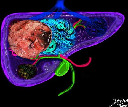
The liver is a vital structure and is considered the metabolic warehouse of the body. The image reflects its 24/7 clockwork nature, processing, churning and producing products for ongoing metabolism, but in this case distorted by a space occupying aggressive tumor throwing a “spanner” in the wheels of production.
Courtesy of: Ashley Davidoff, M.D.
Introduction
The structural changes are characterized by proliferation and space occupation of non functioning tissue, with a variety of macroscopic morphologies, but often characterized at a cellular level by large hyperchromatic nucleii with diminished cytoplasm.
Functionally, the rebel cells and cell group parasitize nutrition and oxygen, and do not contribute to the overall function of the mother organ, in this case the liver, or to the community at large.
The disease is complicated by local spread within the organ and surrounding tissues, displacing well-meaning and well-functioning tissue. Continued uncontrolled growth results in the invasion of blood vessels and lymphatics and spread to distant organs where metastatic disease repeats the pattern of continued advance of rebellious parasitization on normal tissue. Other local complications include bleeding, necrosis, and obstruction. Systemic and non specific complications include fatigue, weight loss, night sweats and pain.
The diagnosis is suspected clinically when a patient presents with unexplained weight loss, or a new mass is felt on clinical examination.
Imaging characteristics include finding a mass, often multiple and multicentric, characteristically round or infiltrating, that shows evidence of local invasion and/or metastatic disease. When cancer is suspected, microscopic confirmation is almost universally indicated to confirm the diagnosis, and to evaluate and classify the type and virulence of the cancer. Types of biopsy include aspiration technique where individual cells are sucked up into a needle, core biopsy, where a cutting needle provides a small sample of tissue, and incisional biopsy, where a part of a lump or a sample is removed using a scalpel.
Staging the disease is essential in the diagnostic workup since treatment plans depend on the extent and location of the disease.
Treatment options depend on staging and type of the cancer. In the early stages of primary liver cancer, the goal of treatment is cure and may include surgical resection. In advanced stages, the role of treatment is control of disease and prolongation of life. This may include surgery, chemotherapy and radiation. Recent advances include localized chemical ablation with alcohol, for example, or thermal or electrical ablation.

The gross pathology specimen shows malignant tissue (white) occupying at least 80% of the normal liver parenchyma (red). The liver has remarkable tolerance for space occupation before showing ill effects, and also has remarkable regenerative capabilities. In this instance the disease was caused by metastatic disease. Even the liver in this instance could not tolerate the degree of invasion and the patient finally succumbed to the disease.
Courtesy of: Ashley Davidoff, M.D.
Hepatic cancer has varied origins including those that arise primarily from liver elements and those that are metastatic deposits. The tumors that arise from liver elements include:
hepatocellular carcinoma
cholangiocarcinoma
hepatoblastoma
liver sarcoma
epithelioid hemangioendothelioma
Metastatic disease most commonly originates from a primary colon, lung, breast or pancreatic cancer, and less commonly from less common tumors such as gastric duodenal, carcinoid malignancies. Capsular metastases usually originate from ovarian carcinoma.
Structural Principles of Liver Cancer
Liver cancer arises from a single cell, that for whatever reason assumes a different and aggressive profile with an agenda and a time cycle that does not conform to the norm, but has the ability to generate new cells of a similarly aggressive nature.
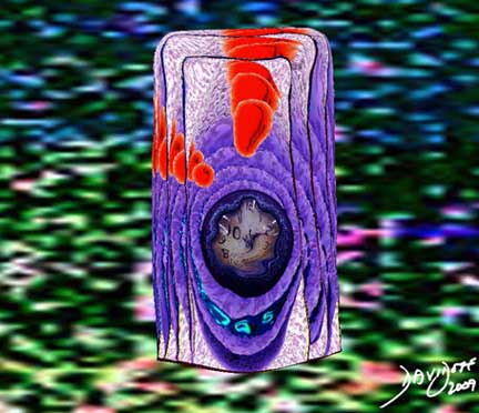
The image represents the life of a single set of columnar cells showing a progression of generations as the cell life dies and is regenerated. The orange secretions of the cell are seen in the background of the pink cytoplasm and the purple nucleus. The nucleus of the newest generation and cell is seen as a clock that has become distorted causing time to become disordered. This is the forerunner of a malignant process.
The abnormal cells also characteristically acquire the capability to invade surrounding structures as well as an ability to spread to distant organs through the bloodstream or lymphatics.
The malignant population of cells compete for nutritional resources and space.
Courtesy Ashley Davidoff MD
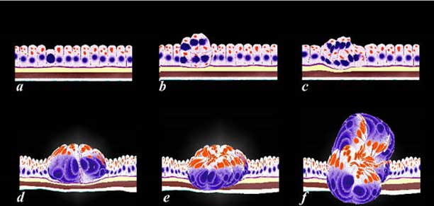
The image represents the evolution of a single cancer cell (a) that fails to conform to the normal time cycle. In image (a), the normal columnar mucosa is composed of rectangular cells supported by a thin purple basement membrane. One cell becomes aberrant, and is characterized by a large blue nucleus, scanty cytoplasm and a change in its shape and overall appearance. The cell grows, multiplies, (b, c, d) and then invades the space of other parts of the tissue like the submucosa (yellow) and muscular layer (maroon) in (e) and finally the serosal layer in (f) (outer white layer).
Courtesy Ashley Davidoff MD
The structure of the liver cell and its organization as a tissue is quite unique so that although the end result of space occupation and uncontrolled proliferation are common to other cancers, it is unique in many of its structural consequences as a result of unique architecture.
Structural Principles of Liver Cancer: Normal Histology and the Cells
The parenchyma of the liver is a glandular epithelium and the cells are aligned as plates or cords, radially arranged and separated by hepatic sinusoids, making up a portal lobule and bounded by portal triads (radicles of the portal venous, hepatic arterial and bile duct systems).
The Cells
The dominant cell of the liver is the hepatocyte. The hepatocytes constitute approximately 70% of the liver by volume. Most of the rest of the cells of the liver derive from the blood cells flowing through the liver and a small proportion represents the Kupffer cells and the cells lining the vessels. It is therefore not surprising that most of the primary liver cancers arise from the hepatocyte.
The hepatocyte is structurally characterized by its large size measuring between 20-30 microns, and the absence of a basement membrane is functionally characterized by its remarkable metabolic and regenerative capability. The cell characteristically responds to injury, whatever its type, by accumulating fat in the cytoplasm, an entity called steatosis.

The cell is the building block of all biological structure. In this image a few, almost hexagonal cells of the liver, are attached together. Each cell has a central dark nucleus which is embedded in a pinkish cytoplasm. The nucleus takes up approximately 1/5 to 1/6 of the volume of the cell and usually lies in the center of the cell. Binucleate cells are normal and account for up to 25% of the normal cell population. There is also considerable variation in the size of the nucleus from cell to cell. The red cells seen in the background are about 5 microns, and the liver cell is about 4 to 5 times their size.
Image courtesy of: Barbara Banner, M.D.
Structural Principles of Liver Cancer: The Tissue
The liver is a compound tubular, serous gland. The cells are aligned in cord formation that branch and anastomose alongside and in parallel with a vascular system called the sinusoids. These plates are arranged like spokes of a wheel around a central vein, which is a tributary of the hepatic veins. Kupffer cells are found within the space of Disse, between the plates of cells and the sinusoids. They have macrophage function and act as a defense against foreign material and organisms that may be absorbed into the mesenteric circulation from the gastrointestinal tract.

The cells of the liver are organized in cords and plates and are distributed like spokes of a wheel around the central vein. The plates and cords (red overlay in b) are lined by the sinusoids (white channels between the cords) which are the vessels which carry blood to and through the liver. Just below the sinusoids, between the wall of the sinusoid and the capsule of the liver, there is a space called the space of Disse which carries the lymphatic fluid of the liver. Spindle shaped cells, that are macrophages called Kupffer cells, (ringed in blue) line the sinusoids and are related and integrated into the space of Disse. The intimate relationship of the cells with the blood vessels and their characteristic involvement has relevance to when the cells undergo malignant change.
The liver lobule is a structural and functional unit of the liver. At its center is the central vein and it is bounded by several portal triads consisting of a hepatic arterial tributary, a portal venous branch, and a biliary radicle. The hepatic arteriole and portal venule bring blood to the lobule and join to form the sinusoid that runs between the plates of hepatocytes. The biliary radicle takes bile away from the smaller branches of bile ductules that lie between the hepatocytes. The lobule is polyhedral in shape and has a wheel-like formation as described above, with spokes of hepatocytes and sinusoids radially positioned around the central vein, and supported by connective tissue. It is functionally characterized by its ability to synthesize metabolically active substances and also by its ability to break down products (catabolic activity), and by its ability to participate in defense mechanisms and filtering mechanisms of the body.
The primary blood flow to the liver is via the portal vein which receives mesenteric blood flow, which enters the lobules as the portal vein radicle in the portal triad. The radicles of the hepatic artery bring oxygenated blood to the liver, and bile ducts take bile to the duodenum via the gallbladder and common bile duct, to facilitate fat digestion. Metastatic deposits that are hematogenously spread are therefore brought to the liver as a first stop, if they are transported by the mesenteric veins. It is therefore no surprise that the liver is the most common site for metastatic, colon carcinoma.
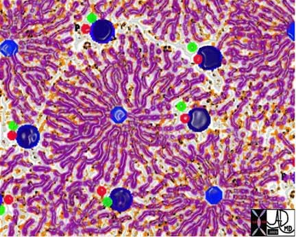
The sinusoids and hepatic cords combine to form a liver lobule, which is a functional and structural unit of the liver. At the center of the lobule is the central vein (royal blue) from which emanate many cords of liver tissue. At the periphery of the lobule there are 4-5 groups of portal triads, consisting of distal branches of the portal vein (dark blue), hepatic artery (red) and biliary radicle (green). They create the border of the lobule.
Courtesy of: Ashley Davidoff, M.D.
Structural Principles of Liver Cancer: Tissue Continued
There are three zones within a liver lobule.
Zone 1 is closest to the periphery of the lobule and portal triad.
Zone 2 is the intermediate zone.
Zone 3 is in the center of the lobule abutting the terminal hepatic venule.
Zone 3 is the most vulnerable since it is furthest away from the oxygenated blood.
Each lobule measures 1-2mm in size, is hexagonal in shape, is metabolically very active, and has tremendous regenerative capacity. The cells are exposed to gastrointestinal products of digestion that are absorbed into the blood and transported to the liver by the superior mesenteric vein, which empties into the portal vein. Alcohol, for example, is transported to the liver via this system and when taken in excess for long periods, exposes the liver cells to its toxic effects, causing cirrhosis and eventually, in some patients, resulting in dysplastic and finally malignant, neoplastic change.
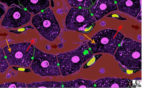
The liver cells (purple) are aligned in plates, with bile canaliculi (green) running between them.· The Kupffer cells (yellow nuclei and black cytoplasm) run alongside the hepatocytes, and between the sinusoids (maroon) and the liver cells.· As the blood enters the sinusoids (maroon arrow), the cells absorb the products of digestion (yellow arrow), metabolize the products, and then export the new product back into the blood (orange arrow).· The Kupffer cell acts as a macrophage and engulfs any foreign material, organisms, or “debris” in the blood and acts as a defense mechanism.
Courtesy of: Ashley Davidoff, M.D.
The sinusoids terminate at the central vein, each a part of the liver lobule. They converge with other central veins to form the three major hepatic veins, which transport the nutritionally rich blood to the right atrium via the inferior vena cava.
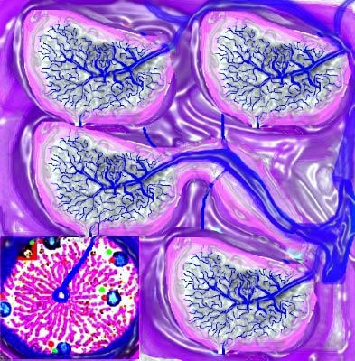
Lobules There are a multiplicity of lobules, each with a central vein and each delivering the “goods” to venules, which collectively join to form the hepatic veins and then into the IVC. Destination? The heart, from where it will be distributed to the body wide system.
Image courtesy of Ashley Davidoff, M.D.
13009%20W17.jpg
Structural Principles of Liver Cancer: The Connective Tissue
The mesenteric extension of the lesser omentum to the liver is called the gastrohepatic ligament and it contains extensive lymphatic networks. The liver capsule also extends toward the porta hepatic to surround the portal triad. As they converge on the porta hepatics, they create a sheath around the portal triad and incorporate the lymphatic with them. As the vessels divide, they carry with them a sheath or envelope of connective tissue.
Tumor that is within the lymphatics, such as that from a primary gastric carcinoma, will be swept within the lymphatics into the liver. Additionally, it is also the route of spread of cancers that have spread into the peritoneum. Ovarian cancer is the primary and most common malignancy to spread in this manner.
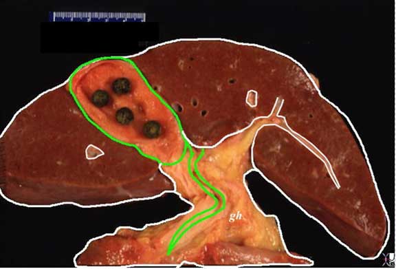
The gastrohepatic ligament (gh) connects the liver and the stomach not only as a ligamentous connection but also with lymphatics, as well as the portal triad which runs on its free edge. In this diagram only the bile duct is shown (green). It extends and connects to the liver capsule (white covering) around the liver, but also into and around the portal triads within the liver. These ligaments, with lymphatics are an important route of spread of disease such as gastric and ovarian malignancy which tends to spread via the transperitoneal route.
The connective tissue of the liver consists of fine reticular networks that bind the liver cells together in a delicate weave.
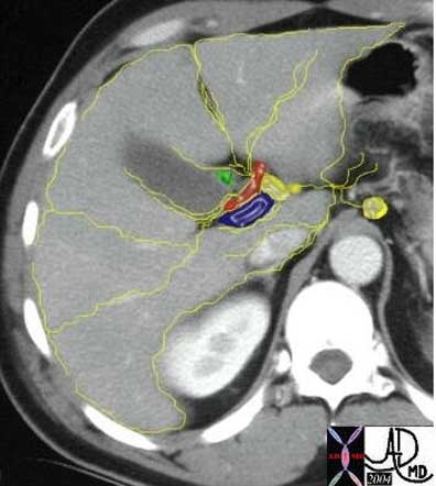
The lymphatic drainage of the liver and connective tissue is intimately related, and is another map of how cancer of the ovary spreads into and around the liver from the peritoneal cavity.
Images courtesy of: Ashley Davidoff, M.D.
Structural Principles of Liver Cancer: Segments of the Liver
It is important to be familiar with the surgical anatomy of the liver, since accurate positional descriptions of liver lesions are needed for the mapping of surgery and for determining operability.
There have been numerous methods of dividing and naming the parts of the liver. The earliest methods divided the liver into the left lobe, the quadrate lobe, the right lobe and the caudate lobe. Subsequently, the liver divisions were based on the venous anatomy. The right lobe was separated from the left by the middle hepatic vein. The falciform ligament was used to separate the left lobe into the medial segment (closest to the middle hepatic vein) and the lateral segment. The right lobe was divided by the right hepatic vein into an anterior segment and a posterior segment. The next several diagrams outline the current segmental nomenclature which is still based on the distribution of the hepatic veins:
Segment I = caudate lobe
Segments II, III & IV = left lobe
Segments V, VI, VII & VIII = right lobe
The above described classification of Couinaud is the most universally accepted.

This coronal image shows the division of the liver into right and left lobes, the division of the right lobe into its superior and inferior segments and the division of the left by the green falciform ligament into its segments.
Image courtesy of Ashley Davidoff, M.D.
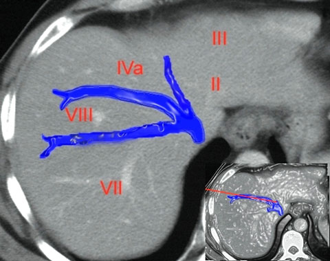
The following cross sectional image is viewed through the superior aspect of the liver with the hepatic venous system in color overlay. The right hepatic vein divides the right lobe into a posterior segment VII and an anterior segment VIII. The left hepatic vein divides the left lobe in the expected location of the falciform ligament into a rightward sub segment IVa and leftward II and III.
Image courtesy of Ashley Davidoff, M.D.
Structural Principles of Liver Cancer: Segments Continued
The following image reveals the cross sectional, segmental pattern at the level of the porta hepatis.
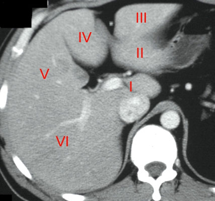
Note that the falciform ligament between segment III and segment IV, divides the left lobe. Note also that segments I, II, III, and IV form the shape of a ?backward? C.
Image courtesy of Ashley Davidoff, M.D.
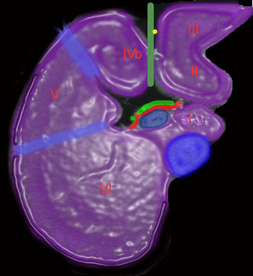
This image demonstrates the royal blue IVC, posterior to the caudate lobe (I), the dark navy blue portal vein anteriorly, the green line and yellow dot indicating the position of the falciform ligament, the thick blue line of the middle hepatic vein and the thin blue line of the right hepatic vein. Since we are seeing the inferior aspect of the liver we are thus seeing segments V and VI of the right lobe, subsegment IVb and segments II and III of the left.
Image courtesy of Ashley Davidoff, M.D.
There are thus two C?s described; Segment I, II, III, and IV describe a backward ?C? in the transverse plane and V, VI, VII, and VIII describe a ?C? in the sagittal plane.

This image shows a vascular hepatocellular carcinoma (yellow arrows) in a cirrhotic liver, situated in segment VIII of the right lobe of the liver, between the middle hepatic vein and the right hepatic vein.
ages courtesy of: Ashley Davidoff, M.D.
The caudate lobe (also known as segment I) is positioned medial to the right lobe and posterior to the left lobe. It is bounded inferiorly by the porta hepatis, posteriorly by the inferior vena cava, anteriorly by the portal vein and more superiorly and anteriorly by the ductus venosus. The caudate lobe is a rather curious structure, situated almost as an appendage to the liver. A unique characteristic is that its venous drainage flows directly and independently to the inferior vena cava below the diaphragm. As previously stated, it belongs neither to the left nor the right lobe and it is considered as an “independent” segment.
Hepatocellular Carcinoma (HCC)
Hepatocellular (HCC)·carcinoma is a primary malignancy of hepatic parenchymal cells, characterized by space occupation and malignant proliferation of aberrant liver cells, with a tendency to invade veins, with a predisposition to multicentricity that usually develops in patients with risk factors of alcohol abuse, viral hepatitis, and metabolic liver disease.
The disease is often clinically silent in the early stages unless it involves the capsule when it will result in pain.
Systemic symptoms include malaise, fatigue, weight loss, fever of unknown origin, and hepatomegaly. Less common presentations include hepatic rupture with hemoperitoneum.
Patients who have a history of alcoholic cirrhosis or a history of hepatitis B or C need continued surveillance. These patients should be routinely screened for the development of hepatocellular carcinoma.
The diagnosis is made by elevated serum alpha fetoprotein or by imaging studies which include ultrasound, computed tomography (CT) and magnetic resonance imaging (MRI).
Surgical resection or liver transplantation remains the mainstay of curative therapy. The results of the various medical treatments (chemotherapy, chemoembolization, ablation, and proton beam therapy) remain disappointing. Untreated,HCC carries a poor prognosis and is directly related to tumor stage and degree of cirrhosis.
Hepatocellular Carcinoma (HCC): Overview
Hepatocellular carcinoma (HCC) develops by a step-wise process of differentiation from regenerative nodules to malignant HCC involving a gradual increase in size and cellular atypia.
Current nomenclature (IWP 1995 – the International Working Party of the World Congress of Gastroenterology) divides hepatic nodular lesions into large regenerative nodules, low grade dysplastic nodules, high grade dysplastic nodules and HCC. The lesions corresponding to low grade dysplastic nodules and high grade dysplastic nodules are classified as adenomatous hyperplasia (AH) and atypical AH, respectively, in the WHO classification.
Large regenerative nodules: They represent well-circumscribed areas of parenchyma (>3mm in diameter) showing enlargement as a response to various hemodynamic stimuli.
Low Grade Dysplastic lesions/nodules: Macroscopically they appear as vaguely nodular lesions, usually 1cm in diameter with a peripheral fibrous scar and slightly more yellowish than the surrounding tissue.
High grade Dysplastic lesions/nodules: Macroscopically they are similar to low-grade dysplastic lesions but are larger. The diagnostic distinction between high grade dysplastic lesion and early HCC is sometimes difficult. An important clue for the differentiation of high-grade dysplastic lesion from well-differentiated HCC is the absence of ?stromal invasion?, i.e. there is tumor cell invasion into the portal tracts in the nodule.
HCC: Macroscopically HCC can be divided into small HCC (<2cm) and advanced HCC (>2cm). Small HCC?s are further divided into vaguely nodular (Early HCC ) and distinctly nodular (Progressed HCC) types . Advanced HCC may be classified based on the Eggel’s classification into a nodular type, massive type, and diffuse type. The nodular type consists of a single tumor or multiple nodular tumors with good demarcation. The massive type consists of a large tumor with an unclear or irregular boundary and occupying almost the entire right or left lobe. The diffuse type is characterized by the diffuse miliary infiltration of the entire liver.
Hepatocellular Carcinoma (HCC): Prevalence
The prevalence rates vary in populations and cultures often reflecting dietary habits. In the US, it comprises only 2% of all malignancies and it is highest among the men of Chinese descent. Its incidence is rising probably because of the rising incidence of hepatitis C infection. Internationally, it is the 5th most common cancer in men and the 8th most common cancer in women. It is most common in Japan, Asia and Africa, with common predisposing factors being hepatitis B, hepatitis C and aflatoxin exposure.
Hepatocellular Carcinoma (HCC): Cause and Predisposing Factors
Cirrhosis from any cause is the seed-bed of HCC. The risk varies according to the etiology of the cirrhosis.
Hepatitis B
Chronic infection with this virus is the most common cause of HCC worldwide. There are about 350 million people world wide that have this disease. In the patients with chronic infection and cirrhosis the incidence is increased 1000 times, probably caused by incorporation of the virus into the genome of the host cells. The introduction of the hepatitis B vaccine has resulted in the reduction of HCC in a pediatric population in Taiwan (Chang) and is being introduced in world wide programs.
Hepatitis C
Hepatitis C is as potent an oncogenic virus as HBV but is less prevalent. It usually requires 25 years of chronic infection to cause tumor. There are about 170 million people worldwide who suffer from hepatitis C and it is the most common form of HCC in Japan and Europe. In the US it accounts for about 30% of the cases of HCC. The risk of HCC increases if the patient, in addition, abuses alcohol or has associated hepatitis B infection.
Alcoholic Liver Disease
Oncogenic potential is probably mediated by inflammation and cirrhosis. This may be augmented by concurrent viral infection. Chronic alcohol use for more than 10 years increases the risk of developing HCC by about 5 times. In the USA about 30% of patients with HCC have alcohol as an underlying cause.
Hemochromatosis – Iron Overload
Untreated genetic hemochromatosis is an especially severe, premalignant state.
Tyrosinaemia
HCC occurs in 37% of patients who survive to 2 years of age and may occur in patients who have had successful liver transplants.
Oral Contraceptives
The absolute risk of developing HCC is very small.
Anabolic Steroids
Case-reports suggest a small increased risk for HCC.

This patient was a middle-aged man with a history of hemochromatosis, who developed a 2.5cm nodule in the right lobe of the liver, which was shown to be a HCC after resection.
Courtesy of: Barbara Banner, M.D.
Hepatocellular Carcinoma (HCC): Genetic
Chromosomal aberrations have been frequently reported in HCC. Amplifications of the chromosomes are noted in 1q, 8q, 6p, and 17q. Among the chromosomes most frequently lost in HCC are 8p, 16q, 4q, 17p, and 13q. These chromosomal regions contain key players in hepatocarcinogenesis such as p53 (chromosome 17p) or Rb (chromosome 13q).
Genes involved in important regulatory pathways are affected by these mutations. Four main pathways can be distinguished.
-
- The p53 pathway involved in DNA damage response and apoptosis,
- The b-catenin/APC pathway involved in intercellular interactions and signal transduction
- The TGF-b pathway involved in growth inhibition and hepatocyte death
- The RB1 pathway involved in cell cycle control.
Hepatocellular Carcinoma (HCC): Carcinogens
Plant Carcinogens
The liver is the main target organ for Aflatoxins produced by the fungi Aspergillus flavus and A. parasitans. These fungi grow readily on grains, peanuts, and food products in the humid subtropical and tropical regions. Higher HCC mortality rates have also been found in people who drink pond-ditch water contaminated with the blue-green algal toxin microcystin. The toxin also causes hepatic hemorrhage and necrosis.
Chemical Carcinogens
DDT, Nitrites, hydrocarbons, solvents, organochlorine pesticides, primary metals, and polychlorinated biphenyls have been implicated as potential carcinogens.
Hepatocellular Carcinoma (HCC): Pathophysiology and Pathogenesis
Hepatocellular carcinoma most frequently arises in a setting of cirrhosis, usually presenting 20-30 years after the initial insult and injury to the liver. The disease, as a result, is most commonly multifocal but has a variety of structural manifestations and clinical presentations.
HCC often arises from a dysplastic nodule and is well differentiated in the early phases. Multiple foci may be present. These subsequently may evolve into well-differentiated tumors or may dedifferentiate and become more aggressive. Within a single nodule both well differentiated and poorly differentiated cells can exist. In general, when the nodules are small they tend to be well differentiated (<1.5cms) and as they grow larger dedifferentiation takes place. As the tumor grows beyond 3cm, the percentage of dedifferentiated cells dominates.
The general response of liver cells to injury is by accumulating fat in the cytoplasm. HCC often contains fat within the tumor, which is highly characteristic of the disease. The close proximity of the cells to the sinusoids, as discussed in the section on structural principles, results eventually in the invasion by the tumor into the portal or hepatic venous radicles. The tumor undergoes significant neogenesis, is usually fed by the hepatic artery with neovascular and hypervascular changes and the development of arteriovenous channels.
Hepatocellular Carcinoma (HCC): Gross Pathology
On gross pathology HCC may show three different patterns:
Nodular form: single or multiple with or without a capsule.
Massive form: appears as an infiltrative mass in noncirrhotic livers
Diffuse form: miliary infiltration of the liver parenchyma.

This gross specimen of HCC involves almost the entire right lobe of the liver and is associated with satellite nodules separate from the main tumor (green nodules in b) which is a characteristic feature Within the left portal vein a small tumor thrombus is noted (white arrow).
Ashley Davidoff MD
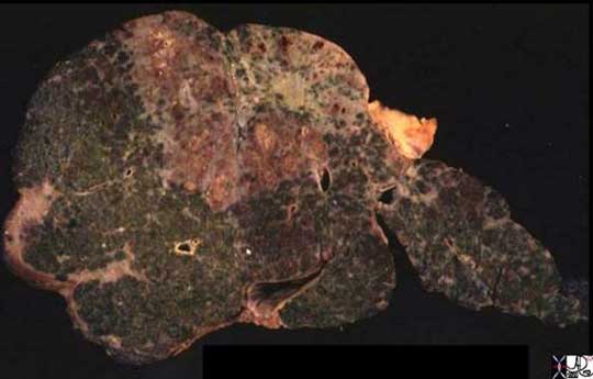
The gross pathology specimen shows the infiltrative form of HCC with relatively straight borders, usually with the base on the surface of the liver and the apex pointing centrally.
Ashley Davidoff MD

Miliary Form
This gross pathology specimen exemplifies the miliary form of HCC characterized by innumerable small nodules scattered throughout the liver. Note the characteristic features of cirrhosis with a micronodular, irregular surface, best seen over the enlarged left lobe, a prominent caudate lobe, and a relatively small right lobe.
Images courtesy of: Barbara Banner, M.D.
Histologically, there are four different architectural types of HCC.
Trabecular type – is the most common type
Pseudo glandular type
Solid
Scirrhous or sclerosing type
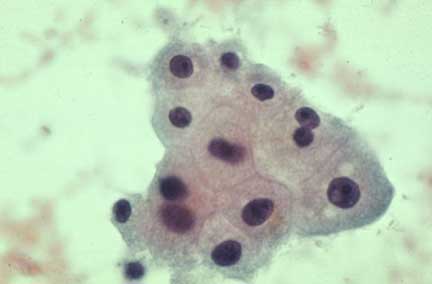
The volume ratio of nucleus to cytoplasm is 1:4 or even 1:6 and the cytoplasm is a light pink (eosinophillic) The shape of the cell is polygonal.
Courtesy Barbara Banner MD

In this cytopathology specimen the nuclear cytoplasmic ratio is closer to 1:1 and sometimes as in the cell in the lower right hand corner the nucleus is 4 or 5 times larger than the cytoplasm. This appearance is characteristic of malignant tissue. This is an example of a hepatocellular carcinoma.
Courtesy Barbara Banner, M.D.
Hepatocellular Carcinoma (HCC): Complications
Complications include advancing cachexia, variceal bleeding, tumor rupture, hemorrhagic necrosis and hepatic failure.

Large well circumscribed HCC, with a capsule (white overlay in b), a focal area of hemorrhage (h), and necrosis (yellow overlay). Courtesy of: Barbara Banner, M.D.
These tumors more frequently invade portal or hepatic veins. Portal vein invasion is much more common than hepatic vein involvement, but hepatic vein involvement is more specific. Tumors invading the hepatic vein may extend to the IVC or the right atrium. Tumor invasion into the hepatic duct or common bile duct can be seen in 2-6% of advanced HCC’s. Obstructive jaundice can occur from direct invasion of the biliary tree or by compression by the tumor or lymph nodes.
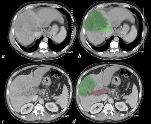
Courtesy of: Ashley Davidoff, M.D.
Same patient as above
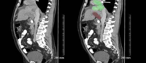
Courtesy of: Ashley Davidoff, M.D.

The CT of the patient shown above, which was performed at a slightly higher axial level, shows the invasion into the right atrium (light green in (b) and (d), with the tumor shown in dark green in the liver).
Courtesy of: Ashley Davidoff, M.D.

This study represents a hepatic artery angiogram. The coiled catheter (white loop) has been placed in the hepatic artery (bright red). The normal flow of blood should be from the artery forward through the sinusoids of the liver and consequently into the hepatic veins and right atrium. In this instance, the blood is shunted through a diffuse HCC (not obvious on the angiogram) into the portal vein and then flows backward into the portal vein (maroon) filling the superior and inferior mesenteric veins in reverse direction (red arrows to maroon arrows). Tumor thrombus is identified in the portal vein (green).
Courtesy of: Ashley Davidoff, M.D.
Hepatocellular Carcinoma (HCC): Natural History
The natural history of the tumor is variable. Patients with HCV infection are more often screened and thus tend to present with signs and symptoms of cirrhosis and earlier stage HCC tumors. Patients with HBV infection or no serological evidence of hepatitis infection tend to present with larger tumors and less cirrhosis.
In general, small nodules HCC starts off as dysplastic nodules that may be low grade or high grade based on cytologic features and about 30% of these will develop into HCC in two years and about 80% will develop into HCC in 5 years (Grisham). The degree of tumor dedifferentiation and the presence of vascular invasion are strong indicators of shortened survival times. In the early stages of the cancer the cell line is mostly well differentiated.
The tumor size is not necessarily a predictor of the course of a particular tumor. A small HCC has a doubling time that may vary from 1 -20 months (Okazaki). In addition, slow growing tumors are more commonly seen in Caucasians and Asian patients when compared to the South African counterparts. (Anthony).
In general, lesions that are 2cm or less are typically well differentiated and as they advance to the 3cm range, vascular invasion and metastatic disease becomes more likely. Once vascular invasion has occurred, successful treatment is unlikely.
Diagnosis and Clinical Approach
Presentation depends on the stage of disease. In countries with systematic screening programs (Taiwan, Hong Kong, Japan and Korea) HCC is often diagnosed at an early stage when patients are asymptomatic or have symptoms due only to the underlying disease. In the United States, there is no systematic screening for HCC. Patients usually present at a late stage, often with abdominal pain, palpable RUQ mass, weight loss, weakness, abdominal fullness and swelling – ascites, anorexia, malaise, weakness, nausea and vomiting. An isolated, self limiting episode mimicking biliary colic may at times be the presenting symptom of HCC. Suspicion for HCC should be heightened when patients present with sudden and rapid decompensation of their liver disease. Jaundice may be due to external compression or tumor invasion of the intrahepatic ducts or rarely due to hemobilia. Abdominal swelling may occur as a consequence of ascites due to the underlying chronic liver disease or may be due to a rapidly expanding tumor. In fewer than 5% of cases, central necrosis or acute hemorrhage into the peritoneal cavity leads to death. Hematemesis may occur due to esophageal varices from the underlying portal hypertension. Bone pain is seen in 3-12% of patients, but necropsies show bone metastases in ~20% of patients.
The triad of abdominal pain, weight loss and an abdominal mass is the most common clinical presentation in the United States.
History taking in HCC
The history is important in evaluating putative predisposing factors like hepatitis, blood transfusion, or use of intravenous drugs, family history of hemochromatosis and other familial diseases. HCC in a first degree relative is associated with increased risk of development of HCC. Social history must include lifetime occupational history and the use of contraceptive hormones.
Diagnosis and Clinical Approach: Physical Signs
A palpable liver is the most common physical sign, occurring in 50-90% of patients. Abdominal bruits reflecting arterio-venous fistulae are noted in 6-25%, and ascites occurs in 30-60% of patients. Splenomegaly, if present, is secondary to portal hypertension. Weight loss and muscle wasting are common, particularly with rapidly growing or large tumors. Fever is found in 10-50% of patients, from unclear cause. The stigmata of chronic liver disease may be present. Budd-Chiari syndrome can occur due to HCC invasion of the hepatic veins; it should be suspected in patients with tense ascites and a large tender liver. Pedal edema may be noted in patients with tumors obstructing the IVC. Extension of tumor to the diaphragm causes shoulder pain and pleural effusion.

This 3D rendering of a CT scan is from a patient with liver failure who is suffering from intractable ascites. The abdomen is distended and a permanent catheter placed in the right upper quadrant allows for intermittent drainage of the ascites. In a patient who has cirrhosis and develops new ascites, HCC is a differential consideration.
Courtesy of: Ashley Davidoff, M.D.
Diagnosis and Clinical Approach: Surveillance for Hepatocellular Carcinoma (HCC)
There is a group of patients who have a higher risk for developing HCC including those with:
- Hepatitis B (patients and carriers)
- Cirrhosis and
- Hepatitis C
- Alcoholic cirrhosis
- Familial hemochromatosis
- Primary Biliary Cirrhosis
These patients require surveillance by ultrasound every 6-12 months.
Nodules between 1-2 cm. require a follow up every 3-6 months and if there is no growth over a two year period, then surveillance every 6-12 months is recommended.
In nodules that are in the 1-2 cm range, contrast-enhanced CT, or contrast-enhanced MRI is recommended. If the criteria for HCC are met in two of the studies, the lesion is considered HCC and no biopsy is needed. If the lesion is not typical for HCC then biopsy is recommended.
For lesions that are larger than 2cm, if typical for HCC by contrast enhanced studies, or if alpha-fetoprotein is >200 ng/ml then a diagnosis of HCC can be made without biopsy . On the other hand a nodule of this size without an elevated AFP or without typical contrast enhancement pattern, biopsy is indicated.
(Practice Guidelines Committee, American Association for the Study of Liver Diseases. Bruix)
Diagnostic Criteria
In general ? imaging and serum alpha-fetoprotein are the hallmarks of diagnosis for HCC, since the clinical presentations are non specific.
Imaging
Imaging has numerous roles including screening, diagnosis, staging, and follow up of HCC. Several imaging modalities can be used to diagnose HCC including ultrasound, helical CT scanning, and MRI. Angiography had a significant diagnostic role in the past. More recently it has played a role in the therapy using chemoembolization.
The structural basis for diagnosis of HCC on imaging is based on the following:
Blood supply
-
- Regenerative nodules are supplied by the portal vein
- Malignant nodules have an arterial supply
Presence of fat
-
- Malignant nodules may contain fat
Presence of a capsule
-
- Malignant nodules may have a fibrous capsule
Vascular invasion
-
- Malignant nodules invade portal and hepatic veins
Imaging: Fat Within the Lesion
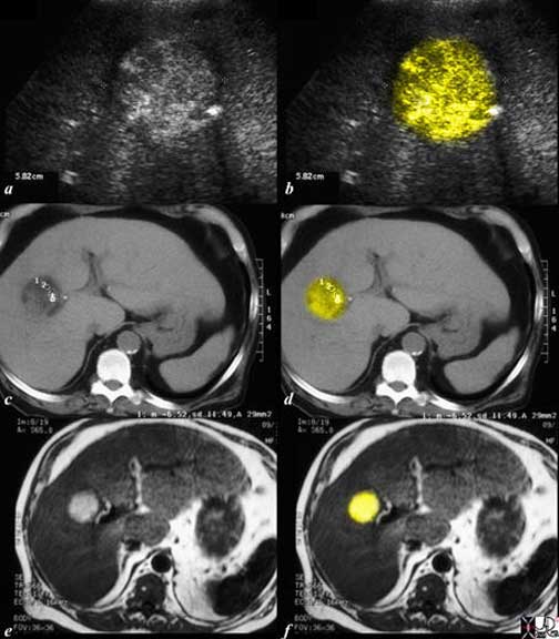
The series of Ultrasound (a, b) CT scan (c, d) and T1-weighted MRI (e, f) all reveal the same finding ? a fat containing lesion, characteristic and pathognomonic for practical purposes of an HCC. The ultrasound shows an echogenic mass, the CT shows a density of -6Hu and the MRI shows T1 bright lesion. All these features are characteristic of fat within the lesion. Note also the prominent artery seen at 3 o’clock on CT and MRI sequences. Surgical removal of this almost 3 cm lesion proved to be a fat containing HCC.
Courtesy of: Ashley Davidoff, M.D.
Imaging: Fibrous Capsule
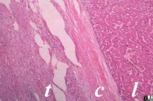
A fibrous capsule (c) is seen in this histopathological section between the tumor (t) and the normal appearing liver (l). The capsule is characteristic of hepatocellular carcinoma.
Ashley Davidoff, M.D.

The CT scan shows a dark ring around an HCC in the dome of the liver, characteristic of the fibrous capsule.
Ashley Davidoff, M.D.

This MRI is from a patient who had a surgically resected HCC. The T1-(a, b) and T2-weighted (c, d) images show a faint hypointense rim on the periphery of the lesion (overlaid in white) characteristic of the fibrous capsule.
Images courtesy of: Ashley Davidoff, M.D.
Imaging: Vascular Invasion


Courtesy of: Ashley Davidoff, M.D.
Imaging: Arterial Supply and Arteriovenous Shunting
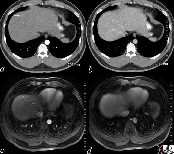
The early arterial feed and rapid wash out as a result of the arteriovenous characteristics allows the earliest detection of HCC. In this instance, both the CT (a, b) and the MRI (c, d) are sensitive to this characteristic. The early arterial phase (wash in) shows the lesion (image (a), CT and (c), MRI), and the lesion disappears rapidly (b and d).
Courtesy of: Ashley Davidoff, M.D.
Imaging: Ultrasound
Ultrasound (US) plays a major role in the diagnosis of HCC, in its role as the modality that is used to screen patients with high risk. It provides a relatively inexpensive way to follow patients, without the use radiation, and is relatively inexpensive.
It is, however, operator-dependant and since differentiated lesions are isoechoic they may be difficult to identify. In addition, the liver is coarsened by cirrhosis, limiting optimal visualization and thus sensitivity is reduced. The addition of color flow Doppler and when possible contrast-enhanced ultrasound, increases sensitivity.
When nodules or masses are identified and do not fulfill the criteria for HCC, then it is the procedure of choice for guided biopsy.
Sonographically, the appearance of HCC is variable. The masses are usually solid and may be hypoechoic, hyperechoic or of mixed echogenicity . The presence of fat makes them hyperechoic, the capsule when present is usually fibrous and hypoechoic. On Doppler sonography, HCC may demonstrate internal vascularity and basket pattern of vascularity (a fine blood flow network surrounding the tumor nodule). Contrast-enhanced ultrasound reveals the characteristic pattern of rapid wash in and rapid wash out. Neovascularity is demonstrated by tortuous, non geometric and sometimes corkscrew or “s” shaped arteries and veins. With necrosis, vascular lakes may be present. Invasion of the venous system can be identified as well.

A 5 cm solid, echogenic mass in this 73-year-old male with cirrhosis was shown to be a fat containing HCC. The echogenic nodule at 9 o’clock (arrows) ? likely represents an area of fat.
Courtesy of: Ashley Davidoff, M.D.
Imaging: CT Scanning
The structural principles of HCC described above apply to both Computed Tomography (CT) imaging and Magnetic Resonance Imaging (MRI). Characteristics include rapid wash in wash out of contrast, (most consistent characteristic), multicentricity, and presence of fat and fibrous capsule. In order to document the wash in wash out characteristic, the technique has to include an arterial phase, a portal venous phase, and a delayed phase.
Non contrast phase is helpful to identify the high density liver of hemochromatosis (HU usually >70) as well as calcification. In well differentiated disease the lesion may be similar to liver and is isodense. It may be hypodense as it becomes dedifferentiated or undergoes necrosis.
Arterial phase imaging is the most useful for the detection of HCC as the predominant blood supply of the tumor is from the hepatic artery. However, it is less sensitive for the detection of small HCC and for dysplastic nodules which appear isodense to the liver parenchyma due to their predominant blood supply from the portal vein. Additionally, other lesions such as FNH and adenomas are also supplied by the hepatic artery. In the setting of cirrhosis, HCC is the favored diagnosis. Necrosis is revealed by avascularity.
In the portal venous phase, lesions may be isodense or hypodense (wash-out).
In the delayed phase the capsule may become more apparent as a ring or part ring on the circumference.
It is important to use high injection rates (3-4cc/sec) and appropriate bolus timing. Sensitivity of dual- or triple-phase CT for the detection of patients with tumors is 60-70%.
CT can also evaluate complications of HCC such as portal venous or hepatic venous invasion. In addition, other complications such as bleeding within the tumor and hemoperitoneum can also be recognized by CT.

Vague heterogeneity in the right lobe of the liver, in the presence of a cirrhotic liver with portal vein invasion (maroon in b and d) and cavernous transformation (red overlay in b, d, and f) typify an infiltrative HCC.
Courtesy of: Ashley Davidoff, M.D.
Imaging: Magnetic Resonance Imaging
Magnetic resonance imaging (MRI) is extremely useful in the detection and characterization of regenerating and dysplastic nodules and HCC. The various techniques of MRI are useful and more sensitive than other modalities in the early detection and characterization of HCC in the cirrhotic liver and in differentiating it from dysplastic nodules and pseudo lesions.
Regenerative nodules are fed by the portal vein and not the hepatic artery, and so are characterized by a lack of enhancement in the arterial phase and usual enhancement in the portal phase. They may contain iron.
Dysplastic nodules may be hyperintense on T1 (an unusual feature) and also fed by the portal vein and not by the artery, and therefore enhance during the portal phase and with siderois (iron within the lesion) have a nodule in a nodule appearance.
As the lesion advances to a small HCC it starts to become fed by the artery, and therefore has early wash in and wash out by the portal phase. The lesion retains water and therefore is bright on T2-weighted images.
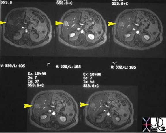
The dynamic, contrast enhanced MRI shows a pre-contrast image (top left) that rapidly accumulates contrast in the wash in phase (middle image top) and rapidly loses by washout of the contrast in the portal venous phase and delayed images. In the presence of cirrhosis, this is highly characteristic of HCC.
Courtesy of: Ashley Davidoff, M.D.
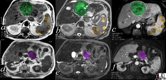
The MRI is from a 71-year-old male with chronic alcoholism and cirrhosis. Image (a), is a T1-weighted image through a large well circumscribed tumor (green) in the left lobe. Image (b) is a T2-weighted showing a small, white necrotic area (white arrow) and minimal hyperintensity revealing water content in the anterior half of the tumor. Splenic metastases (gold) with heterogeneous pattern are noted in the spleen. The tumor shows only minimal enhancement (unusual) in image (c), but with intense rim enhancement. It also reveals multicentric changes in the rest of the liver with satellite nodules. Images (d, e, and f) are taken at a more caudal level and are also in the following order from left to right: T1, T2 and contrast enhancement respectively. A metastasis in the pancreatic body (purple) is seen as a rounded structure between the liver and spleen.
Courtesy of: Ashley Davidoff, M.D.
Imaging: Angiography
The hypervascular nature of HCC made angiography a natural technology as the diagnostic study of choice in the pre CT and MRI era. As technology has advanced, diagnostic angiography has been replaced but now plays a role in the treatment of HCC using chemoembolization.
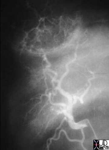
The middle-aged man with hemochromatosis presented with hemoperitoneum. An angiogram with injection into the right hepatic artery shows a mass in the dome of the liver, with evidence of odd -shaped vessels that characterize the neovascularity. The study was performed in about 1981 when alcohol embolization was being explored and used in this patient.
Courtesy of: Ashley Davidoff, M.D.
Biopsy
Biopsy is required only in lesions that are indeterminate for HCC. Core biopsy is favored over fine needle aspiration since larger amounts of tissue, often with normal surrounding parenchyma, can be obtained. It is sometimes useful to take a biopsy of ?normal liver? in the region to help differentiate well differentiated tumors from normal liver. Biopsy is generally obtained percutaneously under ultrasonographic or CT guidance.
Staging
The goal of tumor staging is to separate patients into different groups based on their predicted survival to help determine the most appropriate treatment modality.
Staging involves:
1. Identifying the number, size and location of the lesion
2. Presence or absence of vascular invasion
3. The presence of extrahepatic spread.
To best assess the prognosis, a staging system that takes the tumor stage, liver function and physical status is best equipped to offer a meaningful evaluation.
The currently available staging systems for HCC include the pathologic tumor-node-metastasis (pTNM), Okuda, Cancer of the Liver Italian Program (CLIP), and Barcelona Clinic Liver Cancer.
The staging of liver cancer and the treatment of liver cancer also requires knowledge of the degree of liver damage, and since cirrhosis is commonly present, incorporation of background liver status is important in staging.
The Child-Pugh score for cirrhosis is a well known classification system of cirrhosis. It takes into account serum bilirubin, serum albumin, prothrombin time, the presence of ascites, and level of mental status.
Other classifications for staging liver cancer have developed that incorporate liver disease and physical status and include the Cancer of the Liver Italian program (CLIP), Barcelona-Clinic Liver Cancer (BCLC) system and the Okuda system. The CLIP system tends to aid in prognostication while the BCLC is helpful as a treatment algorithm.
Management
The initial clinical evaluation is aimed at assessing the extent of the tumor, the underlying functional compromise of the liver by cirrhosis and the general condition of the patient. Most HCC patients have two liver diseases: cirrhosis and HCC, each of which is an independent cause of death.
The two major treatment modalities are surgical and liver-directed therapies.
Surgery – Hepatic Resection
The reported overall 5-year survival after resection of HCC is 30%-60%. However, only a minority of patients with HCC are candidates for surgical resection due to the large size of the tumor with insufficient hepatic reserve after liver resection. In general, patients with Child-Pugh class A tolerate resections of up to 50% of liver parenchyma, whereas Child-Pugh class B patients tolerate resections up to 25%. Child-Pugh class C cirrhosis is considered as an absolute contraindication for liver resection. Other contraindications to resection include extensive and multifocal bilobar tumors, extrahepatic metastases of the disease, and tumors with main portal vein tumor thrombus, hepatic vein, or inferior vena cava involvement.
Surgery represents the only hope for potential cure, but in view of the nature of the disease, with multicentricity in a background of cirrhosis, only 15-30% of patients are eligible for tumor resection. For some patients with associated liver disease, transplant is an alternative.
Surgical resection is of two basic types: wedge resection and lobar resection where one or more are resected. Right hepatic lobectomy infers resection of segments 5-8, an extended right lobectomy or trisegmentectomy which infers the removal of segments 4-8.
A left hepatectomy infers resection of segments 2-4, a left lateral segmentectomy infers resection of segments 2 and 3, while a left trisegmentectomy infers resection of segments 2, 3, 4, 5, and 8. The caudate lobe is treated independently but its proximity to the porta creates additional technical challenges for the surgeon. However, it can be removed in isolation or as part of the variety of resections described.
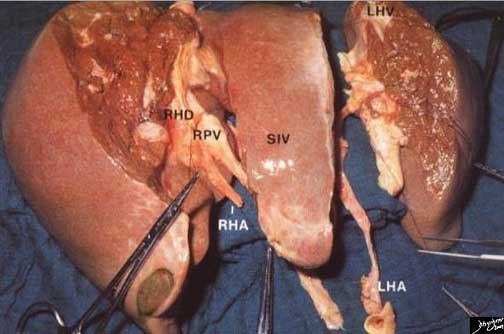
This is a surgical specimen of the liver divided into the right lobe (RHD – right hepatic duct, RPV = right hepatic vein, and RHA = right hepatic artery) segment IV of the left lobe (aka quadrate lobe aka medial segment of the left lobe), and segments II and II (aka the lateral segment of the left lobe) showing the left hepatic vein (LHV). The left hepatic artery is shown between the medial and lateral segment of the left lobe.
Courtesy of: Eli Katz, M.D.
Indications include technical feasibility, tolerance based on patients well being, and Child?s classification, and tumor size of less than 5 cm. Despite apparent potential eligibility for resection, the chances of recurrence are high and up to 75% of patients recur. The proximity of the tumor to the blood supply and sinusoids, and the multicentricity of the disease are plausible explanations for this high rate of recurrence. The observed recurrence rates of HCC after resection are 50 -80 % at 5 years.
Surgery – Hepatic Resection: Liver Transplant
Liver transplant offers the best chance for long-term survival because it removes the tumor, the underlying diseased liver, and it cures portal hypertension. Based on the Milan criteria of solitary HCC < 5cm or no more than three lesions each <3cm, the 4-year overall and disease-free survival rates are 85% and 92%, respectively.
Indications for the procedure include patients with cirrhosis, a single lesion less than 5cm, or up to three lesions that are no larger than 3cm each. 5-year survival rates have been shown (Bismuth, Llovet, Jonas, Philosophe) to be up to 75%.
The 5-year survival rate for untreated HCC is 0% and for in patients undergoing surgery it is 20-40%. Only 5-15% of patients are potentially resectable.
Alternative treatments are used when resection is contraindicated, in the hope of extending life or potentially downstaging to enable transplantation.
Alternative Treatments
Alternative treatments include:
PEI: Percutaneous Ethanol Injection
RFA: Radiofrequency ablation
TAE: Trans arterial embolization
TAC: Trans arterial chemoembolization
TACE (TAC +TAC): Transcatheter arterial embolization
TARE: Trans arterial radio embolization
Brachyhytherapy
MCT: Microwave coagulation therapy
Cryotherapy
Microwave ablation
Laser
High Intensity Focused Sonography
New Chemotherapeutic Agents
Alternative Treatments: Percutaneous Ethanol Injection
Percutaneous Ethanol Injection (PEI) was one of the first techniques used for ablation. Indications include a tumor of less than 3cm. Repeated sessions, usually up to six sessions are required and US as well as CT scanning have been used for guidance. Percutaneous ethanol injection involves the slow injection of ethanol (absolute or 95%) via a needle introduced percutaneously into the liver tumor. Complete ablation has been attained in up to 70% of patients. High recurrence rate has led, in the US, to use other techniques such as radiofrequency ablation.
Alternative Treatments: Radiofrequency Ablation
Radiofrequency ablation (RFA) is a minimally invasive technique used to ablate tumors in the liver, bone, breast, kidney and lung. It uses thermal energy of high-frequency, alternating current applied via electrodes placed within the tissue introduced via a needle electrode to destroy tumor cells. It induces desiccation and coagulative necrosis of the tissue. RFA can be applied percutaneously, laparoscopically, or at open surgery.
The heat is generated from an external generator, and cell death is accomplished based on the RF power used, duration of the heating and distance of the tumor from the electrode. Liver surgery is the treatment of choice for HCC, but is limited for the reasons described above. For those patients who are not eligible for surgery, alternative therapies are considered for palliation purposes. RFA is particularly helpful in those patients who are awaiting transplant.
Indications for RFA include lesions less than 8cm in size, and less than 8 in number. Contraindications for HCC include metastatic disease outside the liver, liver failure, and sepsis.
Since the RFA destroys all tissue in its range, lesions should be remote from important structures such as the liver capsule diaphragm, gallbladder, colon, and porta hepatis. Blood vessels close to the lesion become a heat sink, and so better results are achieved when the lesion is remote from large blood vessels.
The procedure is completed in one session.
The patient should be NPO before the procedure, and is admitted for an overnight stay after the procedure.
Conscious sedation is utilized, and guidance is usually by US, or CT.
The technique requires accurate placement of the electrode, usually in the center of the lesion, and attempts are made to attain necrosis, by heating the tissue to greater than 60 degrees Celsius, 1-2cms beyond the margin if possible.
Radio frequency ablation is a thermoablative technique that has been demonstrated to be superior (increased tumor cure and long term survival) to percutaneous ethanol injection (PEI).

The RFA probe (white prongs) in this instance is in a liver metastasis in a patient with an unknown primary. The bubbles seen in the lesion represent necrosis following ablation. It is not common to see immediate macroscopic results. This lesion is about 7 cm in diameter.
Courtesy of: Ashley Davidoff, M.D.
Alternative Treatments: Trans Arterial Embolization & Trans Arterial Chemoembolization
Transarterial embolization (TAE) involves the use of gelfoam, stainless steel coils, or polyvinyl alcohol sponge to embolize the hepatic artery feeding the tumor.
Trans arterial chemoembolization involves injection of chemotherapeutic agents into the hepatic artery or emulsification of the anticancer drug with lipiodol.
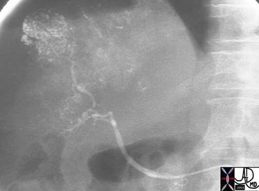
Lipiodol and a chemotherapy agent were selectively introduced into the hepatic branch feeding the tumor.
The catheter has been selectively placed in a branch of the right hepatic artery that feeds the HCC and a mixture of lipiodol and chemotherapeutic agent is injected. The lipiodol is retained selectively in the HCC which is seen as the round mass in the dome of the liver.
Courtesy of: Ashley Davidoff, M.D.
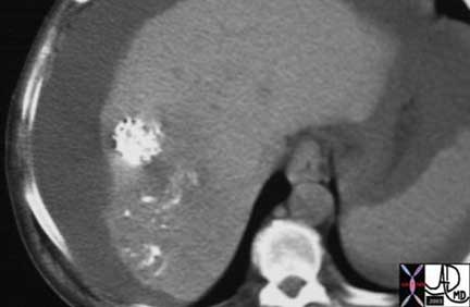
The CT shows a rounded mass in the dome of the liver that has accumulated the radiodense lipiodol. In addition, as is the character of HCC, its multicentricity is shown by the accumulation in multiple other sites near the tumor. The cirrhotic liver is characterized by the irregular edge, and Child Pugh C status is suggested by the ascites.
Courtesy of: Ashley Davidoff, M.D.
Alternative Treatments: Other Treatments
Transcatheter Arterial Embolization (TACE) (TAC +TAE) involves intra-arterial administration of some form of chemotherapy combined with arterial embolization. It is a minimally invasive therapy for HCC where selective injection of the artery feeding the tumor is catheterized and high doses of chemotherapy are delivered locally. The procedure is contraindicated in patients who have advanced cirrhosis or hepatic failure. Once delivered, the vessel is occluded by embolization technique.
TARE: Trans Arterial Radio Embolization
Trans arterial radio embolization involves the regional delivery of radioisotopes (I-lipiodol/ Yttrium 90) intra arterially to the tumor.
Brachytherapy
Brachytherapy involving the delivery of transarterial radioactive yttrium loaded into 30-40 micron glass beads has been a recently developed concept.
MCT: Microwave Coagulation
Microwave coagulation therapy involves microwave therapy following the ultrasound-guided placement of an electrode into the lesion. Coagulation necrosis and hemostasis result, destroying tissue in the treated area.
New Chemotherapeutic Agents
Sorafenib (Nexavar) is an oral agent that inhibits angiogenesis, and has pro-apoptotic activity. A recent study by Llovett on 602 patients was encouraging, improving survival, with toxicity considered minimal.
Prevention and Prognosis
In high risk regions of the world, the introduction of hepatitis B vaccine may have a significant impact on the disease. In Taiwan, hepatitis B vaccine given to newborns and children has shown to reduce the incidence of HCC by 75%, (Chang) and the vaccine is being introduced into vaccination programs of high risk regions.
The natural history of HCC is highly variable. Patients presenting with advanced tumors (vascular invasion, symptoms, extrahepatic spread) have a median survival of ~4 months, with or without treatment.
Conclusion
HCC is an aggressive tumor with a high case fatality rate. Successful treatment is dependent on early detection through widespread use of surveillance of patients at risk for HCC development. Imaging plays an essential role in both the diagnosis and alternative therapies.
References
Block The Practice Of Ultrasound: A Step-By-Step Guide to Abdominal Scanning By Berthold Block Published by Thieme, 2004
Brink JA, Semin MD. Biliary stone disease. In: Gazelle GS, Saini S, Mueller PR, eds. Hepatobiliary and pancreatic radiology: imaging and intervention. New York, NY: Thieme, 1998; 590-630.
Friedman AC, Sachs L. Embryology, anatomy, histology and radiologic anatomy. In: Friedman AC, eds. Radiology of the liver, biliary tract, pancreas and spleen. Baltimore, MD: Williams & Wilkins, 1987; 305-332.
Moore Clinically Oriented Anatomy By Keith L. Moore, Arthur F. Dalley, A. M. R. Agur Published by Lippincott Williams & Wilkins, 2006
Netter FH. The Ciba collection of medical illustrations Vol 111. Digestive system. Part III. Liver, biliary tract and pancreas. Summit, NJ: Ciba Pharmaceutical, 1957; 22-24.
K?jiro Masamichi Pathology of Hepatocellular Carcinoma Blackwell Publishing Malden Mass 2006
Kumar, Vinay Abbas, Abul K., Fausto, Nelson, Robbins, Stanley Leonard, Cotran, Ramzi S. Robbins and Cotran Pathologic Basis Of Disease 7th Edition Philadelphia Elsevier 2005
Gourtsoyiannis, N. C., Ros, P (Editors) Radiologic-Pathologic Correlations from Head to Toe: Understanding the Manifestations of Disease Springer Verlag

