Applied Anatomy of the Adrenal Glands
Learning Objectives[ps2id id=’01’ target=”/]
Recognize the basic anatomy of the adrenal glands.
Be able to locate this organ within the abdominal cavity and identify the main structures of the adrenal gland utilizing cross sectional medical imaging.
Describe the adrenal gland’s function and its relation to the other organs in the abdominal region.
Differentiate the role of radiographic examination, CT, US, and MRI in imaging studies of the adrenal glands.
Introduction:

Imagine two young boys with straw hats fishing by the river.
The boy in red is napping and has his hat on his forehead ? the left adrenal.
The other boy in blue is wide awake and has his hat atop his head ? the right adrenal.
This is how the adrenals are positioned relative to the superior poles of the kidneys.
Ashley Davidoff MD 2018
39535
adrenals-0015
Definition
The adrenals are paired endocrine glands secreting catecholamines (norepinephrine and epinephrine) from the inner medulla, and steroid hormones from the outer cortex. The medulla is controlled by the neuroendocrine axis, while the cortex is controlled mostly by the pituitary gland. The adrenals are located retroperitoneally at the upper poles of the kidneys. They mainly help the body in its response to stress as well as play a role in maintaining blood pressure and salt balance. When faced with a stressful situation, the medulla of the adrenal glands secrete epinephrine (adrenaline) causing an “adrenaline rush” preparing the body for “fight or flight”.
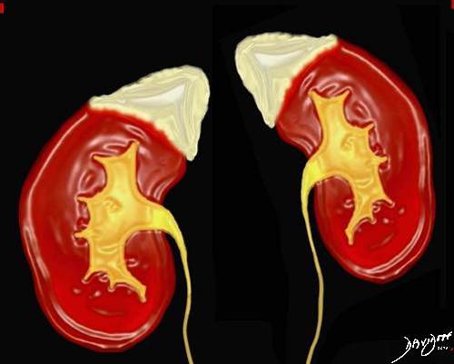
Courtesy of: Ashley Davidoff, M.D.
The anatomy of the adrenal glands appears to have been described first in 1563 by Bartholomeo Eustachius as the “glandulae renis incumbentes” in his Tabulae Anatomicae. Ideas about the function of the adrenal glands lagged behind. Thomas Bartholin proposed that these “capsulae atrabilariae” purified black bile that eventually drained into the renal veins.In 1714 Lancisius Emil Huschke first differentiated the two layers of the adrenal gland, the cortex and the medulla, anatomically. Edme F.A. Vulpian demonstrated the differential staining of the two regions histologically.
In 1716 the Academie des Sciences de Bordeaux offered a prize for the answer to the question, “What is the purpose of the suprarenal glands?” Charles de Montesquieu, judging the responses, found the essays so unsatisfactory that he was unable to award the prize, concluding that “Perhaps some day chance will reveal what all of this work was unable to do.”
Evidence for a central physiological role for adrenal glands came from clinical observation. On March 15, 1849, Thomas Addison presented a paper to the South London Medical Society entitled, “On anaemia: disease of the supra-renal capsules,” a result of his interest in idiopathic, or pernicious, anemia. Three of the patients he described had adrenal disease at autopsy, and it was the only abnormality that was identified in two of them.
Harvey Cushing began his career at the Massachusetts General Hospital and is considered a pioneer of Neurosurgery, making several fundamental discoveries about the pituitary gland and its relation to the adrenals. Due to his extensive work, Cushing’s disease and syndrome are named after him.

Anatomy and Physiology of the Adrenal Glands: Overview
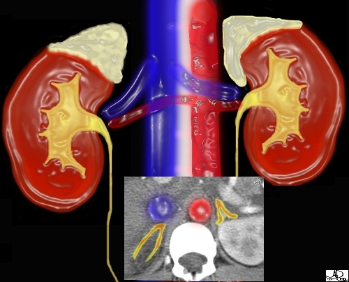
The adrenal glands are small and mighty. They are tucked away in the back of the abdomen, just below the chest cavity, and literally have their finger on the pulse as they sit alongside the highway systems of the aorta, vena cava, and autonomic nerve chains. They sit on top of the waterworks in close contact with the kidneys, and within the functional loop of the renin – angiotensin – aldosterone hormonal system that controls blood pressure, volume and salt metabolism. Some of the most dramatic responses of the body originate in these small and sometimes hidden glands.The adrenals have a reputation in the imaging world of being hard and difficult. Neither of these statements is true. First, they are so soft that they get pushed around by all the other organs that surround them. Secondly, like so many things that appear difficult initially, with appropriate attention, the structural complexity and eccentricities unfold.
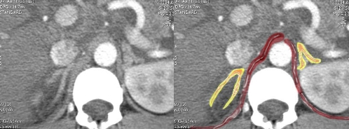
This image combines the coronal view with the axial view and reflects the intimate relationships that the adrenals have with the kidneys as well as the great vessels of the abdomen. They literally have their fingers on the pulse of the aorta (red overlay)and the inferior vena cava (IVC) (blue overlay).
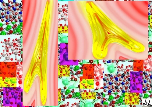
Images courtesy of: Ashley Davidoff, M.D.
Anatomy and Physiology of the Adrenal Glands: Normal Frontal View
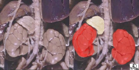
The adrenal glands are located retroperitoneally in the abdomen. They sit on the upper poles of the kidneys on the posterior abdominal wall and extend roughly from the level of the 11th thoracic rib to the first lumbar vertebrae.The right adrenal is slightly more lateral than the left. Since the adrenals sit on top of kidneys, and the right kidney is usually lower than the left, the right adrenal is usually more inferior.

Anatomy and Physiology of the Adrenal Glands: Normal Transverse View
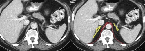
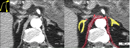
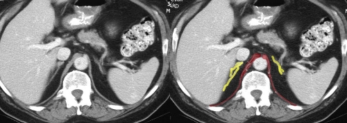
The adrenals can be found just lateral to the crus of the diaphragm. They have a ‘wishbone’ shape, or upside down ‘y’ shape and are surrounded by fat. In general the right adrenal is the long and thin one of family, and the left the short and chubby one.Often the lateral limb of the right adrenal gland is pressed against the liver and cannot be clearly seen. Since the limbs are soft, compressible, and malleable, they often are distorted by normal surrounding structures producing “bends” in them. This sometimes results in difficulty distinguishing cases of abnormal nodules and hyperplasia (increased size) from normal distortion.
Anatomy and Physiology of the Adrenal Glands: Size
The adrenals can vary in weight anywhere between 4 to 14 grams, but the average weight is between 3 to 6 grams. Only about 10% of the gland’s weight is from the medulla (inner portion).The average adrenal dimensions are:
· 20-30 mm. in width
· 40-60 mm. in length
· 3-6 mm. in thickness
The left adrenal is slightly larger than the right.

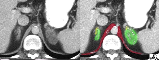
Anatomy and Physiology of the Adrenal Glands: Size – small and large

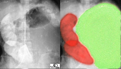
Anatomy and Physiology of the Adrenal Glands: Shape
In general the adrenals have a ‘wishbone‘, or upside down ‘y’ shape, but they are not usually symmetrical. Although they are naturally asymmetric structures, part of their shape discrepancy relates to the variability in their surroundings. Since they are in different relative positions they have different surroundings.When there is a paucity of fat or the surrounding structures push on them, they tend to distort. The right adrenal gets pushed against the liver, and elongates, while the left becomes irregular and “knobbly”. Thus in general, the right is long and thin and the left short and stout. When there is a large amount of retroperitoneal fat they tend to look more alike in cross section.


Position

Imagine two young boys with straw hats fishing by the river.
The boy in red is napping and has his hat on his forehead ? the left adrenal.
The other boy in blue is wide awake and has his hat atop his head ? the right adrenal.
This is how the adrenals are positioned relative to the superior poles of the kidneys.
Ashley Davidoff MD 2018
39535
adrenals-0015
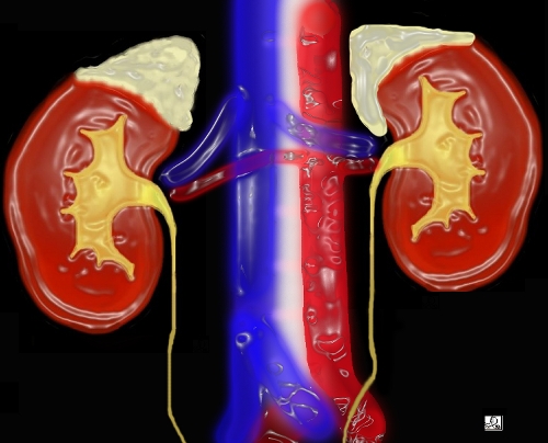
The adrenal glands are usually difficult to find. Since the left kidney is slightly more superior than the right, the left adrenal is usually more superior. The left adrenal can be found when the splenic vein crosses behind the pancreas from the spleen to the portal vein, and the right adrenal can be found when the IVC frees itself from its intrahepatic portion inferiorly. The gland must be identified superiorly and inferiorly until no part of the gland remains, since exophytic tumors off the gland are not uncommon.The adrenals or adrenal like tissue can be found in places other than its normal position. This is termed ‘ectopic’ adrenal tissue.

Structural
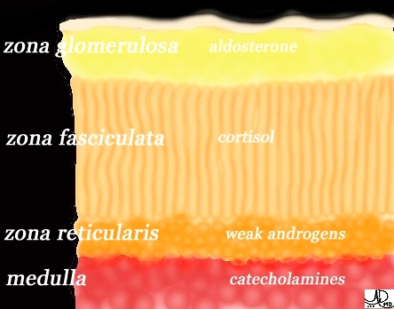
The adrenal gland consists of 2 main parts:1. an outer cortex which secretes several classes of steroid hormones;
2. an inner medulla which is the source of the catecholamines epinephrine and norepinephrine.The adrenal also has a thin outer fibrous capsule which can vary in thickness from gland to gland, and in some cases, even within the same gland.
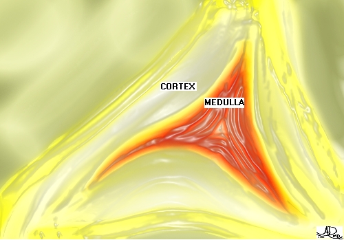
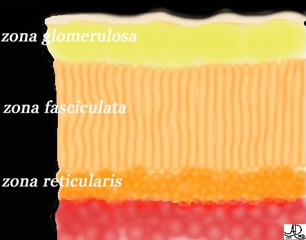
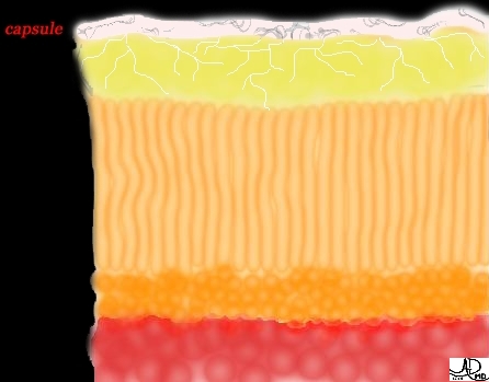
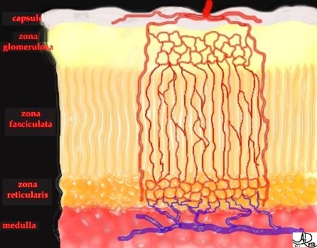
When the adrenal gland is reviewed under the microscope, three different layers are identified in the cortex and one layer in the medulla. In the following images the yellow and orange layers represent the cortical layers and the red represents the medulla.

The cortex is divided into the 3 zones based on the arrangement of cells.
zona glomerulosa is the outer most rounded groups of cell.
zona fasciculata, is the middle column of cells and arranged radially.
zona reticularis is the innermost irregularly arranged cylindrical masses of cells.
“GFR” is a good pneumonic to remember. It stands for glomerular filtration rate but also stands for the three layers, from outer to inner, – glomerulosa, fasciculata, and reticularis.
Anatomy and Physiology of the Adrenal Glands: Parts – Functional

Anatomy and Physiology of the Adrenal Glands: Parts – Imaging
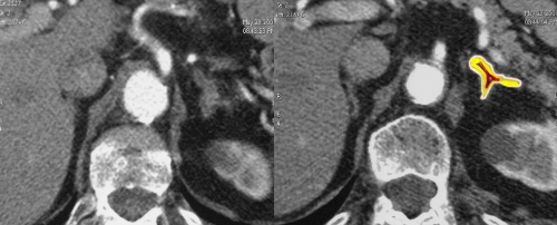
In imaging it is usually not possible to differentiate between the cortex and the medulla, and we divide the adrenal into the medial and lateral limbs which are joined together at the apex of the gland.

Anatomy and Physiology of the Adrenal Glands: Character-Normal
The normal adrenal gland is soft and friable. It is usually surrounded by fatty tissue and is encapsulated. The surface appears corrugated or nodular and is usually not smooth.
The normal color is a golden yellow color which can be distinguished from the more pale surrounding fat. The capsule is a thin fibrous structure surrounding the gland attached by many fibrous bands which penetrate into the gland.
When the adrenal is cut, the outer cortex and the inner medulla can be distinguished by color. The cortical (outer) layer appears golden yellow while the inner medulla appears more flattened and a darker reddish/brown.


Anatomy and Physiology of the Adrenal Glands: Capsule
Each gland is encased in a thin layer of loose areolar connective tissue and a thick fibrous capsule. The capsule is attached by fibrous bands extending into the substance of the gland.

Anatomy and Physiology of the Adrenal Glands: Arterial Supply – Extra – Adrenal Anatomy
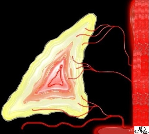
The adrenal gland is very vascular, receiving an estimated 6 to 7 ml/g per minute, or about 25ml/minute in the resting state. Why does it have such a large blood supply? The body’s second to second need to respond to crisis situations requires enough blood to pass by the adrenals to circulate important and necessary hormones.
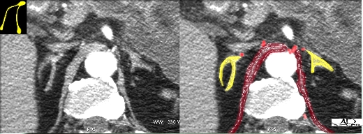
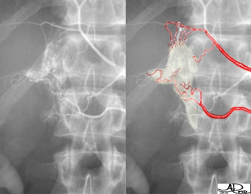
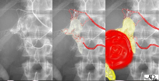
The blood supply of the adrenal gland is derived from three adrenal arteries: the superior artery is a branch of the inferior phrenic artery; the middle artery arises directly from the aorta; and the inferior artery arises from the renal artery.
The primary blood supply of the right adrenal comes from the superior and inferior adrenal arteries, whereas the left adrenal is supplied primarily by the middle and inferior adrenal arteries. The three arteries branch, and each gland may have up to 50 small arterial branches enter the perimeter, supplying a gland that obviously commands a tremendous blood supply.

Courtesy of: Ashley Davidoff, M.D.
.
Courtesy of: Ashley Davidoff, M.D.
Anatomy and Physiology of the Adrenal Glands: Arterial Supply – Intra- Adrenal Anatomy
Up to 50 to 60 small feeder vessels penetrate the anterior and posterior surfaces of the glands and form a plexus beneath the capsule. Cortical arteries supply the cortex from the subcapsular plexus, which drains centripetally toward the medulla. Medullary arteries pass through the cortex and supply the medulla directly. In the zona reticularis, the capillaries coalesce to form progressively larger venous sinuses that drain centrally.
Medullary capillaries form venous channels, which eventually forms a single adrenal vein that usually drains into the vena cava on the right and into the renal vein on the left.
Anatomy and Physiology of the Adrenal Glands: Arterial Supply – Imaging

Anatomy and Physiology of
Courtesy of: Ashley Davidoff, M.D.
Adrenal Glands: Arterial Supply – Abnormal
Anatomy and Physiology of the Adrenal Glands: Venous Drainage
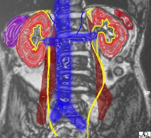
The venous drainage of the adrenals occurs via a single vein that emerges from the anterior surface of each gland.

Adrenal venous drainage is usually through the right and left adrenal veins.
The right adrenal vein exits the apex of the gland and enters the posterior surface of the inferior vena cava. This vein is short (1-5 mm.), fragile, and the most common source of troublesome bleeding during right adrenalectomy.
The left adrenal vein is a bit longer (2-4 cm. in length) and usually drains into the left renal vein, either directly or after being joined by the left inferior phrenic vein. Smaller emissary veins may drain into the inferior phrenic, renal, and rarely the hepatic portal veins.
Not well recognized is the left inferior phrenic vein, which usually communicates with the adrenal vein which courses medially. This can be injured during dissection of the medial edge of the gland.

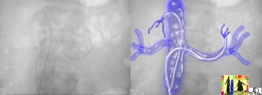
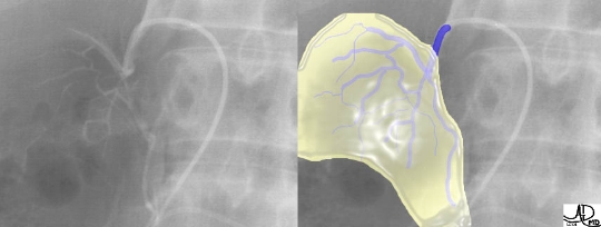
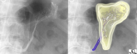
Anatomy and Physiology of the Adrenal Glands: Venous Drainage – Venography
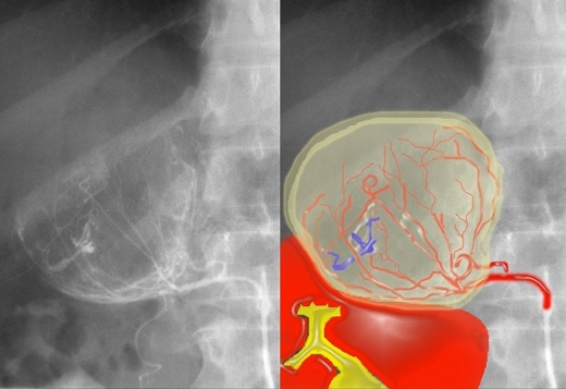
This series of three images reflects a study called “adrenal vein sampling” which requires the simultaneous catheterization of the adrenal veins. This procedure is used to identify relative and absolute concentrations of hormone secretion from the glands to distinguish between normal, bilateral hyperplasia, and unilateral adenoma.

Courtesy of: Ashley Davidoff, M.D.
Anatomy and Physiology of the Adrenal Glands: Venous Drainage – Imaging CT
The left adrenal vein is almost always seen in cross sectional imaging lying at the apex of the gland. In this case both adrenal veins are identified (blue overlay). We have reviewed this case before which represents an aldosteronoma of the left adrenal gland. (green nodule)Courtesy of: Ashley Davidoff, M.D.
Anatomy and Physiology of the Adrenal Glands: Lymphatics

Courtesy of: Ashley Davidoff, M.D.
Anatomy and Physiology of the Adrenal Glands: Lymphatics
Lymphatic vessels arise from a plexuses deep within the capsule and in the medulla only. The lymph vessels follow the central vein and the main venous tributaries throughout the medulla of the adrenal gland. Lymphatic plexuses drain to para-aortic and renal lymph nodes.
There is no lymphatic supply to the cortex of the adrenal.
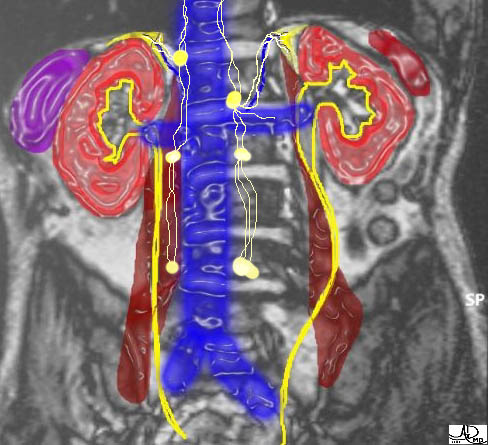
Anatomy and Physiology of the Adrenal Glands: Nerve Supply
The adrenal glands receive nerves from the lower thoracic (T10 – T12) and upper lumbar (L1 – L2) nerve plexuses. The nerves travel with the arteries. The nerve supply forms a plexus of medullated and non medullated nerves on the capsule of the gland primarily on the posterior aspect. The nerves enter the gland with the arterioles and help regulate the high adrenal blood flow.
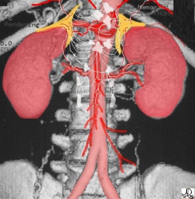
Courtesy of: Ashley Davidoff, M.D.
Anatomy and Physiology of the Adrenal Glands: Relations
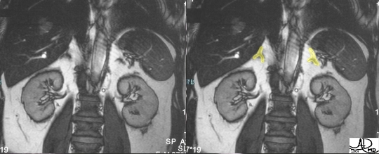
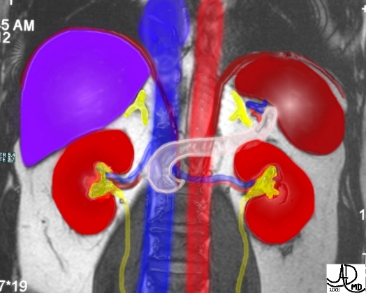
The anatomic location of the adrenals, sandwiches them between several organs, and on the border between two major cavities. The adrenals are positioned differently in relation to the kidneys and to other asymmetric organs such as the liver, pancreas, and spleen.
Anatomy and Physiology of the Adrenal Glands: Relations – Right adrenal
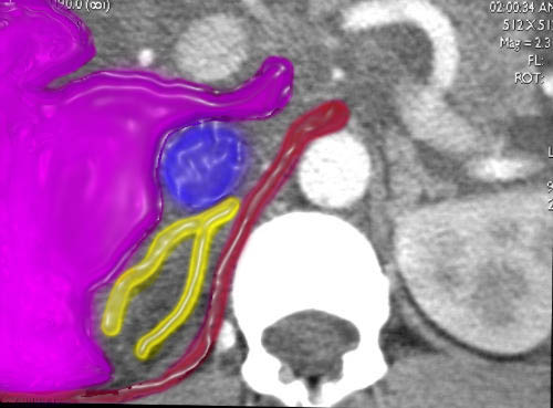
The right adrenal is framed by the following:· anteriorly – the inferior vena cava and the caudate lobe of the liver
· posteriorly – the diaphragm
· superiorly – the bare area of the right lobe of the liver, diaphragm and chest cavity
· inferiorly – the superior pole of the right kidney
· medially – the crus of the diaphragm and the spine
· laterally – the bare area of the right lobe of the liver
Anatomy and Physiology of the Adrenal Glands: Relations – Left adrenal
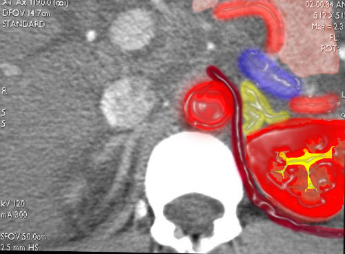
The left adrenal is framed by the following:· anteriorly – the end of stomach above and the pancreas below and branches of the splenic artery and vein
· posteriorly – the left kidney, spleen, and the diaphragm
· superiorly – the diaphragm and chest cavity
· inferiorly – left kidney
· medially – left crus of the diaphragm, aorta, and spine
· laterally – the spleen and left kidney

Anatomy and Physiology of the Adrenal Glands: Embryology
Each adrenal consists of two functionally distinct endocrine glands within a single capsule. The cortex derives from mesenchymal cells of the coelomic cavity lining, adjacent to the urogenital ridge. The fetal adrenal gland is recognizable by the second month of gestation when it is invaded by neuroectodermal cells that will form the medulla.The adrenal becomes quite vascular, increases rapidly in size and is actually larger than the kidney at midgestation. By the second trimester, the thin, outer definitive zone that will form the adult cortex becomes distinct. The inner fetal zone comprises most of the adrenal mass and still represents three quarters of the cortex at birth.
The fetal zone degenerates rapidly after birth, accounting for only one quarter of the cortical mass at 2 months and vanishing by 1 year. The total adrenal weight declines until age 2 to 3 months, and the growth from this point on parallels growth of the body. The zona reticularis develops during the first year of life.
Anatomy and Physiology of the Adrenal Glands: Abnormal Gland
Adrenal gland abnormality can vary from subtle thickening of the limbs as seen in hyperplasia, to small subcentimeter adenomatous nodules, to multiple tiny nodules of hyperplasia, to 6cm. (or more) masses. Attention to detail and imaging technique is key to the diagnosis of the abnormal gland.


Masses can be classified according to their size.
Conditions producing small masses less than 4 cm. include:
adenoma
hyperplasia
metastasis
pheochromocytomas
Conditions with larger masses greater than 4 cm. include:
cysts
hematomas
metastasis (especially from breast and lung cancers)
myelolipomas
pheochromocytomas belonging to the MEN syndrome
When a mass is greater than 6cm., the most likely diagnosis is a primary carcinoma.
Anatomy and Physiology of the Adrenal Glands: Incidentaloma
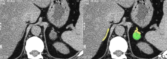
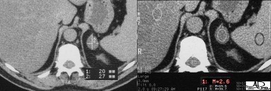
An “incidentaloma” is the most common adrenal tumor. It represents a small benign adenoma, (“oma” = tumor) of no functional significance, which is found incidentally, and hence its name. A nodule that is less than 3cm. with a density of less than 10 HU on CT, is diagnostic of the “incidentaloma”. The characteristic finding on MRI is a darkening in the “out of phase” T1 sequence of the tumor .
Anatomy and Physiology of the Adrenal Glands: Bilateral Adrenal Masses
In a patient with a known malignancy, particularly in lung or breast carcinomas, the finding of bilateral adrenal masses most likely represents metastatic disease. However, documenting the entity of metastatic adrenal disease is sometimes a decision focus for the type of treatment a patient may receive. In these circumstances, an adrenal biopsy must be performed .
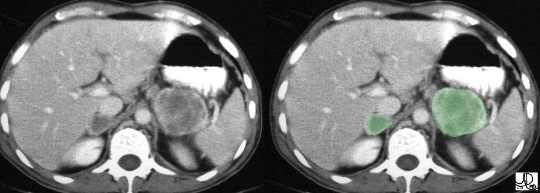
The finding of bilateral masses (green overlay), in a patient with a known malignancy most likely represents metastatic disease. This patient has known lung carcinoma and bilateral masses with peripheral enhancement, which almost certainly represents metastatic disease.Courtesy of: Ashley Davidoff, M.D.
The most common causes of bilateral adrenal enlargement include:
metastases (especially lung and breast)
bilateral adenomas,
hemorrhage; spontaneous,: (particularly in infants),
traumatic, and bleeding disorders.
Less common causes include:
· histoplasmosis and tuberculosis
· neuroblastoma
· pheochromocytoma
Additionally, rare causes include:
· Addison’s disease
· amyloidosis
· lymphoma

This is a rare case of bilateral adrenal enlargement in a patient with end stage lymphoma.Courtesy of: Ashley Davidoff, M.D.
Calcification in the Adrenal Glands
Courtesy of: Ashley Davidoff, M.D.
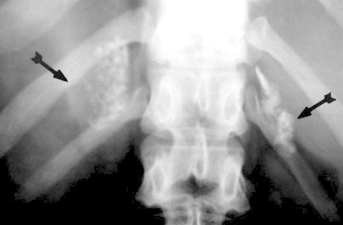
The most common cause of adrenal calcification is:
hemorrhage (occurring in the perinatal period) or
trauma.
Other less common entities producing calcifications in the adrenal include:
· histoplasmosis
· neuroblastomas
· tuberculosis
Uncommon causes include:
· Addison’s disease
· amyloidosis
· Cushing’s syndrome
· cysts
· neoplasms
· pheochromocytomas
The idiopathic causes have no known etiology.
Anatomy and Physiology of the Adrenal Glands: Cyst in the Adrenal
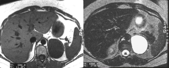
Cysts in the adrenal are not uncommon and they may be quite large. Adrenal cysts are fluid containing structures enclosed by a thin smooth wall. In general, adrenal cysts have no functional or clinical significance. MRI is the best method of confirming their make up and morphology.
CT Imaging of the Adrenal Glands: Overview
The adrenal gland is surrounded by a natural contrast agent – retroperitoneal fat. The soft tissue density (grey) of the adrenal is easily identified in marked contrast to the black background of the fat. CT is a powerful tool that is well equipped to define the morphology of the gland, but it falls short of MRI in the characterization of a few lesions including cystic lesions and pheochromocytoma.Intravenous contrast is generally not required to define the morphology of the gland. It is particularly important not to use contrast when there is a question of pheochromocytoma, since the contrast can induce a fatal hypertensive crisis.
Optimal collimation is key when studying the adrenal. The adrenals are best visualized with contiguous 3mm. sections (sometimes retrospectively viewed at 1.5mm collimation)through the entire gland.Adrenal CT is included in the staging evaluation of a variety of tumors, particularly bronchogenic carcinoma. Thus the technologist should ensure that both the adrenals are visualised with appropriate collimation in their entirety in cancer staging examinations. In fact, if you “pick up” an unsuspected lung mass on any CT scan, it would be very helpful to include the adrenals using 3mm collimation.
It is also important to know that some tumors are exophytic, meaning they hang off the edges of the main gland. Seeing a normal gland on one cut does not mean the gland is normal. The glands have to be examined from top to bottom.
CT Imaging of the Adrenal Glands: Normal Character on CT Scan
The adrenal gland is of soft tissue density on non contrast scan, measuring between 10 and 30 HU on the non contrast study. Because of its rich blood supply, a contrast study will enhance the gland significantly, particularly in stressful situations where blood flow to the gland is increased.
CT Imaging of the Adrenal Glands: Abnormal Adrenal on CT scan
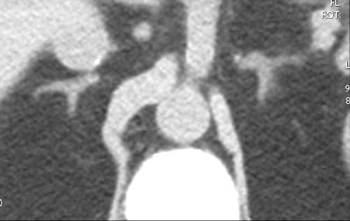
CT is a highly sensitive modality, although slightly less specific than MRI. The CT may be able to see a disorder, but often cannot exactly define the disorder. However, the CT can easily identify fat, which is present in conditions such as incidentaloma and myelolipoma. If an incidentaloma is suspected, non-contrast study is indicated. CT is more sensitive to the presence of calcification than MRI.It is very important for the radiologist to make a distinction between unilateral disease and bilateral disease. Cushing?s disease, for example, affects both adrenal glands and treatment may relate to surgical excision of a tumor of the pituitary gland. A unilateral nodule in Conn’s syndrome would require excision only of the affected adrenal gland.
In adrenal hemorrhage, serial CT examination will reveal evolution and usually resorption, and hence a progressive decrease in the size of the gland. The issue of adrenal mass or hemorrhage arises in the neonatal setting as well as following trauma in the adult.
MR Imaging of the Adrenal Glands: Overview
MRI is slightly more specific than CT but less sensitive to morphologic disorder. It is well suited to characterize fat, hemorrhage, and cystic changes, and MRI is the study of choice in patients with suspected pheochromocytoma or adrenal cysts. The advantage of being able to acquire images in many planes enables the radiologist and referring physicians to have a different perspective that sometimes provides relevant clinical information.MRI of the pituitary is superior to CT imaging, which has relevance in adrenal disease that is secondary to pituitary disease. Hence, in planning trans-sphenoidal surgery of the pituitary in patients with pituitary adenoma and resultant Cushing’s disease, it would be important to visualise the optic chiasm, the optic nerves, and the cavernous sinuses. This evaluation is best performed by MRI.
If a study of the adrenals is required during pregnancy, MRI is the diagnostic option of choice.
Visualization of calcification and bony structures is limited on MRI. On the other hand, MRI is very useful for defining vascular structures and the perfusional nature of adrenal lesions.
MR Imaging of the Adrenal Glands: Normal Character on MRI
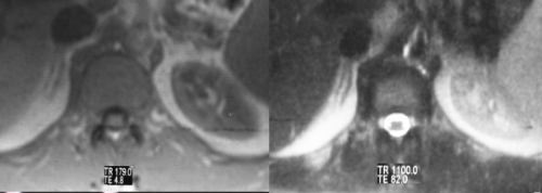
Because the normal adrenal gland is surrounded by retroperitoneal fat, the relatively low intensity of the gland is seen in sharp contrast to the fat, particularly on T1 weighted images. MRI is best used in the characterization of adrenal gland masses.
The T1-weighted sequence is performed to optimize the morphology of the gland and the “in-phase” and “out-of-phase” techniques are also useful in defining the incidentaloma. The T2-weighted sequence enables the characterization of lesions with high water content as well as pheochromocytomas.


MR Imaging of the Adrenal Glands: Abnormal Adrenal on MRI
MRI is very useful in the relatively frequently asked indication to “rule out pheochromocytoma”. It is said that the adrenal gland is like a “lightbulb” on T2-weighted imaging in patients with pheochromocytoma – meaning of course that it is very bright. This is not always the case in pheochromocytoma, but if you really want to see a lightbulb, take a look at the adrenal cyst. It will hurt your eyes.
“In-phase” and “out-of-phase” T1 sequences are sensitive to the presence of steroidal fat present in the incidentaloma, and MRI competes well with CT for the diagnosis of this condition. Characterization of another fatty lesion called the myelolipoma is also easily solved by fat sensitive MRI sequences.

US Imaging of the Adrenal Glands: Overview
Ultrasound is quick, and a relatively inexpensive technology, but it has significant limitations in adrenal scanning. The adrenal glands are difficult to examine by ultrasound because of their small size, their location high in the abdomen under the rib cage, and the presence of retroperitoneal fat and bowel gas. In thin patients, the normal adrenal may be identified.An anterior or lateral approach may be necessary, particularly for the right adrenal gland, which occasionally can be seen through the liver. Bowel gas often impairs visualization of the left adrenal gland. A variety of patient positions and scanning windows are often required to adequately examine the glands.
Since CT and MRI have so many diagnostic advantages, ultrasound is not used as the primary modality for the adrenal glands. Ultrasound can help in the characterization of large adrenal masses, as well as to define whether a suprarenal mass originates from the kidney or the adrenal.

This image represents a solid mass that is half the size of the kidney below the mass. A mass of this size typically indicates a primary carcinoma, however, this case represents a large pheochromocytoma.Courtesy of: Ashley Davidoff, M.D.
Conclusion: Final Thoughts
The adrenal glands are small and mighty. We started this module with that as the opening statement and we end with the same statement. Hidden in the depths of the body – these seemingly insignificant structures play an essential role. One only has to witness or read of the dire consequences of an “adrenal crisis”, the manifestations of the Waterhouse-Frederichsen syndrome or the “Addisonian crisis” to know and understand the part these two little glands play and what happens when they fail.The glands are often difficult to find on imaging. The left gland is best found on the axial cuts showing the splenic vein, and the right is found on the axial cuts showing the IVC emerging from the liver. Once a wisp of the gland is seen it should be followed up and down till it can no longer be seen. Nodules and masses of the adrenals are notorious for hanging out of the gland (exophytic) and a gland cannot be considered normal until its full extent has been scanned up and down.
The incidentaloma, or benign adenoma, is the most common mass of the adrenal and proof of a lipid substrate in the tumor makes a significant contribution to diagnosis and patient management. A pheochromocytoma crisis may be precipitated by administration of iodinated contrast, and although CT is our preferred modality for the adrenals, pheochromocytoma is best evaluated by MRI or MIBG nuclear scan.
The distinction between the cortex and medulla has eluded conventional imaging. Multidetector scanning is enabling better vascular imaging, and since the cortex is an arterial part of the gland and the medulla, dominantly a venous structure, the arterial phase should enable identification of the enhanced cortex and unenhanced medulla. We are looking forward to the possible potential of the advancing technology to unfold the mysteries that still hide in the adrenals.
References: Reference Material
· Abeloff. (2000). Clinical Oncology (2nd ed.). New York: W. B. Saunders.
· Brenner, & Rector. (2000). The Kidney (6th ed.). New York: W. B. Saunders.
· Cotran. (1999). Robbins Pathologic Basis of Disease (6th ed.). New York: W. B. Saunders.
· Goldman. (2000). Cecil Textbook of Medicine (21th ed.). New York: W. B. Saunders.
· Gray’s Anatomy (38th ed.). (1995). Churchill Livingstone.
· Moore. (1999). Clinically Oriented Anatomy (4th ed.). Lippincott, Williams & Wilkins.
· Townsend. (2001). Sabiston Textbook of Surgery (16th ed.). New York: W. B. Saunders.
· Walsh. (1998). Campbell’s Urology (7th ed.). New York: W. B. Saunders.
· Wilson. (1998). Williams Textbook of Endocrinology (9th ed.). New York: W. B. Saunders.

