effusion pleural (small, large, subpulmonic, decubitus films, supine and upright)
ground glass opacity (GGO)
mass solid lung mass
nodule solitary pulmonary (spn)
pneumothorax
pneumothorax tension
silhouette sign
white out opacification distinguishing causes of hemithorax (effusion, vs vs pneumonia vs pneumonectomy)
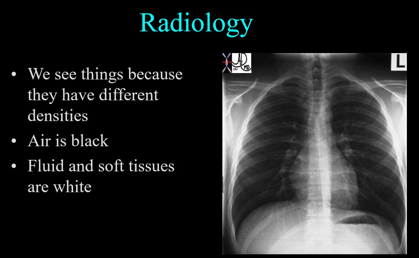
Why We See Things?
Contrasts Differences and Visibility Ashley Davidoff thecommonvein.net How many squares Ashley Davidoff thecommonvein.net How many squares Ashley Davidoff thecommonvein.net
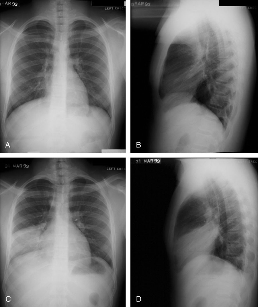
Silhouetting
NORMAL AND SILHOUETTING OF THE LEFT DIAPHGRAGM Silhouette sign, right middle lobe pneumonia. Initial frontal (A) and lateral (B) radiographs in a patient with clinical suspicion of pneumonia demonstrate obliteration of the right heart border. Follow-up radiographs the next day (C, D) illustrate dense opacification on the lateral view and persisting loss of the right heart border, confirming the presence of a right middle lobe pneumococcal pneumonia. Source Signs in Thoracic Imaging Journal of Thoracic Imaging 21(1):76-90, March 2006.
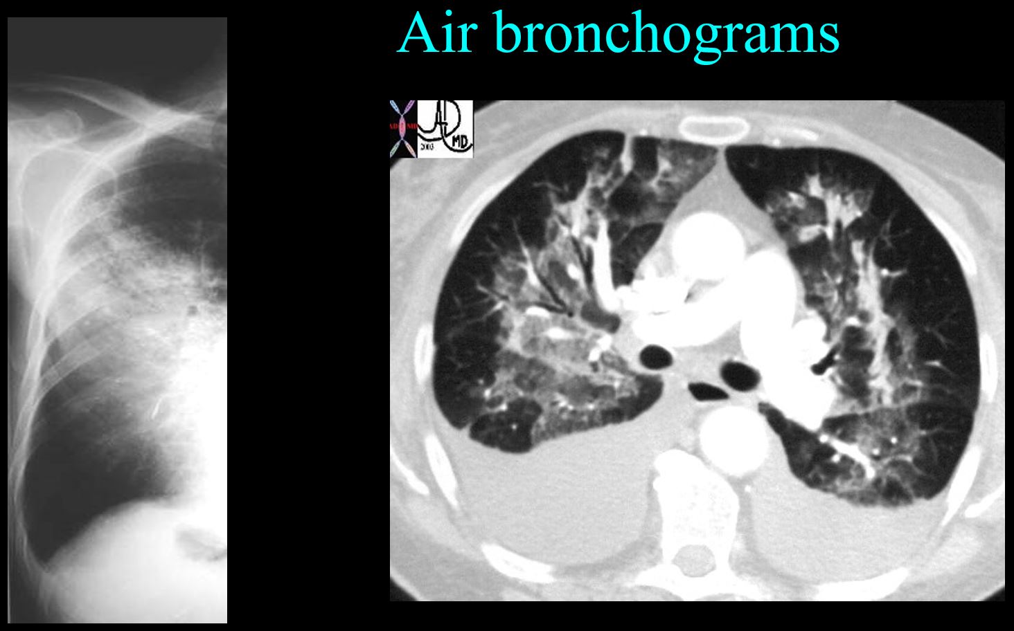
Air Bronchograms Ashley Davidoff thecommonvein.net
How to Approach the Field of Radiology
My Story – Curiosity
Sherlock Holmes Not invisible but unnoticed Watson…. Ashley Davidoff thecommonvein.net
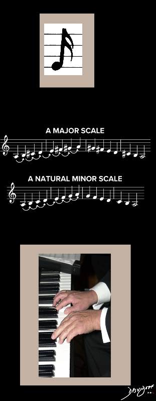
In the pursuit of excellence of piano playing … principles are
Notes Scales and Music
THE ART OF PERFECTING MUSIC Ashley Davidoff Scales from MusicNotes.com thecommonvein.net
THE ART OF PERFECTING DANCE Ashley Davidoff thecommonvein.net THE ART OF PERFECTING TENNIS Ashley Davidoff thecommonvein.net
TIGER WOODS AND TOM BRADY
NOTES SCALES AND MUSIC IN THE PURSUIT OF PERFECTION
Both images are in the public domain
Story of Dr Jerry Balikian
Application to Radiology
Where to look, how to look and
what to look for
Ashley Davidoff thecommonvein.net
Notes
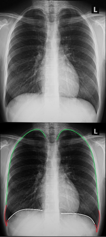
Anatomy of the Lungs
Frontal Examination of the Lungs
THE HEART, LUNGS, PLEURA, DIAPHRAGM, ARTERIES, VEINS, PROXIMAL TRACHEOBRONCHIAL TREE and SKELETON
Ashley Davidoff MD
Parts of the Lungs- Basics
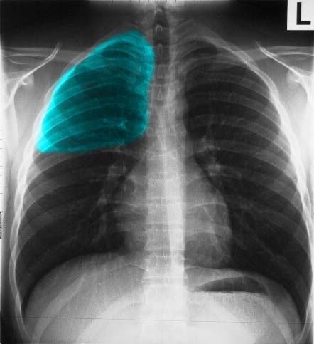
Left Lung ? Left Upper Lobe ? Frontal Projection
LEFT UPPER LOBE IN THE FRONTAL PROJECTION
Ashley Davidoff MD
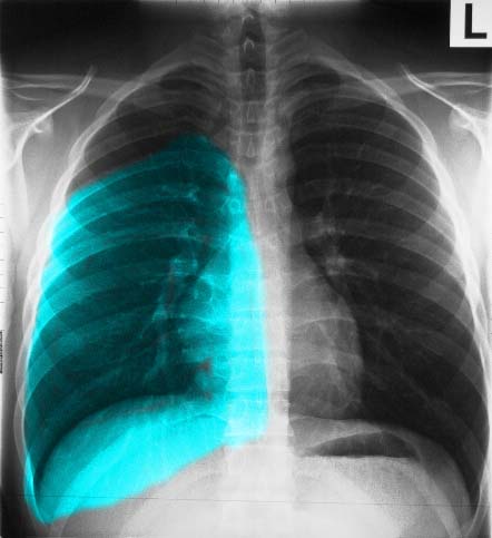
Frontal Projection Left Lower Lobe
LEFT LOWER LOBE IN THE FRONTAL PROJECTION
Ashley Davidoff MD
Right Lung ? Right Upper Lobe ? Frontal Projection
RIGHT UPPER LOBE IN THE FRONTAL PROJECTION
Ashley Davidoff MD
Right Middle Lobe ? Frontal Projection
RIGHT MIDDLE LOBE IN THE FRONTAL PROJECTION
Ashley Davidoff MD
Right Lower Lobe ? Frontal Projection
RIGHT MIDDLE LOBE IN THE FRONTAL PROJECTION
Ashley Davidoff MD
Lateral Examination of the Lungs
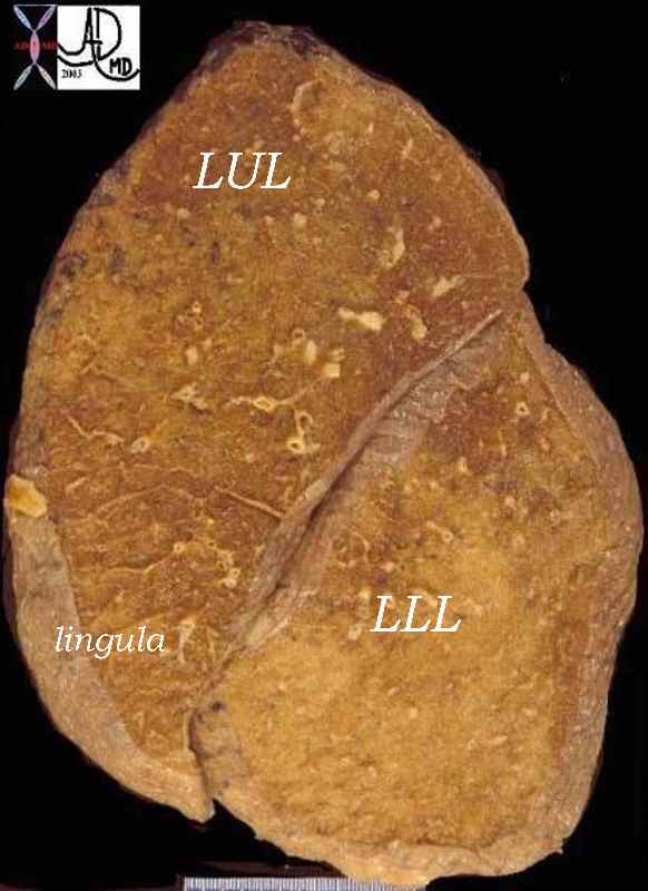
Major Fissure on the Left
LATERAL PROJECTION ? FISSURE DIVIDES THE LEFT UPPER LOBE (INCLUDING LINGULA) AND LEFT LOWER LOBE
Ashley Davidoff MDMAJOR FISSURE ON THE LEFT
Ashley Davidoff MD
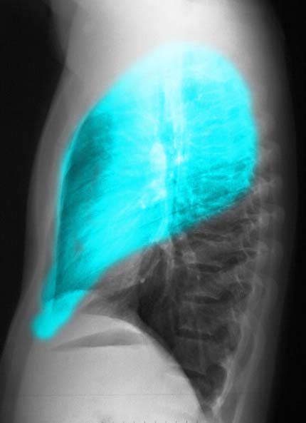
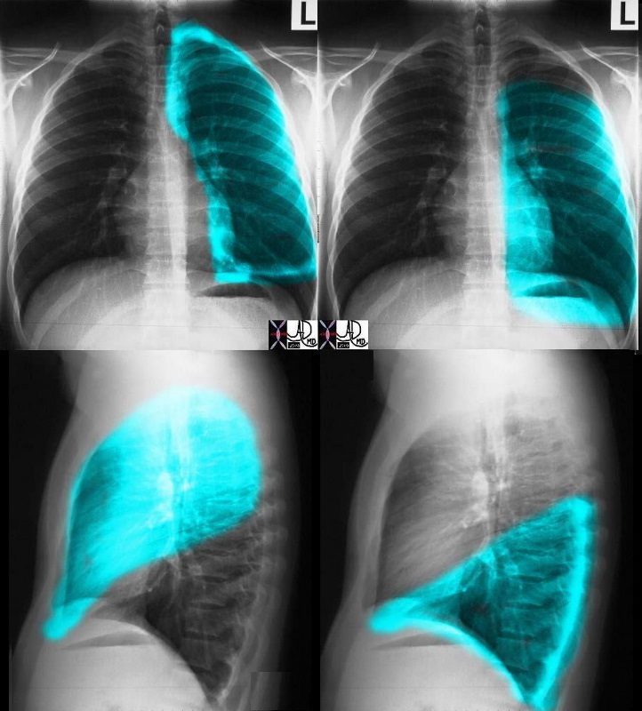
Lateral Projection ? Left Upper Lobe
LATERAL PROJECTION, LEFT UPPER LOBE
Ashley Davidoff MD
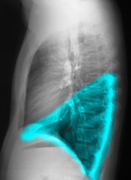
Lateral Projection left Lower Lobe
LATERAL CXR SHOWING LEFT LOWER LOBE
Ashley Davidoff MD
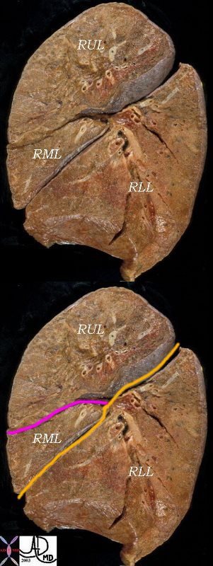
Major and Minor Fissures on the Right
ANATOMIC SPECIMEN SHOWING MAJOR and MINOR FISSURES DIVIDING THE RIGHT LUNG INTO 3 LOBES
The right lung has a small right upper lobe (RUL) separated from the middle lobe (RML) by the minor fissure (pink,lower image) . Both the RUL and RML are anterior and are separated from the lower lobe by the major fissure (orange line)
Ashley Davidoff MDLATERAL X-RAY SHOWING MAJOR and MINOR FISSURES DIVIDING THE RIGHT LUNG INTO 3 LOBES
The right lung has a relatively small right upper lobe (RUL) separated from the middle lobe (RML) by the minor fissure (pink,lower image). Both the RUL and RML are anterior and are separated from the lower lobe by the major fissure (orange line)
Ashley Davidoff MD
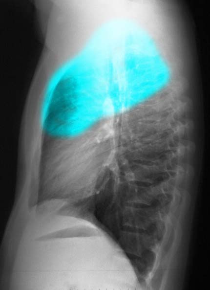
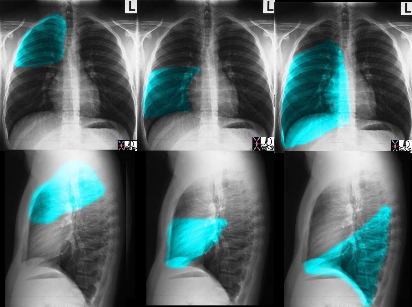
Right Upper Lobe ? Lateral Projection
LATERAL CXR SHOWING RIGHT UPPER LOBE
Ashley Davidoff MD
Right Middle Lobe ? Lateral Projection
LATERAL CXR SHOWING RIGHT MIDDLE LOBE
Ashley Davidoff MD
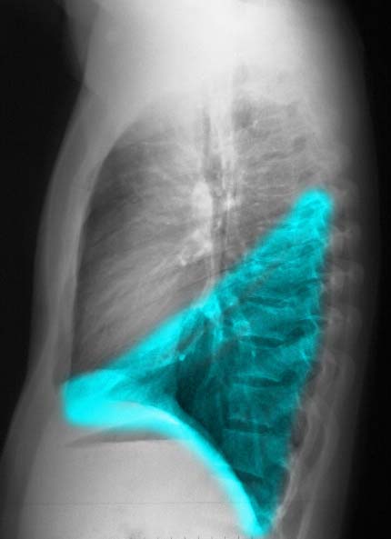
Right Lower Lobe ? Lateral Projection
LATERAL CXR SHOWING RIGHT LOWER LOBE
Ashley Davidoff MD
Summary
CXR of LEFT LUNG
Ashley Davidoff MD
CXR of RIGHT LUNG
Ashley Davidoff MD
Trachea, Main Stem Bronchi, and Carina
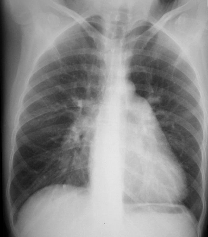
NORMAL FRONTAL CXR NORMAL ASYMMETRIC BRANCHING OF MAINSTEM BRONCHI The normal CXR shows the characteristic asymmetric branching of the main stem bronchi. The right is short and stout and slightly more vertical while the left is long and thin and slightly more obtuse. The normal carinal angle is between 40-80 degrees. Ashley Davidoff MD
ASYMMETRIC BRANCHING PATTERN ? RIGHT SHORT AND STOUT AND THE LEFT LONG AND THIN
CARINAL ANGLE ? 40-80 degrees
Ashley Davidoff MDTHE RIGHT ? SHORT STOUT AND CUTE
THE LEFT ? TALL THIN AND GRACILE
The carinal angleOLIVER HARDY AND STAN LAUREL IN THE 1939 FILM ? THE FLYING DEUCESSCREW IN THE RIGHT MAIN STEM
http://www.wikiradiography.net/N-G TUBE IN RIGHT MAIN STEM BRONCHUS
Courtesy Radiopaedia
ET TUBE IN RIGHT MAIN STEM BRONCHUS
Anatomy of the Heart
Chambers that are Border Forming on the PA Examination
FRONTAL CXR AND PARTS OF THE HEART
If we were to ?crack open? the chest of the chest X-ray, the structures that would dominate this bloody, black and white scene, would be the right sided chambers. The right ventricle (RV) would be the dominant anterior chamber, and would form the dominant interface with the diaphragm. The right atrium (RA) would form the border with the right lung. The RA would of course be slightly posterior to the RV. The left border would be formed by the left ventricle. Most the left ventricle is hidden posteriorly in this view. The left anterior descending artery would be visible from this anterior view. It marks the position of the interventricular septum.
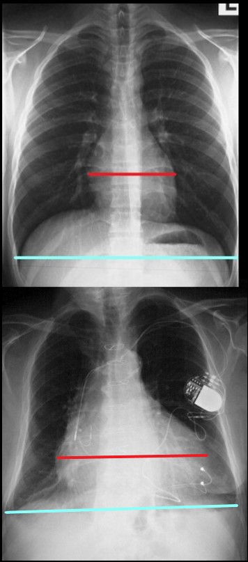
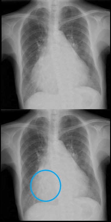
Ashley Davidoff MD CARDIOMEGALY – THE CARDIOTHORACIC RATIO The maximum transverse length of the heart is expressed as a percentage of the maximum length of the internal diameter of the chest. When this ratio – the cardiothoracic ratio (c t r) is greater than 50% cardiomegaly is present. The top image is normal and the bottom reflects cardiomegaly Ashley Davidoff MD
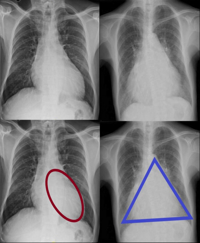
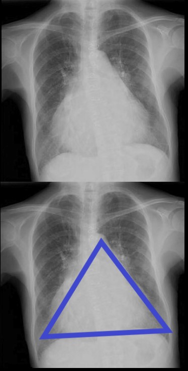
There are Two Basic Common Shapes of Cardiomegaly
CARDIOMEGALY ? TWO BASIC TYPES -OVOID and TRIANGULAR The ovoid form which suggests left ventricular dominance and triangular form which suggests right ventricular dominance. Ashley Davidoff MD
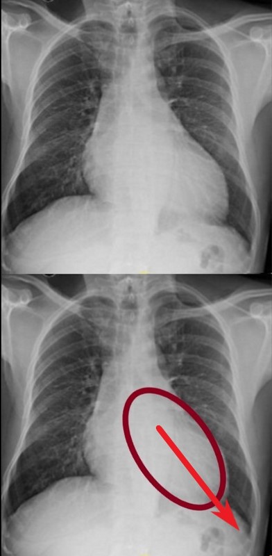
The Ovoid Form of Cardiomegaly ? Consider LVE
SHAPE OF THE LEFT VENTRICLE
The enlarged LV (a,b) is shaped like an oval and it is likened to a rugby ball or an American football placed on the field at kick off time. LVE on CXR is mostly assessed by an increased cardiothoracic ratio as well as the accentuation of the ovoid shape. (lower images c, d,e, f)
Ashley Davidoff MD
Vector of the Enlarging LV ? Rotate Down and to the Left
VECTOR FOR LV ENLARGEMENT DOWN AND OUT The left ventricle (LV) enlarges in a posterior, downward and lateral direction resulting in the characteristic changes of LVE on CXR Ashley Davidoff MD
CLINICAL EVALUATION OF LVE ? DOWN AND OUT
The left ventricle (LV) enlarges in a downward and lateral direction resulting in the apical impulse displacement and increase forcefulness of the apical tap.
Ashley Davidoff MD
ISCHEMIC CARDIOMYOPATHY S/P RCA OCCLUSION 62 year old female with acute chest pain atrial fibrillation, hypotension admitted to ICU. Clinical evaluation was considered to be non-ischemic cardiomyopathy with EF by echo of about 20%. She was hypotensive and, in the ICU, and CXR showed acute CHF with cardiomegaly. The TEE was more in keeping with segmental dyssynergy, Cardiac cath showed occluded RCA bot good collateralization from the LAD. MRI showed subendocardial LGE in the inferior and inferolateral portions of the LV consistent with a prior infarction and EF of 20% Ashley Davidoff MD
The Triangular Form of Cardiomegaly
Consider RVE or any disease from the left side that may cause RVE eg mitral stenosis
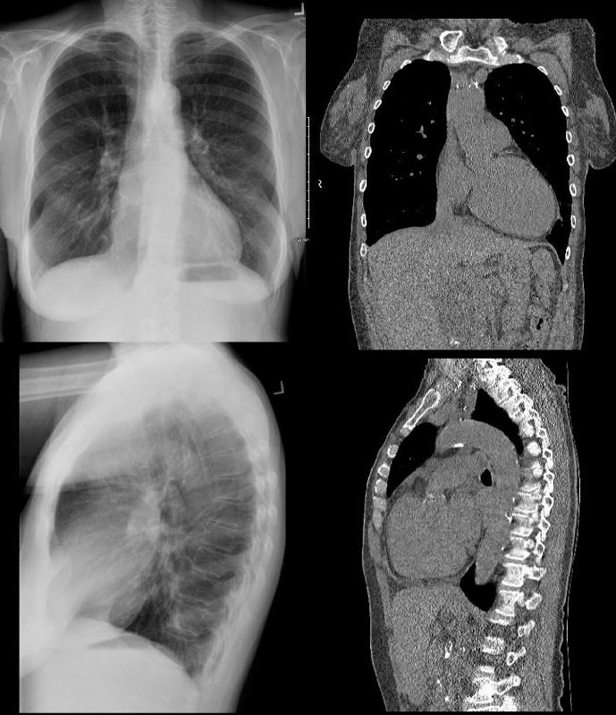
TRIANGULAR SHAPED HEART ? SUGGESTING RIGHT VENTRICULAR DOMINANCE. MITRAL STENOSIS PULMONARY HYPERTENSION 71 year old Asian female with rheumatic heart disease dominated by calcific mitral stenosis mild MR, moderate tricuspid regurgitation and secondary pulmonary hypertension. Ashley Davidoff MD
Note the Basic Shape of the RV is Triangular in almost all Views
SHAPE OF THE LEFT VENTRICLE
The enlarged LV (a,b) is shaped like an oval and it is likened to a rugby ball or an American football placed on the field at kick off time. LVE on CXR is mostly assessed by an increased cardiothoracic ratio as well as the accentuation of the ovoid shap. (lower images c, d,e, f)
Ashley Davidoff MD
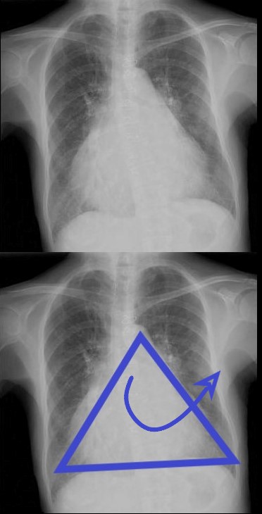
Vector of the Enlarging RV ? Rotate Laterally to the Right
VECTOR FOR RV ENLARGEMENT
ANTERIOR LEFTWARD
The right ventricle (RV) enlarges with a clockwise rotation resulting in an upward turning of the apex and enlargement in a anterior and leftward lateral direction.
Ashley Davidoff MD
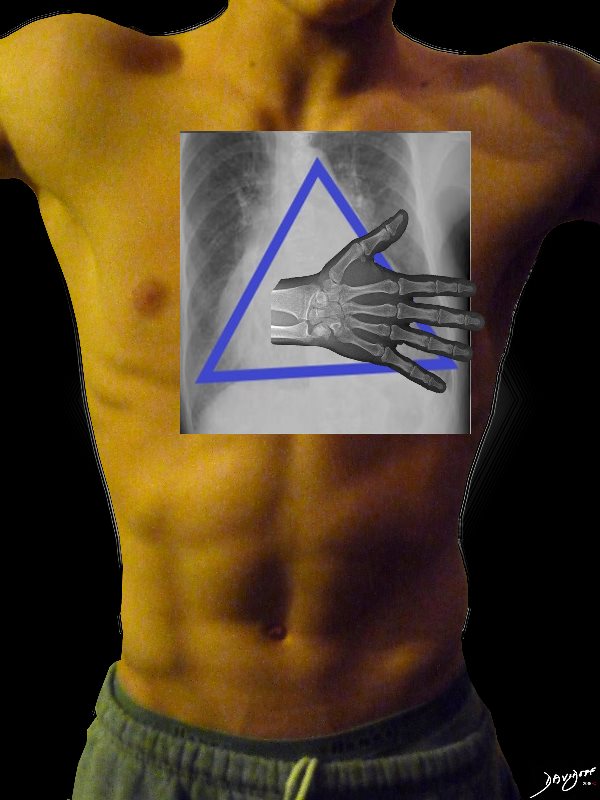
Clinical Exam of the RV ? Diffuse Anterior Parasternal Heave
CLINICAL EVALUATION OF RVE ? PARASTERNAL HEAVE
The right ventricle (RV) enlarges in a anterior, upward and lateral direction resulting in the broad based parasternal pulsation which on clinical examination is identified as a parasternal heave identified with the base of the extended hand
Ashley Davidoff MD
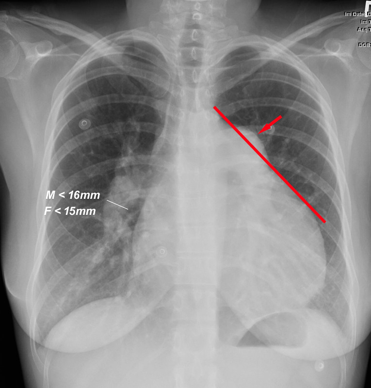
Normal Main Pulmonary Artery When a line is drawn from the aortic knob to the left edge of the heart, (red line) the pulmonary artery should lie medial to that line (ie along the line drawn to 1.5cms medial to the line) Ashley Davidoff MD TheCommonVein.net
Pulmonary Hypertension
Pulmonary Arteries in Pulmonary Hypertension When a line is drawn from the aortic knob to the left edge of the heart, (red line) the pulmonary artery lies lateral to that line indicating an enlarged pulmonary artery most commonly caused by hypertension . In this instance the size of the descending right pulmonary artery is greater than 15 mms confirming the presence of pulmonary hypertension Ashley Davidoff MD TheCommonVein.net PULMONARY HYPERTENSION Frontal x-ray with triangular shaped heart due to pulmonary hypertension with enlarged MPA and enlarged descending RPA . Ashley Davidoff MD
What about the Right Atrium (RA) and Right Heart Border?
The RA does not make much a statement on the frontal CXR unless very large
FRONTAL CXR AND PARTS OF THE HEART
If we were to ?crack open? the chest of the chest X-ray, the structures that would dominate this bloody, black and white scene, would be the right sided chambers. The right ventricle (RV) would be the dominant anterior chamber, and would form the dominant interface with the diaphragm. The right atrium (RA) would form the border with the right lung. The RA would of course be slightly posterior to the RV. The left border would be formed by the left ventricle. Most the left ventricle is hidden posteriorly in this view. The left anterior descending artery would be visible from this anterior view. It marks the position of the interventricular septum.
Ashley Davidoff MD
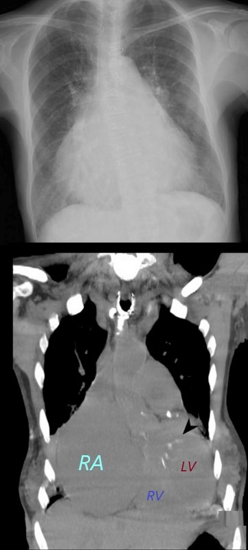
The Enlarged Right Atrium
The right atrium is the most difficult chamber to assess unless it is very large in which case it will manifest on the frontal CXR with a very large right paravertebral border.
enlarged, globular heart
narrow pedicle
gross enlargement of the right atrial shadow, i.e. increased convexity in the lower half of the right cardiac border
right atrial convexity is more than 50% of the cardiovascular height
right atrial margin is more than 5.5 cm from the midline
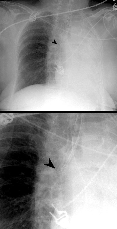
RIGHT ATRIAL ENLARGEMENT ON FRONTAL X-RAY
The right atrium is the most difficult chamber to assess unless it is very large in which case it will present on the frontal CXR with a very large right paravertebral border. This is a 71 year old female person with rheumatic heart disease with pulmonary hypertension and tricuspid regurgitation hence resulting in a large right atrium (RAE)
Ashley Davidoff MDRIGHT ATRIAL ENLARGEMENT ON FRONTAL X-RAY
The right atrium is the most difficult chamber to assess unless it is very large in which case it will present on the frontal CXR with a very large right paravertebral border. The frontal CXR and coronal CT through the RA is from a 71 year old female with rheumatic heart disease with pulmonary hypertension and tricuspid regurgitation resulting in a giant right atrium (RAE). The RA accounts for the large bulge of the right border of the cardiac silhouette. The black arrowhead in the loer image points to the calcified mitral valve.
Ashley Davidoff MD
NORMAL FRONTAL CXR NORMAL ASYMMETRIC BRANCHING OF MAINSTEM BRONCHI
The normal CXR shows the characteristic asymmetric branching of the main stem bronchi. The right is short and stout and slightly more vertical while the left is long and thin and slightly more obtuse.
The normal carinal angle is between 40-80 degrees.
Ashley Davidoff MD
ASYMMETRIC BRANCHING PATTERN ? RIGHT SHORT AND STOUT AND THE LEFT LONG AND THIN
CARINAL ANGLE ? 40-80 degrees
Ashley Davidoff MD
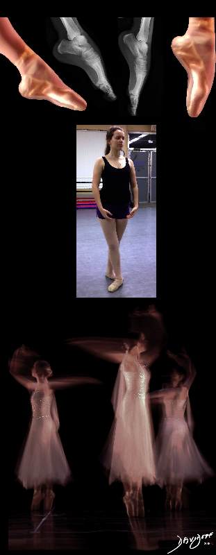
The Abnormal Carinal Angle
NORMAL AND WIDENED CARINAL ANGLE
A dancer demonstrates a normal carinal angle (upper image) and as she continues to extend her left leg, (lower images) the angle becomes greater than 80 degrees and in terms of the carinal angle becomes abnormal.
Ashley Davidoff MD
MITRAL STENOSIS WITH ENLARGED LEFT ATRIUM ? WIDENED CARINAL ANGLE DOUBLE DENSITY ENLARGED LEFT ATRIAL APPENDAGE
The frontal CXR demonstrates findings consistent with mitral stenosis including a widened carinal angle (teal blue and black arc), a double density (red arc) and an enlarged left atrial appendage (maroon arc).
The overall shape of the heart is triangular suggesting right ventricular enlargement. A mitral valve prosthesis is in position
Courtesy of Radiopaedia
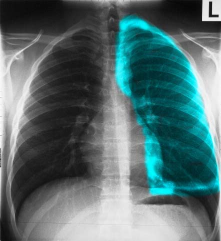
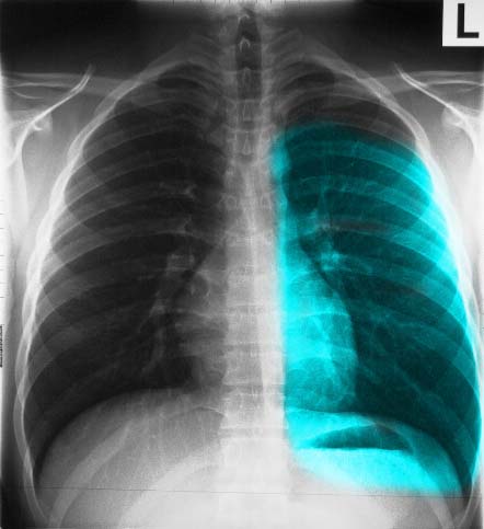
SMALL APICAL PNEUMOTHORAX IN AN UPRIGHT CXR TAKEN IN EXPIRATION. In an upright position a pneumothorax rises to the apex of the lung and assumes the shape of the apex because it exerts pressure on the lung apex which yield to the greater pressure. The expiration film accentuates the pneumothorax because it further reduces the pressure in the lungs and increases the pressure difference between the PTX and the intraparenchymal pressure. The PTX is barely seen in (a) and is better seen in the magnified views (b and c) and with increasing contrast (c) the faint line of the the pleura becomes better visualized (white arrowheads). Ashley Davidoff MD
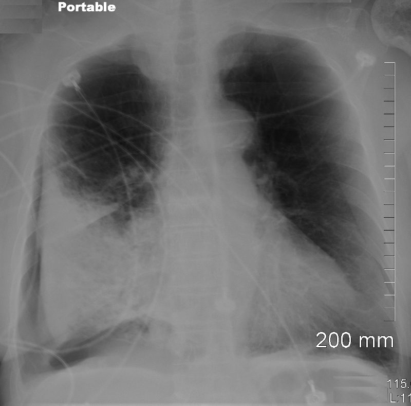
DEEP SULCUS SIGN Portable supine examination in the ICU shows a pneumothorax in the right subpulmonic region Ashley Davidoff MD Spontaneous Tension Pneumothorax 49 year old male with a cough presents for a Chest Xray which showed a tension pneumothorax. Chest tube was placed emergently in the radiology department. Ashley Davidoff MD TheCommonVein.net 117300c
Pleural Effusion
Fluid in the Upright and Supine Projection Ashley Davidoff thecommonvein.net
Small Effusion May Only Seen on the Lateral Exam
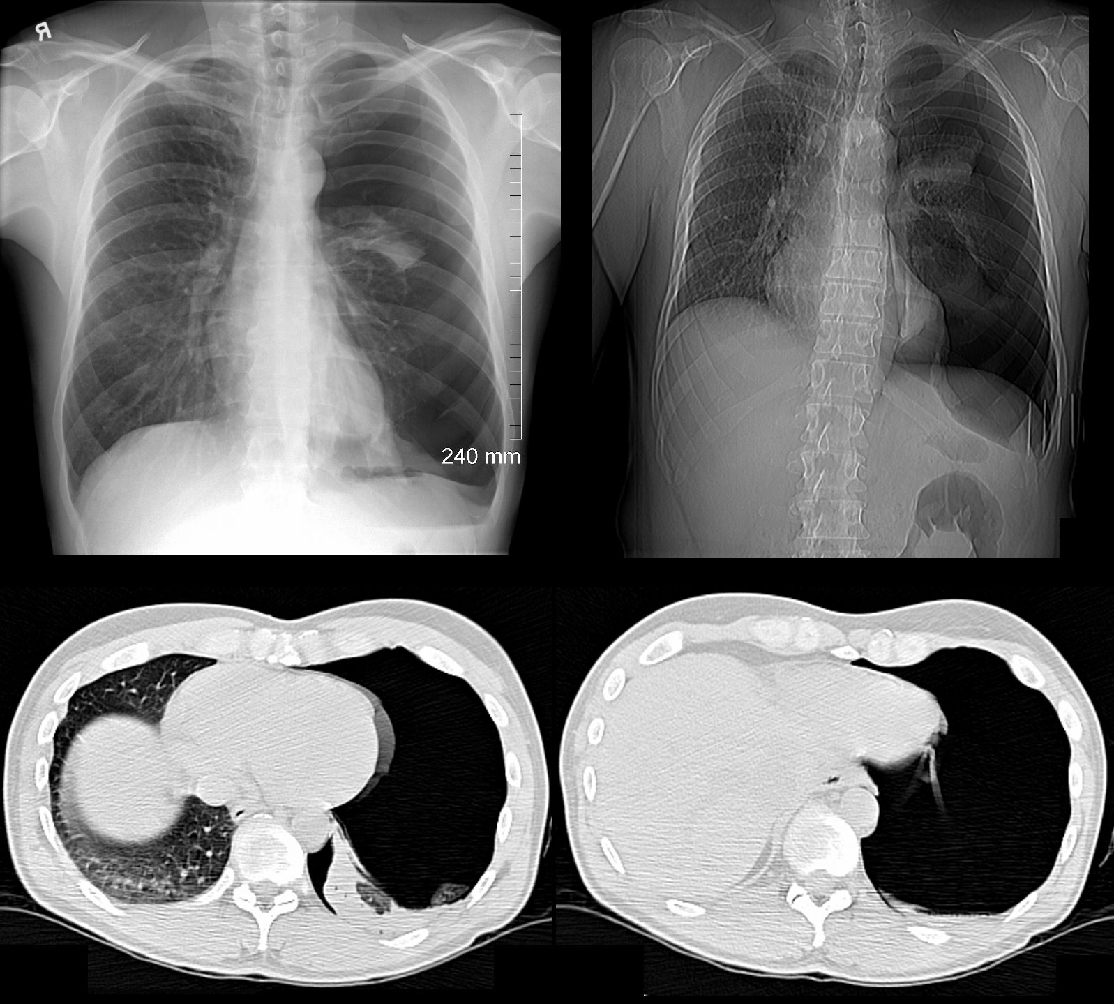
SMALL EFFUSION ONLY IDENTIFIED ON THE LATERAL EXAMINATION A small effusion is not identified on the PA chest since it is hidden by the diaphragm and the effusion first fills the posterior recess because it is most inferior. The lateral examination is required to identify the effusion Courtesy How to Interpret CXR Strong Medicine https://www.youtube.com/watch?v=wOpDvUO5sD8
Typical shape of a moderately large right pleural effusion Case courtesy of RMH Core Conditions, Radiopaedia.org, rID: 34401
Veiling Opacity
Veiling Opacity left pleural effusion in the supine projection Case courtesy of RMH Core Conditions, Radiopaedia.org, rID: 34401
Scales
Learning how to look so that you can see
How to look and what to look for Ashley Davidoff thecommonvein.net
Notes Scales and Music
Scales
Have a fluid and logical method of looking (search pattern) and
practice practice practice
SCALES
4 Review Spaces relating to 4 areas of major disease
Pleura – PTX and Effusions
Lungs – Pneumonia and Masses
Hila – Masses and CHF
Heart – Megalies and Failure
First Scale – Pleural Run
PANORAMA WALK FOR PLEURAL ASSESSMENT Ashley Davidoff MD
Pattern – start at the white lines to the right and left of the thoracic vertebra running along the diaphragms into the pleural recesses, up the lateral walls to the apices
2nd Scale – Lung Loops
LUNG LOOPS – UPPER MID AND LOWER Ashley Davidoff MD
Pattern –
Come down the trachea and and
loop the upper lung fields, then the
mid lung fields, and finally the
lower lung fields
looking for
symmetry,
masses,
infiltrates,
interstitial changes
3rd Scale – Skiing on the Moguls
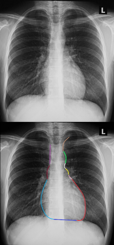
SKI TRIP DOWN THE MOGULS OF THE HEART A methodical approach to evaluation of the cardiac silhouette is likened to skiing down a mogul laden ski slope and then taking a trip on the ski lift back to the top of the mountain. The ski slope starts at the left subclavian artery (light brown) , followed by the mogul of the aortic knob (bright green) at the bottom of which is the A-P window (white) only to be presented with a second mogul of the main pulmonary artery(yellow), and then the bay of the left atrial appendage (pink)and finally free at last of moguls and an exciting and accelerating ski down the LV (red) We then have to take a walk back to the ski lift. At the junction of the LV (red) and the RV (blue) , if we take a right ward look up the mountain we can spot the LAD on top of the interventricular septum. The walk along the border of the RV is terminated at the junction of the RV (dark blue) and the right atrium (light blue). At this point we wait in line to get on to the ski lift. We ride up the right hand border of the right atrium (light blue) a little rough bump over the ascending aorta )maroon) and then straight to the top along the SVC (pink). Ashley Davidoff MD
Pattern of the Ski Run –
Start at the left apex at the left subclavian artery,
jump the aortic mogul and
land in the AP window, and then
jump the PA mogul,
land in the LA appendiceal bay, and then
ski all the way down the LV.
Walk back to the ski lift via the inferior border of the RV and then the RA.
At the bottom of the RA get on to the ski lift as the IVC enters the RA
and then take the lift up the mountain via the RA ,
portion of the ascending aorta and the
the SVC
4th Scale – Hilar Hoops
Pattern of the Hilar Hoops –
Trip down the trachea – Specifically to look at the carinal angle
Upper hoop specifically to look at pa- bronchus ratio
Middle Hoop – Specifically to look at hilar size and shape
Lower Hoop – Specifically to look for redistribution and clarity of RPA
Pericardial Effusion
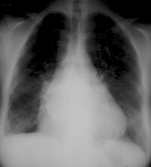
WATER BOTTLE HEART X-ray shows the typical appearance of a “water bottle heart? characteristic of a pericardial of a large pericardial effusion. Ashley Davidoff MD
sn chest integrated 09ep finding br3 imaging
Word First
Definition
air bronchogram
atelectasis
bleb
<1-2cms
bronchiectasis
bulla
>1-2cms
congestive heart failure (CHF)
consolidation
effusion pleural (small, large, subpulmonic, decubitus films, supine and upright)
ground glass opacity (GGO)
mass solid lung mass
>3cms
nodule solitary pulmonary (spn)
<3cms
pneumothorax (small, large, supine and upright, decubitus and expiratory films, tension)
pneumothorax tension
silhouette sign
white out opacification distinguishing causes of hemithorax (effusion, vs vs pneumonia vs pneumonectomy)
DOMElement Object
(
    [schemaTypeInfo] =>
    [tagName] => td
    [firstElementChild] => (object value omitted)
    [lastElementChild] => (object value omitted)
    [childElementCount] => 1
    [previousElementSibling] =>
    [nextElementSibling] => (object value omitted)
    [nodeName] => td
    [nodeValue] => white out opacification distinguishing causes of hemithorax (effusion, vs vs pneumonia vs pneumonectomy)
    [nodeType] => 1
    [parentNode] => (object value omitted)
    [childNodes] => (object value omitted)
    [firstChild] => (object value omitted)
    [lastChild] => (object value omitted)
    [previousSibling] => (object value omitted)
    [nextSibling] => (object value omitted)
    [attributes] => (object value omitted)
    [ownerDocument] => (object value omitted)
    [namespaceURI] =>
    [prefix] =>
    [localName] => td
    [baseURI] =>
    [textContent] => white out opacification distinguishing causes of hemithorax (effusion, vs vs pneumonia vs pneumonectomy)
)
DOMElement Object
(
    [schemaTypeInfo] =>
    [tagName] => td
    [firstElementChild] => (object value omitted)
    [lastElementChild] => (object value omitted)
    [childElementCount] => 1
    [previousElementSibling] => (object value omitted)
    [nextElementSibling] =>
    [nodeName] => td
    [nodeValue] =>
    [nodeType] => 1
    [parentNode] => (object value omitted)
    [childNodes] => (object value omitted)
    [firstChild] => (object value omitted)
    [lastChild] => (object value omitted)
    [previousSibling] => (object value omitted)
    [nextSibling] => (object value omitted)
    [attributes] => (object value omitted)
    [ownerDocument] => (object value omitted)
    [namespaceURI] =>
    [prefix] =>
    [localName] => td
    [baseURI] =>
    [textContent] =>
)
DOMElement Object
(
    [schemaTypeInfo] =>
    [tagName] => td
    [firstElementChild] => (object value omitted)
    [lastElementChild] => (object value omitted)
    [childElementCount] => 1
    [previousElementSibling] =>
    [nextElementSibling] => (object value omitted)
    [nodeName] => td
    [nodeValue] => silhouette sign
    [nodeType] => 1
    [parentNode] => (object value omitted)
    [childNodes] => (object value omitted)
    [firstChild] => (object value omitted)
    [lastChild] => (object value omitted)
    [previousSibling] => (object value omitted)
    [nextSibling] => (object value omitted)
    [attributes] => (object value omitted)
    [ownerDocument] => (object value omitted)
    [namespaceURI] =>
    [prefix] =>
    [localName] => td
    [baseURI] =>
    [textContent] => silhouette sign
)
DOMElement Object
(
    [schemaTypeInfo] =>
    [tagName] => td
    [firstElementChild] => (object value omitted)
    [lastElementChild] => (object value omitted)
    [childElementCount] => 1
    [previousElementSibling] => (object value omitted)
    [nextElementSibling] =>
    [nodeName] => td
    [nodeValue] =>
    [nodeType] => 1
    [parentNode] => (object value omitted)
    [childNodes] => (object value omitted)
    [firstChild] => (object value omitted)
    [lastChild] => (object value omitted)
    [previousSibling] => (object value omitted)
    [nextSibling] => (object value omitted)
    [attributes] => (object value omitted)
    [ownerDocument] => (object value omitted)
    [namespaceURI] =>
    [prefix] =>
    [localName] => td
    [baseURI] =>
    [textContent] =>
)
DOMElement Object
(
    [schemaTypeInfo] =>
    [tagName] => td
    [firstElementChild] => (object value omitted)
    [lastElementChild] => (object value omitted)
    [childElementCount] => 1
    [previousElementSibling] =>
    [nextElementSibling] => (object value omitted)
    [nodeName] => td
    [nodeValue] => pneumothorax tension
    [nodeType] => 1
    [parentNode] => (object value omitted)
    [childNodes] => (object value omitted)
    [firstChild] => (object value omitted)
    [lastChild] => (object value omitted)
    [previousSibling] => (object value omitted)
    [nextSibling] => (object value omitted)
    [attributes] => (object value omitted)
    [ownerDocument] => (object value omitted)
    [namespaceURI] =>
    [prefix] =>
    [localName] => td
    [baseURI] =>
    [textContent] => pneumothorax tension
)
DOMElement Object
(
    [schemaTypeInfo] =>
    [tagName] => td
    [firstElementChild] => (object value omitted)
    [lastElementChild] => (object value omitted)
    [childElementCount] => 1
    [previousElementSibling] => (object value omitted)
    [nextElementSibling] =>
    [nodeName] => td
    [nodeValue] =>
    [nodeType] => 1
    [parentNode] => (object value omitted)
    [childNodes] => (object value omitted)
    [firstChild] => (object value omitted)
    [lastChild] => (object value omitted)
    [previousSibling] => (object value omitted)
    [nextSibling] => (object value omitted)
    [attributes] => (object value omitted)
    [ownerDocument] => (object value omitted)
    [namespaceURI] =>
    [prefix] =>
    [localName] => td
    [baseURI] =>
    [textContent] =>
)
DOMElement Object
(
    [schemaTypeInfo] =>
    [tagName] => td
    [firstElementChild] => (object value omitted)
    [lastElementChild] => (object value omitted)
    [childElementCount] => 1
    [previousElementSibling] =>
    [nextElementSibling] => (object value omitted)
    [nodeName] => td
    [nodeValue] => pneumothorax (small, large, supine and upright, decubitus and expiratory films, tension)
    [nodeType] => 1
    [parentNode] => (object value omitted)
    [childNodes] => (object value omitted)
    [firstChild] => (object value omitted)
    [lastChild] => (object value omitted)
    [previousSibling] => (object value omitted)
    [nextSibling] => (object value omitted)
    [attributes] => (object value omitted)
    [ownerDocument] => (object value omitted)
    [namespaceURI] =>
    [prefix] =>
    [localName] => td
    [baseURI] =>
    [textContent] => pneumothorax (small, large, supine and upright, decubitus and expiratory films, tension)
)
DOMElement Object
(
    [schemaTypeInfo] =>
    [tagName] => td
    [firstElementChild] => (object value omitted)
    [lastElementChild] => (object value omitted)
    [childElementCount] => 1
    [previousElementSibling] =>
    [nextElementSibling] => (object value omitted)
    [nodeName] => td
    [nodeValue] => nodule solitary pulmonary (spn)
    [nodeType] => 1
    [parentNode] => (object value omitted)
    [childNodes] => (object value omitted)
    [firstChild] => (object value omitted)
    [lastChild] => (object value omitted)
    [previousSibling] => (object value omitted)
    [nextSibling] => (object value omitted)
    [attributes] => (object value omitted)
    [ownerDocument] => (object value omitted)
    [namespaceURI] =>
    [prefix] =>
    [localName] => td
    [baseURI] =>
    [textContent] => nodule solitary pulmonary (spn)
)
DOMElement Object
(
    [schemaTypeInfo] =>
    [tagName] => td
    [firstElementChild] => (object value omitted)
    [lastElementChild] => (object value omitted)
    [childElementCount] => 1
    [previousElementSibling] => (object value omitted)
    [nextElementSibling] =>
    [nodeName] => td
    [nodeValue] => >3cms
    [nodeType] => 1
    [parentNode] => (object value omitted)
    [childNodes] => (object value omitted)
    [firstChild] => (object value omitted)
    [lastChild] => (object value omitted)
    [previousSibling] => (object value omitted)
    [nextSibling] => (object value omitted)
    [attributes] => (object value omitted)
    [ownerDocument] => (object value omitted)
    [namespaceURI] =>
    [prefix] =>
    [localName] => td
    [baseURI] =>
    [textContent] => >3cms
)
DOMElement Object
(
    [schemaTypeInfo] =>
    [tagName] => td
    [firstElementChild] => (object value omitted)
    [lastElementChild] => (object value omitted)
    [childElementCount] => 1
    [previousElementSibling] =>
    [nextElementSibling] => (object value omitted)
    [nodeName] => td
    [nodeValue] => mass solid lung mass
    [nodeType] => 1
    [parentNode] => (object value omitted)
    [childNodes] => (object value omitted)
    [firstChild] => (object value omitted)
    [lastChild] => (object value omitted)
    [previousSibling] => (object value omitted)
    [nextSibling] => (object value omitted)
    [attributes] => (object value omitted)
    [ownerDocument] => (object value omitted)
    [namespaceURI] =>
    [prefix] =>
    [localName] => td
    [baseURI] =>
    [textContent] => mass solid lung mass
)
DOMElement Object
(
    [schemaTypeInfo] =>
    [tagName] => td
    [firstElementChild] => (object value omitted)
    [lastElementChild] => (object value omitted)
    [childElementCount] => 1
    [previousElementSibling] => (object value omitted)
    [nextElementSibling] =>
    [nodeName] => td
    [nodeValue] =>
    [nodeType] => 1
    [parentNode] => (object value omitted)
    [childNodes] => (object value omitted)
    [firstChild] => (object value omitted)
    [lastChild] => (object value omitted)
    [previousSibling] => (object value omitted)
    [nextSibling] => (object value omitted)
    [attributes] => (object value omitted)
    [ownerDocument] => (object value omitted)
    [namespaceURI] =>
    [prefix] =>
    [localName] => td
    [baseURI] =>
    [textContent] =>
)
DOMElement Object
(
    [schemaTypeInfo] =>
    [tagName] => td
    [firstElementChild] => (object value omitted)
    [lastElementChild] => (object value omitted)
    [childElementCount] => 1
    [previousElementSibling] =>
    [nextElementSibling] => (object value omitted)
    [nodeName] => td
    [nodeValue] => ground glass opacity (GGO)
    [nodeType] => 1
    [parentNode] => (object value omitted)
    [childNodes] => (object value omitted)
    [firstChild] => (object value omitted)
    [lastChild] => (object value omitted)
    [previousSibling] => (object value omitted)
    [nextSibling] => (object value omitted)
    [attributes] => (object value omitted)
    [ownerDocument] => (object value omitted)
    [namespaceURI] =>
    [prefix] =>
    [localName] => td
    [baseURI] =>
    [textContent] => ground glass opacity (GGO)
)
DOMElement Object
(
    [schemaTypeInfo] =>
    [tagName] => td
    [firstElementChild] => (object value omitted)
    [lastElementChild] => (object value omitted)
    [childElementCount] => 1
    [previousElementSibling] => (object value omitted)
    [nextElementSibling] =>
    [nodeName] => td
    [nodeValue] =>
    [nodeType] => 1
    [parentNode] => (object value omitted)
    [childNodes] => (object value omitted)
    [firstChild] => (object value omitted)
    [lastChild] => (object value omitted)
    [previousSibling] => (object value omitted)
    [nextSibling] => (object value omitted)
    [attributes] => (object value omitted)
    [ownerDocument] => (object value omitted)
    [namespaceURI] =>
    [prefix] =>
    [localName] => td
    [baseURI] =>
    [textContent] =>
)
DOMElement Object
(
    [schemaTypeInfo] =>
    [tagName] => td
    [firstElementChild] => (object value omitted)
    [lastElementChild] => (object value omitted)
    [childElementCount] => 1
    [previousElementSibling] =>
    [nextElementSibling] => (object value omitted)
    [nodeName] => td
    [nodeValue] => effusion pleural (small, large, subpulmonic, decubitus films, supine and upright)
    [nodeType] => 1
    [parentNode] => (object value omitted)
    [childNodes] => (object value omitted)
    [firstChild] => (object value omitted)
    [lastChild] => (object value omitted)
    [previousSibling] => (object value omitted)
    [nextSibling] => (object value omitted)
    [attributes] => (object value omitted)
    [ownerDocument] => (object value omitted)
    [namespaceURI] =>
    [prefix] =>
    [localName] => td
    [baseURI] =>
    [textContent] => effusion pleural (small, large, subpulmonic, decubitus films, supine and upright)
)
DOMElement Object
(
    [schemaTypeInfo] =>
    [tagName] => td
    [firstElementChild] => (object value omitted)
    [lastElementChild] => (object value omitted)
    [childElementCount] => 1
    [previousElementSibling] => (object value omitted)
    [nextElementSibling] =>
    [nodeName] => td
    [nodeValue] =>
    [nodeType] => 1
    [parentNode] => (object value omitted)
    [childNodes] => (object value omitted)
    [firstChild] => (object value omitted)
    [lastChild] => (object value omitted)
    [previousSibling] => (object value omitted)
    [nextSibling] => (object value omitted)
    [attributes] => (object value omitted)
    [ownerDocument] => (object value omitted)
    [namespaceURI] =>
    [prefix] =>
    [localName] => td
    [baseURI] =>
    [textContent] =>
)
DOMElement Object
(
    [schemaTypeInfo] =>
    [tagName] => td
    [firstElementChild] => (object value omitted)
    [lastElementChild] => (object value omitted)
    [childElementCount] => 1
    [previousElementSibling] =>
    [nextElementSibling] => (object value omitted)
    [nodeName] => td
    [nodeValue] => consolidation
    [nodeType] => 1
    [parentNode] => (object value omitted)
    [childNodes] => (object value omitted)
    [firstChild] => (object value omitted)
    [lastChild] => (object value omitted)
    [previousSibling] => (object value omitted)
    [nextSibling] => (object value omitted)
    [attributes] => (object value omitted)
    [ownerDocument] => (object value omitted)
    [namespaceURI] =>
    [prefix] =>
    [localName] => td
    [baseURI] =>
    [textContent] => consolidation
)
DOMElement Object
(
    [schemaTypeInfo] =>
    [tagName] => td
    [firstElementChild] => (object value omitted)
    [lastElementChild] => (object value omitted)
    [childElementCount] => 1
    [previousElementSibling] => (object value omitted)
    [nextElementSibling] =>
    [nodeName] => td
    [nodeValue] =>
    [nodeType] => 1
    [parentNode] => (object value omitted)
    [childNodes] => (object value omitted)
    [firstChild] => (object value omitted)
    [lastChild] => (object value omitted)
    [previousSibling] => (object value omitted)
    [nextSibling] => (object value omitted)
    [attributes] => (object value omitted)
    [ownerDocument] => (object value omitted)
    [namespaceURI] =>
    [prefix] =>
    [localName] => td
    [baseURI] =>
    [textContent] =>
)
DOMElement Object
(
    [schemaTypeInfo] =>
    [tagName] => td
    [firstElementChild] => (object value omitted)
    [lastElementChild] => (object value omitted)
    [childElementCount] => 1
    [previousElementSibling] =>
    [nextElementSibling] => (object value omitted)
    [nodeName] => td
    [nodeValue] => congestive heart failure (CHF)
    [nodeType] => 1
    [parentNode] => (object value omitted)
    [childNodes] => (object value omitted)
    [firstChild] => (object value omitted)
    [lastChild] => (object value omitted)
    [previousSibling] => (object value omitted)
    [nextSibling] => (object value omitted)
    [attributes] => (object value omitted)
    [ownerDocument] => (object value omitted)
    [namespaceURI] =>
    [prefix] =>
    [localName] => td
    [baseURI] =>
    [textContent] => congestive heart failure (CHF)
)
DOMElement Object
(
    [schemaTypeInfo] =>
    [tagName] => td
    [firstElementChild] => (object value omitted)
    [lastElementChild] => (object value omitted)
    [childElementCount] => 1
    [previousElementSibling] => (object value omitted)
    [nextElementSibling] =>
    [nodeName] => td
    [nodeValue] => >1-2cms
    [nodeType] => 1
    [parentNode] => (object value omitted)
    [childNodes] => (object value omitted)
    [firstChild] => (object value omitted)
    [lastChild] => (object value omitted)
    [previousSibling] => (object value omitted)
    [nextSibling] => (object value omitted)
    [attributes] => (object value omitted)
    [ownerDocument] => (object value omitted)
    [namespaceURI] =>
    [prefix] =>
    [localName] => td
    [baseURI] =>
    [textContent] => >1-2cms
)
DOMElement Object
(
    [schemaTypeInfo] =>
    [tagName] => td
    [firstElementChild] => (object value omitted)
    [lastElementChild] => (object value omitted)
    [childElementCount] => 1
    [previousElementSibling] =>
    [nextElementSibling] => (object value omitted)
    [nodeName] => td
    [nodeValue] => bulla
    [nodeType] => 1
    [parentNode] => (object value omitted)
    [childNodes] => (object value omitted)
    [firstChild] => (object value omitted)
    [lastChild] => (object value omitted)
    [previousSibling] => (object value omitted)
    [nextSibling] => (object value omitted)
    [attributes] => (object value omitted)
    [ownerDocument] => (object value omitted)
    [namespaceURI] =>
    [prefix] =>
    [localName] => td
    [baseURI] =>
    [textContent] => bulla
)
DOMElement Object
(
    [schemaTypeInfo] =>
    [tagName] => td
    [firstElementChild] => (object value omitted)
    [lastElementChild] => (object value omitted)
    [childElementCount] => 1
    [previousElementSibling] => (object value omitted)
    [nextElementSibling] =>
    [nodeName] => td
    [nodeValue] =>
    [nodeType] => 1
    [parentNode] => (object value omitted)
    [childNodes] => (object value omitted)
    [firstChild] => (object value omitted)
    [lastChild] => (object value omitted)
    [previousSibling] => (object value omitted)
    [nextSibling] => (object value omitted)
    [attributes] => (object value omitted)
    [ownerDocument] => (object value omitted)
    [namespaceURI] =>
    [prefix] =>
    [localName] => td
    [baseURI] =>
    [textContent] =>
)
DOMElement Object
(
    [schemaTypeInfo] =>
    [tagName] => td
    [firstElementChild] => (object value omitted)
    [lastElementChild] => (object value omitted)
    [childElementCount] => 1
    [previousElementSibling] =>
    [nextElementSibling] => (object value omitted)
    [nodeName] => td
    [nodeValue] => bronchiectasis
    [nodeType] => 1
    [parentNode] => (object value omitted)
    [childNodes] => (object value omitted)
    [firstChild] => (object value omitted)
    [lastChild] => (object value omitted)
    [previousSibling] => (object value omitted)
    [nextSibling] => (object value omitted)
    [attributes] => (object value omitted)
    [ownerDocument] => (object value omitted)
    [namespaceURI] =>
    [prefix] =>
    [localName] => td
    [baseURI] =>
    [textContent] => bronchiectasis
)
DOMElement Object
(
    [schemaTypeInfo] =>
    [tagName] => td
    [firstElementChild] => (object value omitted)
    [lastElementChild] => (object value omitted)
    [childElementCount] => 1
    [previousElementSibling] =>
    [nextElementSibling] => (object value omitted)
    [nodeName] => td
    [nodeValue] => bleb
    [nodeType] => 1
    [parentNode] => (object value omitted)
    [childNodes] => (object value omitted)
    [firstChild] => (object value omitted)
    [lastChild] => (object value omitted)
    [previousSibling] => (object value omitted)
    [nextSibling] => (object value omitted)
    [attributes] => (object value omitted)
    [ownerDocument] => (object value omitted)
    [namespaceURI] =>
    [prefix] =>
    [localName] => td
    [baseURI] =>
    [textContent] => bleb
)
DOMElement Object
(
    [schemaTypeInfo] =>
    [tagName] => td
    [firstElementChild] => (object value omitted)
    [lastElementChild] => (object value omitted)
    [childElementCount] => 1
    [previousElementSibling] => (object value omitted)
    [nextElementSibling] =>
    [nodeName] => td
    [nodeValue] =>
    [nodeType] => 1
    [parentNode] => (object value omitted)
    [childNodes] => (object value omitted)
    [firstChild] => (object value omitted)
    [lastChild] => (object value omitted)
    [previousSibling] => (object value omitted)
    [nextSibling] => (object value omitted)
    [attributes] => (object value omitted)
    [ownerDocument] => (object value omitted)
    [namespaceURI] =>
    [prefix] =>
    [localName] => td
    [baseURI] =>
    [textContent] =>
)
DOMElement Object
(
    [schemaTypeInfo] =>
    [tagName] => td
    [firstElementChild] => (object value omitted)
    [lastElementChild] => (object value omitted)
    [childElementCount] => 1
    [previousElementSibling] =>
    [nextElementSibling] => (object value omitted)
    [nodeName] => td
    [nodeValue] => atelectasis
    [nodeType] => 1
    [parentNode] => (object value omitted)
    [childNodes] => (object value omitted)
    [firstChild] => (object value omitted)
    [lastChild] => (object value omitted)
    [previousSibling] => (object value omitted)
    [nextSibling] => (object value omitted)
    [attributes] => (object value omitted)
    [ownerDocument] => (object value omitted)
    [namespaceURI] =>
    [prefix] =>
    [localName] => td
    [baseURI] =>
    [textContent] => atelectasis
)
DOMElement Object
(
    [schemaTypeInfo] =>
    [tagName] => td
    [firstElementChild] => (object value omitted)
    [lastElementChild] => (object value omitted)
    [childElementCount] => 1
    [previousElementSibling] => (object value omitted)
    [nextElementSibling] =>
    [nodeName] => td
    [nodeValue] =>
    [nodeType] => 1
    [parentNode] => (object value omitted)
    [childNodes] => (object value omitted)
    [firstChild] => (object value omitted)
    [lastChild] => (object value omitted)
    [previousSibling] => (object value omitted)
    [nextSibling] => (object value omitted)
    [attributes] => (object value omitted)
    [ownerDocument] => (object value omitted)
    [namespaceURI] =>
    [prefix] =>
    [localName] => td
    [baseURI] =>
    [textContent] =>
)
DOMElement Object
(
    [schemaTypeInfo] =>
    [tagName] => td
    [firstElementChild] => (object value omitted)
    [lastElementChild] => (object value omitted)
    [childElementCount] => 1
    [previousElementSibling] =>
    [nextElementSibling] => (object value omitted)
    [nodeName] => td
    [nodeValue] => air bronchogram
    [nodeType] => 1
    [parentNode] => (object value omitted)
    [childNodes] => (object value omitted)
    [firstChild] => (object value omitted)
    [lastChild] => (object value omitted)
    [previousSibling] => (object value omitted)
    [nextSibling] => (object value omitted)
    [attributes] => (object value omitted)
    [ownerDocument] => (object value omitted)
    [namespaceURI] =>
    [prefix] =>
    [localName] => td
    [baseURI] =>
    [textContent] => air bronchogram
)
DOMElement Object
(
    [schemaTypeInfo] =>
    [tagName] => table
    [firstElementChild] => (object value omitted)
    [lastElementChild] => (object value omitted)
    [childElementCount] => 3
    [previousElementSibling] => (object value omitted)
    [nextElementSibling] => (object value omitted)
    [nodeName] => table
    [nodeValue] =>


Word First
Definition


air bronchogram


atelectasis


bleb


bronchiectasis


bulla


congestive heart failure (CHF)


consolidation


effusion pleural (small, large, subpulmonic, decubitus films, supine and upright)


ground glass opacity (GGO)


mass solid lung mass


nodule solitary pulmonary (spn)


pneumothorax


pneumothorax tension


silhouette sign


white out opacification distinguishing causes of hemithorax (effusion, vs vs pneumonia vs pneumonectomy)


    [nodeType] => 1
    [parentNode] => (object value omitted)
    [childNodes] => (object value omitted)
    [firstChild] => (object value omitted)
    [lastChild] => (object value omitted)
    [previousSibling] => (object value omitted)
    [nextSibling] => (object value omitted)
    [attributes] => (object value omitted)
    [ownerDocument] => (object value omitted)
    [namespaceURI] =>
    [prefix] =>
    [localName] => table
    [baseURI] =>
    [textContent] =>


Word First
Definition


air bronchogram


atelectasis


bleb


bronchiectasis


bulla


congestive heart failure (CHF)


consolidation


effusion pleural (small, large, subpulmonic, decubitus films, supine and upright)


ground glass opacity (GGO)


mass solid lung mass


nodule solitary pulmonary (spn)


pneumothorax


pneumothorax tension


silhouette sign


white out opacification distinguishing causes of hemithorax (effusion, vs vs pneumonia vs pneumonectomy)


)
DOMElement Object
(
    [schemaTypeInfo] =>
    [tagName] => td
    [firstElementChild] => (object value omitted)
    [lastElementChild] => (object value omitted)
    [childElementCount] => 1
    [previousElementSibling] =>
    [nextElementSibling] => (object value omitted)
    [nodeName] => td
    [nodeValue] => white out opacification distinguishing causes of hemithorax (effusion, vs vs pneumonia vs pneumonectomy)
    [nodeType] => 1
    [parentNode] => (object value omitted)
    [childNodes] => (object value omitted)
    [firstChild] => (object value omitted)
    [lastChild] => (object value omitted)
    [previousSibling] => (object value omitted)
    [nextSibling] => (object value omitted)
    [attributes] => (object value omitted)
    [ownerDocument] => (object value omitted)
    [namespaceURI] =>
    [prefix] =>
    [localName] => td
    [baseURI] =>
    [textContent] => white out opacification distinguishing causes of hemithorax (effusion, vs vs pneumonia vs pneumonectomy)
)
DOMElement Object
(
    [schemaTypeInfo] =>
    [tagName] => td
    [firstElementChild] => (object value omitted)
    [lastElementChild] => (object value omitted)
    [childElementCount] => 1
    [previousElementSibling] => (object value omitted)
    [nextElementSibling] =>
    [nodeName] => td
    [nodeValue] =>
    [nodeType] => 1
    [parentNode] => (object value omitted)
    [childNodes] => (object value omitted)
    [firstChild] => (object value omitted)
    [lastChild] => (object value omitted)
    [previousSibling] => (object value omitted)
    [nextSibling] => (object value omitted)
    [attributes] => (object value omitted)
    [ownerDocument] => (object value omitted)
    [namespaceURI] =>
    [prefix] =>
    [localName] => td
    [baseURI] =>
    [textContent] =>
)
DOMElement Object
(
    [schemaTypeInfo] =>
    [tagName] => td
    [firstElementChild] => (object value omitted)
    [lastElementChild] => (object value omitted)
    [childElementCount] => 1
    [previousElementSibling] =>
    [nextElementSibling] => (object value omitted)
    [nodeName] => td
    [nodeValue] => silhouette sign
    [nodeType] => 1
    [parentNode] => (object value omitted)
    [childNodes] => (object value omitted)
    [firstChild] => (object value omitted)
    [lastChild] => (object value omitted)
    [previousSibling] => (object value omitted)
    [nextSibling] => (object value omitted)
    [attributes] => (object value omitted)
    [ownerDocument] => (object value omitted)
    [namespaceURI] =>
    [prefix] =>
    [localName] => td
    [baseURI] =>
    [textContent] => silhouette sign
)
DOMElement Object
(
    [schemaTypeInfo] =>
    [tagName] => td
    [firstElementChild] => (object value omitted)
    [lastElementChild] => (object value omitted)
    [childElementCount] => 1
    [previousElementSibling] => (object value omitted)
    [nextElementSibling] =>
    [nodeName] => td
    [nodeValue] =>
    [nodeType] => 1
    [parentNode] => (object value omitted)
    [childNodes] => (object value omitted)
    [firstChild] => (object value omitted)
    [lastChild] => (object value omitted)
    [previousSibling] => (object value omitted)
    [nextSibling] => (object value omitted)
    [attributes] => (object value omitted)
    [ownerDocument] => (object value omitted)
    [namespaceURI] =>
    [prefix] =>
    [localName] => td
    [baseURI] =>
    [textContent] =>
)
DOMElement Object
(
    [schemaTypeInfo] =>
    [tagName] => td
    [firstElementChild] => (object value omitted)
    [lastElementChild] => (object value omitted)
    [childElementCount] => 1
    [previousElementSibling] =>
    [nextElementSibling] => (object value omitted)
    [nodeName] => td
    [nodeValue] => pneumothorax tension
    [nodeType] => 1
    [parentNode] => (object value omitted)
    [childNodes] => (object value omitted)
    [firstChild] => (object value omitted)
    [lastChild] => (object value omitted)
    [previousSibling] => (object value omitted)
    [nextSibling] => (object value omitted)
    [attributes] => (object value omitted)
    [ownerDocument] => (object value omitted)
    [namespaceURI] =>
    [prefix] =>
    [localName] => td
    [baseURI] =>
    [textContent] => pneumothorax tension
)
DOMElement Object
(
    [schemaTypeInfo] =>
    [tagName] => td
    [firstElementChild] => (object value omitted)
    [lastElementChild] => (object value omitted)
    [childElementCount] => 1
    [previousElementSibling] => (object value omitted)
    [nextElementSibling] =>
    [nodeName] => td
    [nodeValue] =>
    [nodeType] => 1
    [parentNode] => (object value omitted)
    [childNodes] => (object value omitted)
    [firstChild] => (object value omitted)
    [lastChild] => (object value omitted)
    [previousSibling] => (object value omitted)
    [nextSibling] => (object value omitted)
    [attributes] => (object value omitted)
    [ownerDocument] => (object value omitted)
    [namespaceURI] =>
    [prefix] =>
    [localName] => td
    [baseURI] =>
    [textContent] =>
)
DOMElement Object
(
    [schemaTypeInfo] =>
    [tagName] => td
    [firstElementChild] => (object value omitted)
    [lastElementChild] => (object value omitted)
    [childElementCount] => 1
    [previousElementSibling] =>
    [nextElementSibling] => (object value omitted)
    [nodeName] => td
    [nodeValue] => pneumothorax
    [nodeType] => 1
    [parentNode] => (object value omitted)
    [childNodes] => (object value omitted)
    [firstChild] => (object value omitted)
    [lastChild] => (object value omitted)
    [previousSibling] => (object value omitted)
    [nextSibling] => (object value omitted)
    [attributes] => (object value omitted)
    [ownerDocument] => (object value omitted)
    [namespaceURI] =>
    [prefix] =>
    [localName] => td
    [baseURI] =>
    [textContent] => pneumothorax
)
DOMElement Object
(
    [schemaTypeInfo] =>
    [tagName] => td
    [firstElementChild] => (object value omitted)
    [lastElementChild] => (object value omitted)
    [childElementCount] => 1
    [previousElementSibling] => (object value omitted)
    [nextElementSibling] =>
    [nodeName] => td
    [nodeValue] =>
    [nodeType] => 1
    [parentNode] => (object value omitted)
    [childNodes] => (object value omitted)
    [firstChild] => (object value omitted)
    [lastChild] => (object value omitted)
    [previousSibling] => (object value omitted)
    [nextSibling] => (object value omitted)
    [attributes] => (object value omitted)
    [ownerDocument] => (object value omitted)
    [namespaceURI] =>
    [prefix] =>
    [localName] => td
    [baseURI] =>
    [textContent] =>
)
DOMElement Object
(
    [schemaTypeInfo] =>
    [tagName] => td
    [firstElementChild] => (object value omitted)
    [lastElementChild] => (object value omitted)
    [childElementCount] => 1
    [previousElementSibling] =>
    [nextElementSibling] => (object value omitted)
    [nodeName] => td
    [nodeValue] => nodule solitary pulmonary (spn)
    [nodeType] => 1
    [parentNode] => (object value omitted)
    [childNodes] => (object value omitted)
    [firstChild] => (object value omitted)
    [lastChild] => (object value omitted)
    [previousSibling] => (object value omitted)
    [nextSibling] => (object value omitted)
    [attributes] => (object value omitted)
    [ownerDocument] => (object value omitted)
    [namespaceURI] =>
    [prefix] =>
    [localName] => td
    [baseURI] =>
    [textContent] => nodule solitary pulmonary (spn)
)
DOMElement Object
(
    [schemaTypeInfo] =>
    [tagName] => td
    [firstElementChild] => (object value omitted)
    [lastElementChild] => (object value omitted)
    [childElementCount] => 1
    [previousElementSibling] => (object value omitted)
    [nextElementSibling] =>
    [nodeName] => td
    [nodeValue] =>
    [nodeType] => 1
    [parentNode] => (object value omitted)
    [childNodes] => (object value omitted)
    [firstChild] => (object value omitted)
    [lastChild] => (object value omitted)
    [previousSibling] => (object value omitted)
    [nextSibling] => (object value omitted)
    [attributes] => (object value omitted)
    [ownerDocument] => (object value omitted)
    [namespaceURI] =>
    [prefix] =>
    [localName] => td
    [baseURI] =>
    [textContent] =>
)
DOMElement Object
(
    [schemaTypeInfo] =>
    [tagName] => td
    [firstElementChild] => (object value omitted)
    [lastElementChild] => (object value omitted)
    [childElementCount] => 1
    [previousElementSibling] =>
    [nextElementSibling] => (object value omitted)
    [nodeName] => td
    [nodeValue] => mass solid lung mass
    [nodeType] => 1
    [parentNode] => (object value omitted)
    [childNodes] => (object value omitted)
    [firstChild] => (object value omitted)
    [lastChild] => (object value omitted)
    [previousSibling] => (object value omitted)
    [nextSibling] => (object value omitted)
    [attributes] => (object value omitted)
    [ownerDocument] => (object value omitted)
    [namespaceURI] =>
    [prefix] =>
    [localName] => td
    [baseURI] =>
    [textContent] => mass solid lung mass
)
DOMElement Object
(
    [schemaTypeInfo] =>
    [tagName] => td
    [firstElementChild] => (object value omitted)
    [lastElementChild] => (object value omitted)
    [childElementCount] => 1
    [previousElementSibling] => (object value omitted)
    [nextElementSibling] =>
    [nodeName] => td
    [nodeValue] =>
    [nodeType] => 1
    [parentNode] => (object value omitted)
    [childNodes] => (object value omitted)
    [firstChild] => (object value omitted)
    [lastChild] => (object value omitted)
    [previousSibling] => (object value omitted)
    [nextSibling] => (object value omitted)
    [attributes] => (object value omitted)
    [ownerDocument] => (object value omitted)
    [namespaceURI] =>
    [prefix] =>
    [localName] => td
    [baseURI] =>
    [textContent] =>
)
DOMElement Object
(
    [schemaTypeInfo] =>
    [tagName] => td
    [firstElementChild] => (object value omitted)
    [lastElementChild] => (object value omitted)
    [childElementCount] => 1
    [previousElementSibling] =>
    [nextElementSibling] => (object value omitted)
    [nodeName] => td
    [nodeValue] => ground glass opacity (GGO)
    [nodeType] => 1
    [parentNode] => (object value omitted)
    [childNodes] => (object value omitted)
    [firstChild] => (object value omitted)
    [lastChild] => (object value omitted)
    [previousSibling] => (object value omitted)
    [nextSibling] => (object value omitted)
    [attributes] => (object value omitted)
    [ownerDocument] => (object value omitted)
    [namespaceURI] =>
    [prefix] =>
    [localName] => td
    [baseURI] =>
    [textContent] => ground glass opacity (GGO)
)
DOMElement Object
(
    [schemaTypeInfo] =>
    [tagName] => td
    [firstElementChild] => (object value omitted)
    [lastElementChild] => (object value omitted)
    [childElementCount] => 1
    [previousElementSibling] => (object value omitted)
    [nextElementSibling] =>
    [nodeName] => td
    [nodeValue] =>
    [nodeType] => 1
    [parentNode] => (object value omitted)
    [childNodes] => (object value omitted)
    [firstChild] => (object value omitted)
    [lastChild] => (object value omitted)
    [previousSibling] => (object value omitted)
    [nextSibling] => (object value omitted)
    [attributes] => (object value omitted)
    [ownerDocument] => (object value omitted)
    [namespaceURI] =>
    [prefix] =>
    [localName] => td
    [baseURI] =>
    [textContent] =>
)
DOMElement Object
(
    [schemaTypeInfo] =>
    [tagName] => td
    [firstElementChild] => (object value omitted)
    [lastElementChild] => (object value omitted)
    [childElementCount] => 1
    [previousElementSibling] =>
    [nextElementSibling] => (object value omitted)
    [nodeName] => td
    [nodeValue] => effusion pleural (small, large, subpulmonic, decubitus films, supine and upright)
    [nodeType] => 1
    [parentNode] => (object value omitted)
    [childNodes] => (object value omitted)
    [firstChild] => (object value omitted)
    [lastChild] => (object value omitted)
    [previousSibling] => (object value omitted)
    [nextSibling] => (object value omitted)
    [attributes] => (object value omitted)
    [ownerDocument] => (object value omitted)
    [namespaceURI] =>
    [prefix] =>
    [localName] => td
    [baseURI] =>
    [textContent] => effusion pleural (small, large, subpulmonic, decubitus films, supine and upright)
)
DOMElement Object
(
    [schemaTypeInfo] =>
    [tagName] => td
    [firstElementChild] => (object value omitted)
    [lastElementChild] => (object value omitted)
    [childElementCount] => 1
    [previousElementSibling] => (object value omitted)
    [nextElementSibling] =>
    [nodeName] => td
    [nodeValue] =>
    [nodeType] => 1
    [parentNode] => (object value omitted)
    [childNodes] => (object value omitted)
    [firstChild] => (object value omitted)
    [lastChild] => (object value omitted)
    [previousSibling] => (object value omitted)
    [nextSibling] => (object value omitted)
    [attributes] => (object value omitted)
    [ownerDocument] => (object value omitted)
    [namespaceURI] =>
    [prefix] =>
    [localName] => td
    [baseURI] =>
    [textContent] =>
)
DOMElement Object
(
    [schemaTypeInfo] =>
    [tagName] => td
    [firstElementChild] => (object value omitted)
    [lastElementChild] => (object value omitted)
    [childElementCount] => 1
    [previousElementSibling] =>
    [nextElementSibling] => (object value omitted)
    [nodeName] => td
    [nodeValue] => consolidation
    [nodeType] => 1
    [parentNode] => (object value omitted)
    [childNodes] => (object value omitted)
    [firstChild] => (object value omitted)
    [lastChild] => (object value omitted)
    [previousSibling] => (object value omitted)
    [nextSibling] => (object value omitted)
    [attributes] => (object value omitted)
    [ownerDocument] => (object value omitted)
    [namespaceURI] =>
    [prefix] =>
    [localName] => td
    [baseURI] =>
    [textContent] => consolidation
)
DOMElement Object
(
    [schemaTypeInfo] =>
    [tagName] => td
    [firstElementChild] => (object value omitted)
    [lastElementChild] => (object value omitted)
    [childElementCount] => 1
    [previousElementSibling] => (object value omitted)
    [nextElementSibling] =>
    [nodeName] => td
    [nodeValue] =>
    [nodeType] => 1
    [parentNode] => (object value omitted)
    [childNodes] => (object value omitted)
    [firstChild] => (object value omitted)
    [lastChild] => (object value omitted)
    [previousSibling] => (object value omitted)
    [nextSibling] => (object value omitted)
    [attributes] => (object value omitted)
    [ownerDocument] => (object value omitted)
    [namespaceURI] =>
    [prefix] =>
    [localName] => td
    [baseURI] =>
    [textContent] =>
)
DOMElement Object
(
    [schemaTypeInfo] =>
    [tagName] => td
    [firstElementChild] => (object value omitted)
    [lastElementChild] => (object value omitted)
    [childElementCount] => 1
    [previousElementSibling] =>
    [nextElementSibling] => (object value omitted)
    [nodeName] => td
    [nodeValue] => congestive heart failure (CHF)
    [nodeType] => 1
    [parentNode] => (object value omitted)
    [childNodes] => (object value omitted)
    [firstChild] => (object value omitted)
    [lastChild] => (object value omitted)
    [previousSibling] => (object value omitted)
    [nextSibling] => (object value omitted)
    [attributes] => (object value omitted)
    [ownerDocument] => (object value omitted)
    [namespaceURI] =>
    [prefix] =>
    [localName] => td
    [baseURI] =>
    [textContent] => congestive heart failure (CHF)
)
DOMElement Object
(
    [schemaTypeInfo] =>
    [tagName] => td
    [firstElementChild] => (object value omitted)
    [lastElementChild] => (object value omitted)
    [childElementCount] => 1
    [previousElementSibling] => (object value omitted)
    [nextElementSibling] =>
    [nodeName] => td
    [nodeValue] =>
    [nodeType] => 1
    [parentNode] => (object value omitted)
    [childNodes] => (object value omitted)
    [firstChild] => (object value omitted)
    [lastChild] => (object value omitted)
    [previousSibling] => (object value omitted)
    [nextSibling] => (object value omitted)
    [attributes] => (object value omitted)
    [ownerDocument] => (object value omitted)
    [namespaceURI] =>
    [prefix] =>
    [localName] => td
    [baseURI] =>
    [textContent] =>
)
DOMElement Object
(
    [schemaTypeInfo] =>
    [tagName] => td
    [firstElementChild] => (object value omitted)
    [lastElementChild] => (object value omitted)
    [childElementCount] => 1
    [previousElementSibling] =>
    [nextElementSibling] => (object value omitted)
    [nodeName] => td
    [nodeValue] => bulla
    [nodeType] => 1
    [parentNode] => (object value omitted)
    [childNodes] => (object value omitted)
    [firstChild] => (object value omitted)
    [lastChild] => (object value omitted)
    [previousSibling] => (object value omitted)
    [nextSibling] => (object value omitted)
    [attributes] => (object value omitted)
    [ownerDocument] => (object value omitted)
    [namespaceURI] =>
    [prefix] =>
    [localName] => td
    [baseURI] =>
    [textContent] => bulla
)
DOMElement Object
(
    [schemaTypeInfo] =>
    [tagName] => td
    [firstElementChild] => (object value omitted)
    [lastElementChild] => (object value omitted)
    [childElementCount] => 1
    [previousElementSibling] => (object value omitted)
    [nextElementSibling] =>
    [nodeName] => td
    [nodeValue] =>
    [nodeType] => 1
    [parentNode] => (object value omitted)
    [childNodes] => (object value omitted)
    [firstChild] => (object value omitted)
    [lastChild] => (object value omitted)
    [previousSibling] => (object value omitted)
    [nextSibling] => (object value omitted)
    [attributes] => (object value omitted)
    [ownerDocument] => (object value omitted)
    [namespaceURI] =>
    [prefix] =>
    [localName] => td
    [baseURI] =>
    [textContent] =>
)
DOMElement Object
(
    [schemaTypeInfo] =>
    [tagName] => td
    [firstElementChild] => (object value omitted)
    [lastElementChild] => (object value omitted)
    [childElementCount] => 1
    [previousElementSibling] =>
    [nextElementSibling] => (object value omitted)
    [nodeName] => td
    [nodeValue] => bronchiectasis
    [nodeType] => 1
    [parentNode] => (object value omitted)
    [childNodes] => (object value omitted)
    [firstChild] => (object value omitted)
    [lastChild] => (object value omitted)
    [previousSibling] => (object value omitted)
    [nextSibling] => (object value omitted)
    [attributes] => (object value omitted)
    [ownerDocument] => (object value omitted)
    [namespaceURI] =>
    [prefix] =>
    [localName] => td
    [baseURI] =>
    [textContent] => bronchiectasis
)
DOMElement Object
(
    [schemaTypeInfo] =>
    [tagName] => td
    [firstElementChild] => (object value omitted)
    [lastElementChild] => (object value omitted)
    [childElementCount] => 1
    [previousElementSibling] => (object value omitted)
    [nextElementSibling] =>
    [nodeName] => td
    [nodeValue] =>
    [nodeType] => 1
    [parentNode] => (object value omitted)
    [childNodes] => (object value omitted)
    [firstChild] => (object value omitted)
    [lastChild] => (object value omitted)
    [previousSibling] => (object value omitted)
    [nextSibling] => (object value omitted)
    [attributes] => (object value omitted)
    [ownerDocument] => (object value omitted)
    [namespaceURI] =>
    [prefix] =>
    [localName] => td
    [baseURI] =>
    [textContent] =>
)
DOMElement Object
(
    [schemaTypeInfo] =>
    [tagName] => td
    [firstElementChild] => (object value omitted)
    [lastElementChild] => (object value omitted)
    [childElementCount] => 1
    [previousElementSibling] =>
    [nextElementSibling] => (object value omitted)
    [nodeName] => td
    [nodeValue] => bleb
    [nodeType] => 1
    [parentNode] => (object value omitted)
    [childNodes] => (object value omitted)
    [firstChild] => (object value omitted)
    [lastChild] => (object value omitted)
    [previousSibling] => (object value omitted)
    [nextSibling] => (object value omitted)
    [attributes] => (object value omitted)
    [ownerDocument] => (object value omitted)
    [namespaceURI] =>
    [prefix] =>
    [localName] => td
    [baseURI] =>
    [textContent] => bleb
)
DOMElement Object
(
    [schemaTypeInfo] =>
    [tagName] => td
    [firstElementChild] => (object value omitted)
    [lastElementChild] => (object value omitted)
    [childElementCount] => 1
    [previousElementSibling] => (object value omitted)
    [nextElementSibling] =>
    [nodeName] => td
    [nodeValue] =>
    [nodeType] => 1
    [parentNode] => (object value omitted)
    [childNodes] => (object value omitted)
    [firstChild] => (object value omitted)
    [lastChild] => (object value omitted)
    [previousSibling] => (object value omitted)
    [nextSibling] => (object value omitted)
    [attributes] => (object value omitted)
    [ownerDocument] => (object value omitted)
    [namespaceURI] =>
    [prefix] =>
    [localName] => td
    [baseURI] =>
    [textContent] =>
)
DOMElement Object
(
    [schemaTypeInfo] =>
    [tagName] => td
    [firstElementChild] => (object value omitted)
    [lastElementChild] => (object value omitted)
    [childElementCount] => 1
    [previousElementSibling] =>
    [nextElementSibling] => (object value omitted)
    [nodeName] => td
    [nodeValue] => atelectasis
    [nodeType] => 1
    [parentNode] => (object value omitted)
    [childNodes] => (object value omitted)
    [firstChild] => (object value omitted)
    [lastChild] => (object value omitted)
    [previousSibling] => (object value omitted)
    [nextSibling] => (object value omitted)
    [attributes] => (object value omitted)
    [ownerDocument] => (object value omitted)
    [namespaceURI] =>
    [prefix] =>
    [localName] => td
    [baseURI] =>
    [textContent] => atelectasis
)
DOMElement Object
(
    [schemaTypeInfo] =>
    [tagName] => td
    [firstElementChild] => (object value omitted)
    [lastElementChild] => (object value omitted)
    [childElementCount] => 1
    [previousElementSibling] => (object value omitted)
    [nextElementSibling] =>
    [nodeName] => td
    [nodeValue] =>
    [nodeType] => 1
    [parentNode] => (object value omitted)
    [childNodes] => (object value omitted)
    [firstChild] => (object value omitted)
    [lastChild] => (object value omitted)
    [previousSibling] => (object value omitted)
    [nextSibling] => (object value omitted)
    [attributes] => (object value omitted)
    [ownerDocument] => (object value omitted)
    [namespaceURI] =>
    [prefix] =>
    [localName] => td
    [baseURI] =>
    [textContent] =>
)
DOMElement Object
(
    [schemaTypeInfo] =>
    [tagName] => td
    [firstElementChild] => (object value omitted)
    [lastElementChild] => (object value omitted)
    [childElementCount] => 1
    [previousElementSibling] =>
    [nextElementSibling] => (object value omitted)
    [nodeName] => td
    [nodeValue] => air bronchogram
    [nodeType] => 1
    [parentNode] => (object value omitted)
    [childNodes] => (object value omitted)
    [firstChild] => (object value omitted)
    [lastChild] => (object value omitted)
    [previousSibling] => (object value omitted)
    [nextSibling] => (object value omitted)
    [attributes] => (object value omitted)
    [ownerDocument] => (object value omitted)
    [namespaceURI] =>
    [prefix] =>
    [localName] => td
    [baseURI] =>
    [textContent] => air bronchogram
)


 ET TUBE IN RIGHT MAIN STEM BRONCHUS

 VECTOR FOR LV ENLARGEMENT


 NORMAL FRONTAL CXR NORMAL ASYMMETRIC BRANCHING OF MAINSTEM BRONCHI
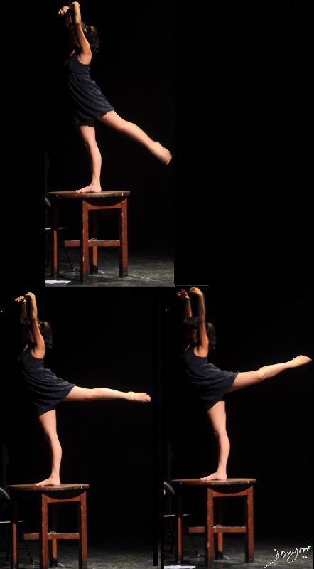


