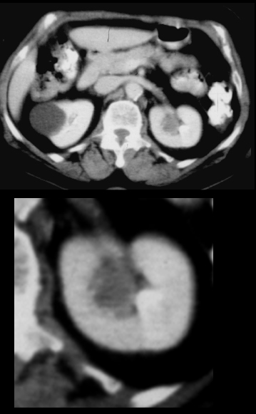
CT Filling Defect in a Calyx Phantom Calyx Sign

77-year-old male with smoking history presents with hematuria. CT with contrast in the axial plane during the excretory shows evidence of an expansile filling defect occupying a left upper pole calyx. Histology showed a transitional cell carcinoma (TCC), The associated imaging sign is called a ?phantom calyx sign? inferring a calyx that completely fails to opacify
Ashley Davidoff MD TheCommonVein.net 06104c
