
38 year old female with Complete duplication of the right kidney and upper pole moiety showing atrophy and partial duplication of the left kidney with prominent column of Bertin
US Partially Duplicated Collecting System and Column of Bertin

US of the left kidney in a 38year old female shows separation of the renal sinus fat by a column of Bertin reminiscent of a duplicated collecting system. The kidney measured 11.8cms which is large for a female.
Ashley Davidoff MD TheCommonVein.net TCV 26K Also see 24K and 25K 135929
Partially Duplicated Left Collecting System
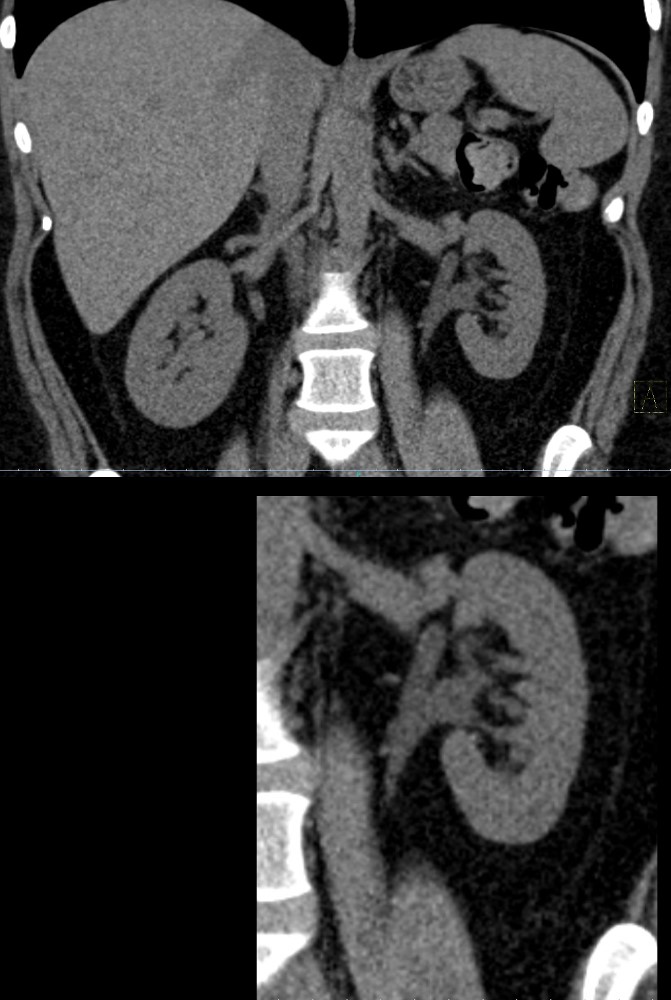
CT of the Kidneys in the coronal plane without contrast in a 38year old female shows a partially duplicated left collecting system. The components join at the renal pelvis
Ashley Davidoff MD TheCommonVein.net TCV 26K Also see 24K and 25K 135929
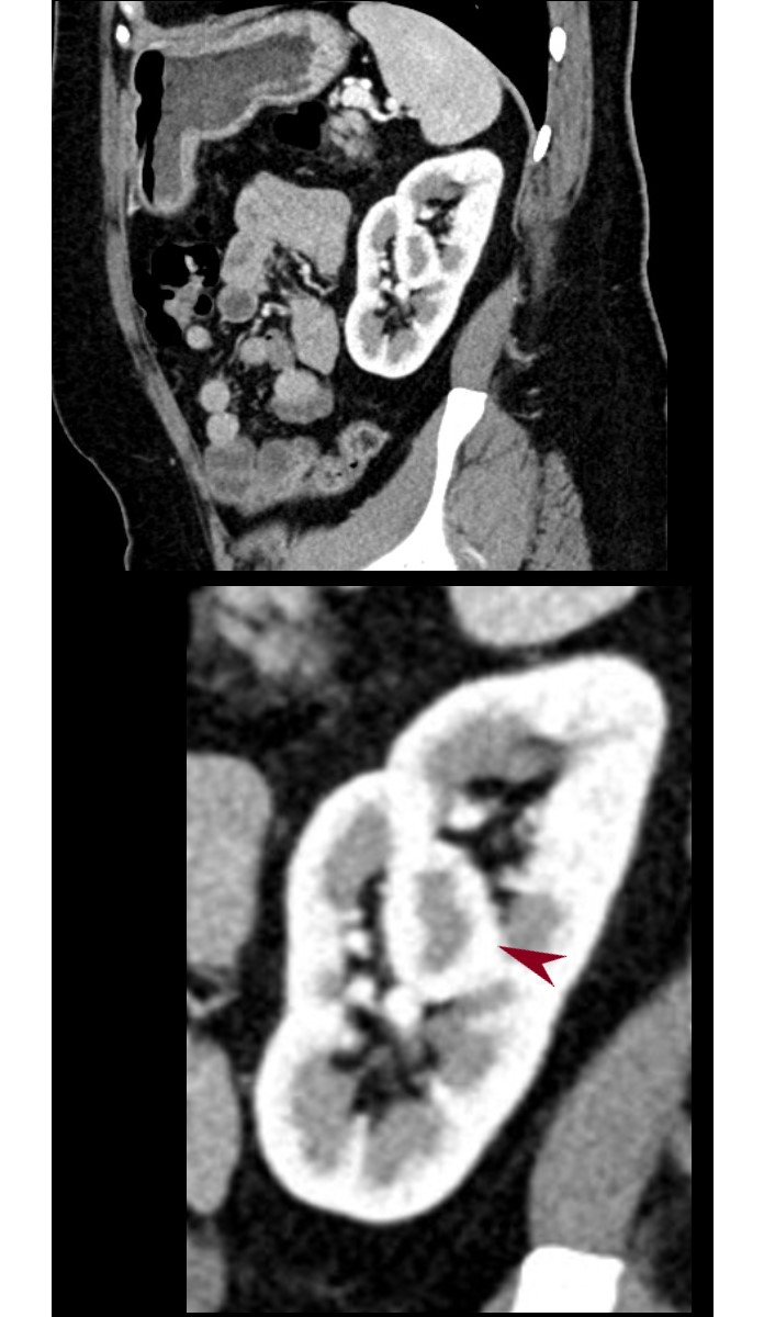
Column of Bertin
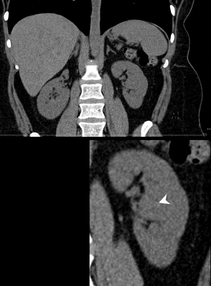
CT of the Kidneys in the coronal plane without contrast in a 38year old female with a complete duplication on the right kidney and partially duplicated left system shows a prominent column of Bertin (white arrowhead lower image). The upper pole moiety on the right is atrophied
Ashley Davidoff MD TheCommonVein.net TCV 26K Also see 24K and 25K 135932c
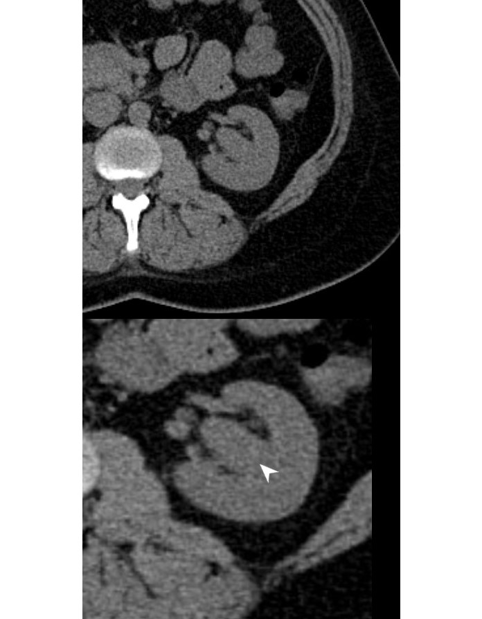
CT of the left kidney in the axial plane without contrast in a 38year old female with a partially duplicated left system, shows a prominent column of Bertin (white arrowhead lower image).
Ashley Davidoff MD TheCommonVein.net TCV 26K Also see 24K and 25K 135931c
Column of Bertin Corticomedullary Phase

CT of the Kidneys in the coronal plane with contrast in a 38year old female with a complete duplication on the right kidney and partial duplication of the left collecting system. The image shows a prominent column of Bertin (maroon arrowhead lower image) during the corticomedullary phase 20 seconds after injection. The column of Bertin contains both cortical and medullary parenchyma exemplified in this image
Ashley Davidoff MD TheCommonVein.net TCV 26K Also see 24K and 25K 135939cL
CT of the Kidneys in the coronal plane with contrast in a 38year old female with a complete duplication on the right kidney and partial duplication of the left collecting system. The image shows a prominent column of Bertin (maroon arrowhead lower image) during the corticomedullary phase 20 seconds after injection. The column of Bertin contains both cortical and medullary parenchyma exemplified in this image
Ashley Davidoff MD TheCommonVein.net TCV 26K Also see 24K and 25K 135934cL
CT of the kidneys in the sagittal plane with contrast in a 38year old female with a complete duplication on the right kidney and partial duplication of the left collecting system. The image shows a prominent column of Bertin (maroon arrowhead lower image) during the corticomedullary phase, 20 seconds after injection. The column of Bertin contains both cortical and medullary parenchyma exemplified in this image
Ashley Davidoff MD TheCommonVein.net TCV 26K Also see 24K and 25K 135936cL
Partially Duplex Left Collecting System Nephrographic Phase

CT of the Kidneys in the coronal plane in the nephrographic phase 70 seconds after injection in a 38year old female shows a partially duplicated left collecting system. The components join at the renal pelvis
Ashley Davidoff MD TheCommonVein.net TCV 26K Also see 24K and 25K 135940c
Bilateral Duplicated Systems Column of Bertin
Nephrographic Phase
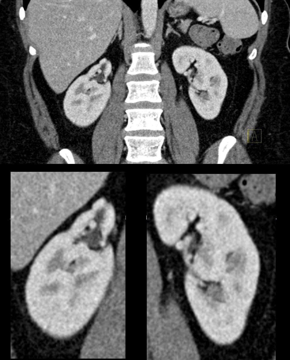
CT of the kidneys in the coronal plane in the nephrographic phase 70 seconds after injection in a 38year old female shows a duplicated right collecting system with atrophy of the upper pole moiety. The right kidney shows a prominent column of Bertin .
Ashley Davidoff MD TheCommonVein.net TCV 26K Also see 24K and 25K 135942c
SH

CT of the kidneys in the coronal plane in the nephrographic phase 70 seconds after injection in a 38year old female shows a duplicated right collecting system with atrophy of the upper pole moiety (yellow arrowhead lower image). The right kidney shows a prominent column of Bertin (maroon arrowhead lower image)
Ashley Davidoff MD TheCommonVein.net TCV 26K Also see 24K and 25K 135942cL
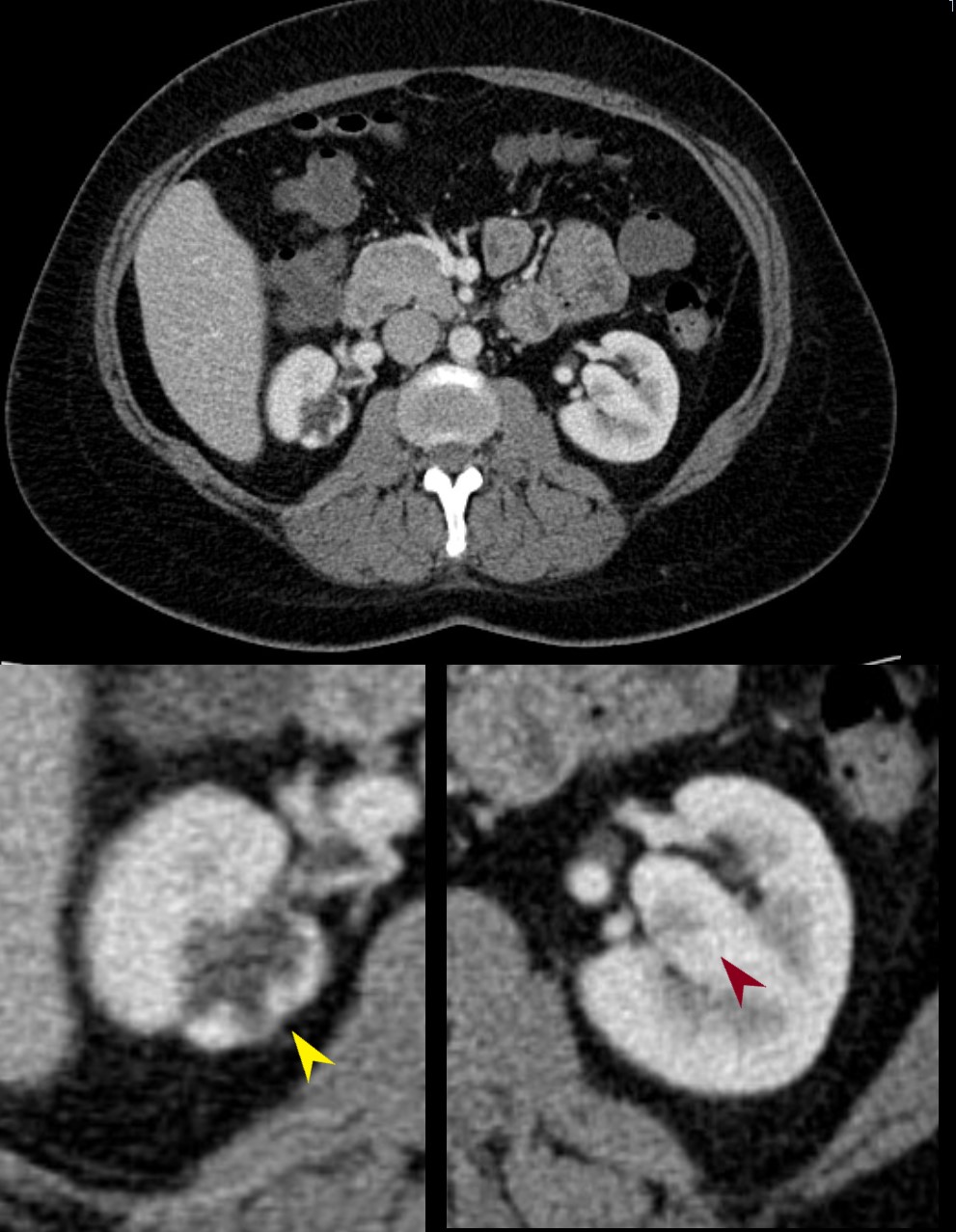
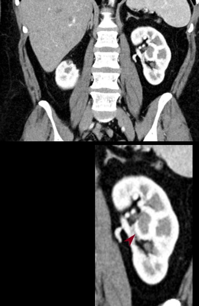
CT of the kidneys in the axial plane in the nephrographic phase 70 seconds after injection in a 38year old female shows a duplex right collecting system with atrophy of the upper pole moiety (yellow arrowhead lower image). The right kidney shows a prominent column of Bertin (maroon arrowhead lower image)
Ashley Davidoff MD TheCommonVein.net TCV 26K Also see 24K and 25K 135941cL
Bilateral Duplicated Systems
Pyelographic (Excretory) Phase and Column of Bertin
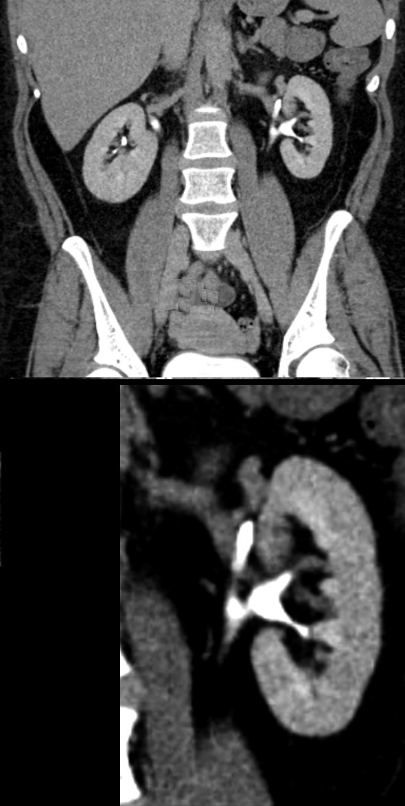
CT of the kidneys in the coronal plane in the excretory phase 10 minutes after injection in a 38year old female shows a partially duplex left collecting system.
Ashley Davidoff MD TheCommonVein.net TCV 26K Also see 24K and 25K 135943c
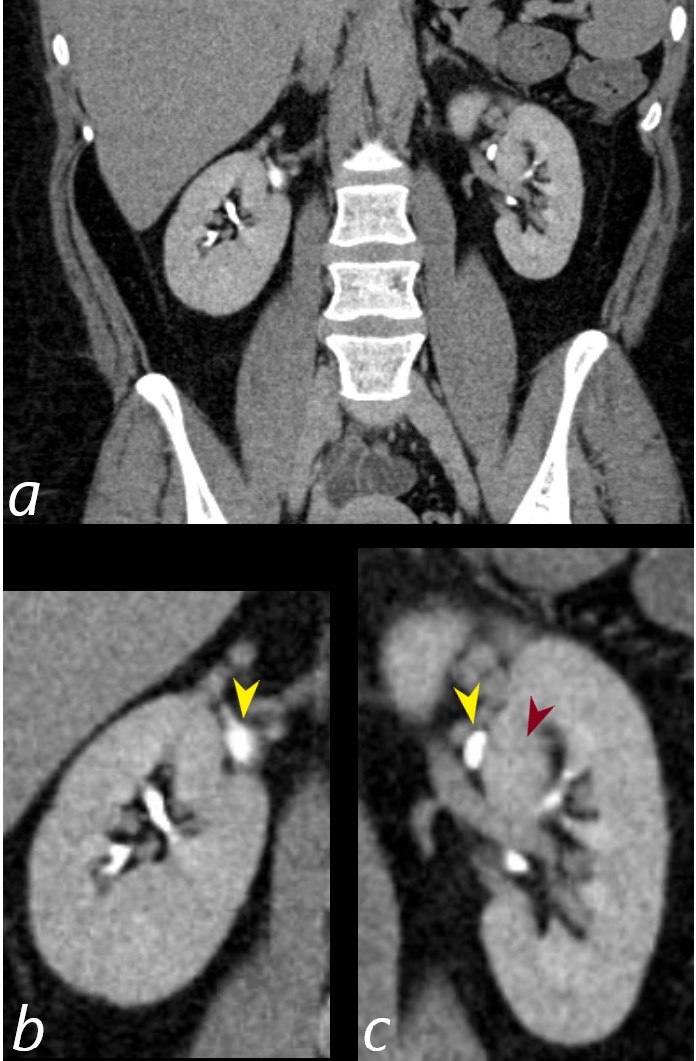
CT of the kidneys in the coronal plane in the excretory phase 10 minutes after injection in a 38year old female shows a collecting systems of the upper pole components of the duplex systems filled with contrast (yellow arrowheads b for the right side and c for the left) The column of Bertin on the left (c maroon arrowhead) separates the upper and lower moieties of the partial duplication on the left .
Ashley Davidoff MD TheCommonVein.net TCV 26K Also see 24K and 25K 135943cL
Bilateral Duplex Systems Pyelographic (Excretory) Phase and Right Hydronephrosis with Post Obstructive Atrophy
Column of Bertin

CT of the kidneys in the axial plane in the excretory phase 10 minutes after injection in a 38year old female shows a hydronephrotic collecting systems of the upper pole components of the duplex systems filled with contrast (yellow arrowheads b) and evidence of post obstructive atrophy. The left extrarenal collecting system is of normal caliber as a common ureter (c yellow arrowhead) The column of Bertin on the left is noted (c maroon arrowhead
Ashley Davidoff MD TheCommonVein.net TCV 26K Also see 24K and 25K 135943cL

CT of the kidneys in the coronal plane in the excretory phase 10 minutes after injection in a 38year old female shows a collecting systems of the upper pole components of the duplex systems filled with contrast (yellow arrowheads b for the right side and c for the left) The column of Bertin on the left (c maroon arrowhead) separates the upper and lower moieties of the partial duplication on the left .
Ashley Davidoff MD TheCommonVein.net TCV 26K Also see 24K and 25K 135943cL