
CT Solid Homogeneous Enhancement
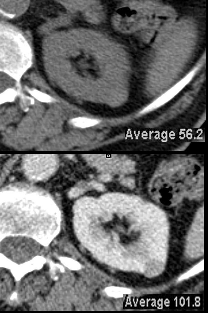
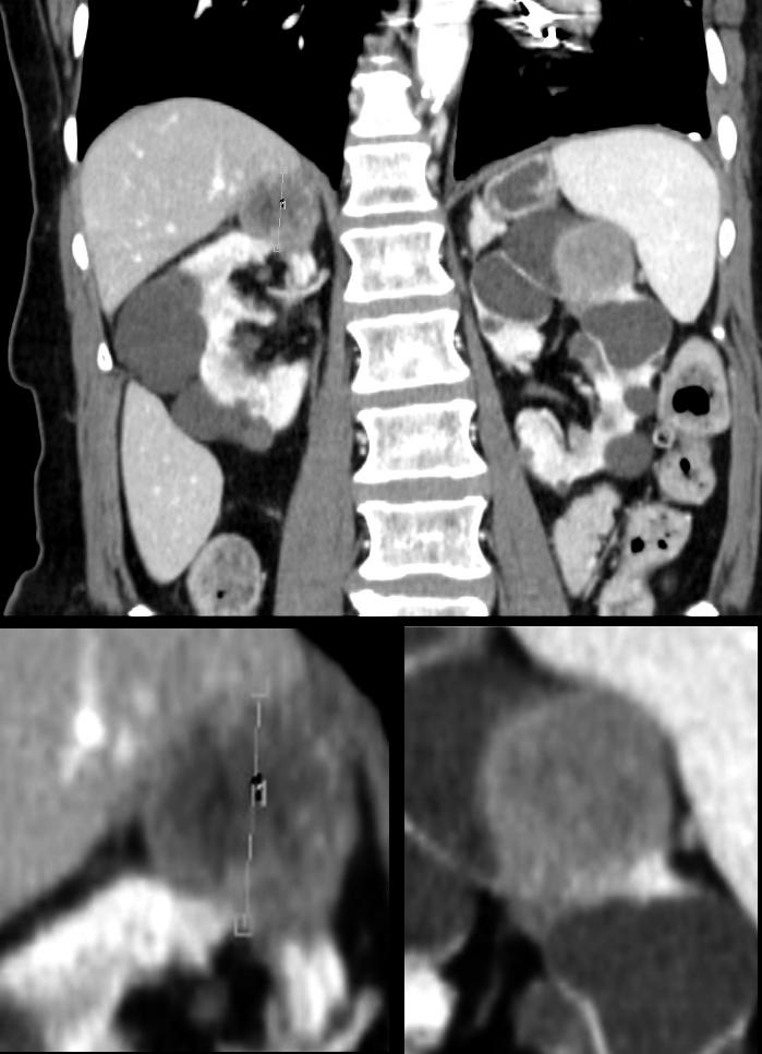
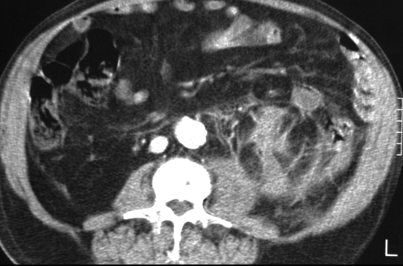
CT through the kidneys reveal a hyperdense nodule that measured 56.HU. Following contrast, the density of the nodule increased to 101.8 HU. An enhancing mass in the kidney is highly suspicious for a renal cell carcinoma
Ashley Davidoff MD TheCommonVein.net 135674
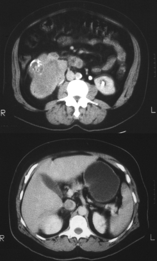
CT Solid Heterogeneous Enhancement

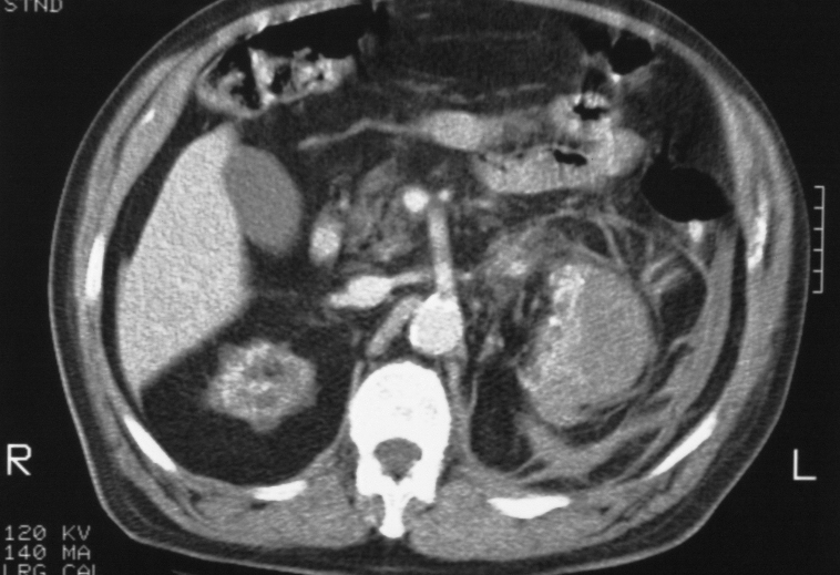
Axial CT through the mid kidneys in a patient with hematuria shows a 2cms heterogeneously enhancing mass on the anterior lip of the right kidney. The most likely diagnosis is a renal cell carcinoma (RCC)
Ashley Davidoff MD TheCommonVein.net 130186
CT Heterogeneous Enhancement and Calcification
CT and US – Solid Mass with Calcification
05675.jpg
Exophytic Renal cell Carcinoma (RCC) with Dystrophic CalcificationAshley Davidoff MD
CT Solid Mass with Heterogeneous Components
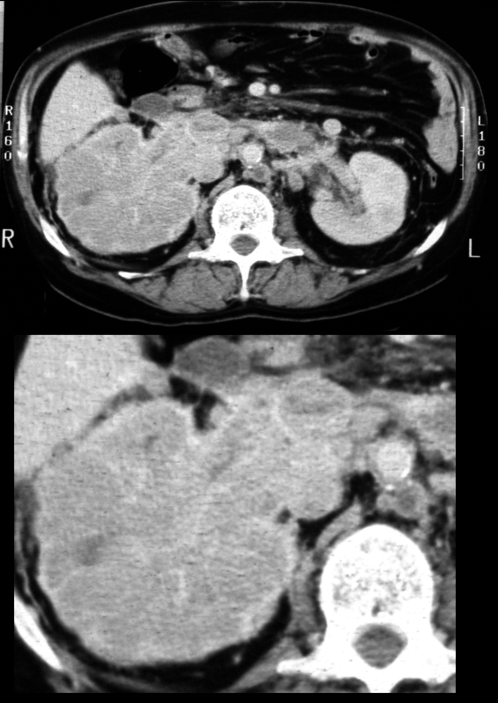
Solid Cystic and Calcified
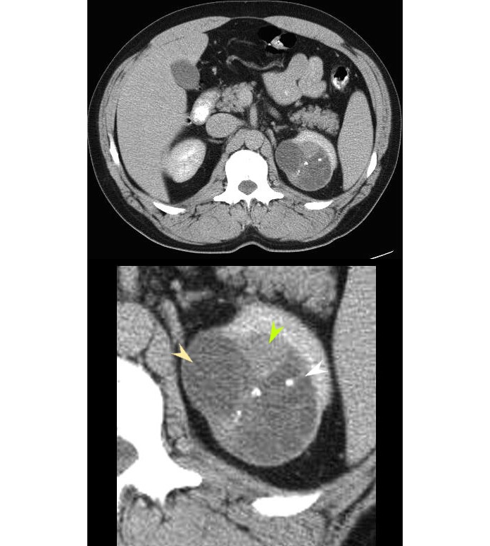
CT of the upper abdomen in axial projection shows a complex cyst in the left upper pole (yellow arrowhead), coarse rounded calcifications along septations (white arrowhead) and a soft tissue nodule (green arrowhead) consistent with a Bosniak IV lesion.
Ashley Davidoff MD TheCommonVein.net 19505bL
CT Hypovascularity Papillary Form

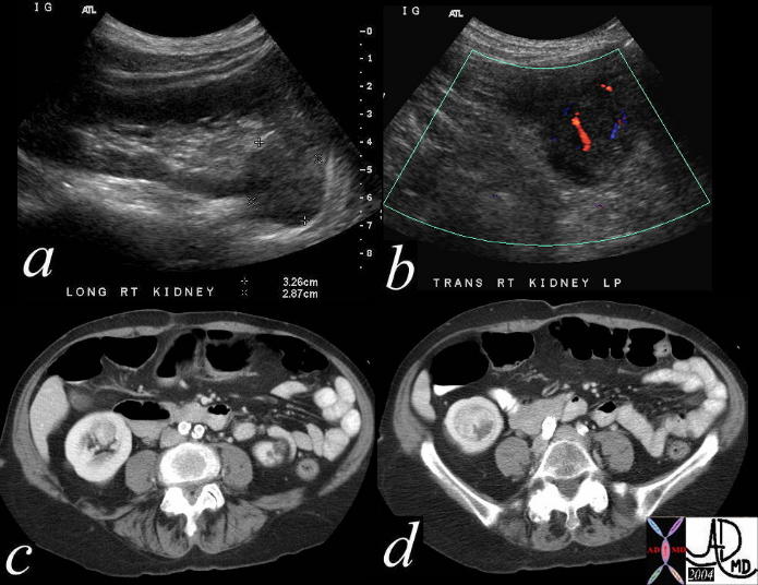
64 year old male
IVP at 20 seconds shows a mass in the right lower pole and 20 minute film conforms the finding.. CT shows a deforming mass and US shows mildly hypoechoic mass. Pathology reveals papillary cell RCC
Ashley Davidoff MD
Ashley Davidoff MD
Cystic Renal Cell Carcinoma CT and Pathology

Cystic renal cell carcinoma with dystrophic calcification in the mid portion of the left kidney. Pathology reveals cystic renal cell carcinoma with clear cell histopathology
Ashley Davidoff MD
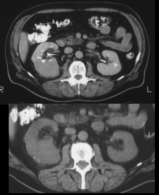
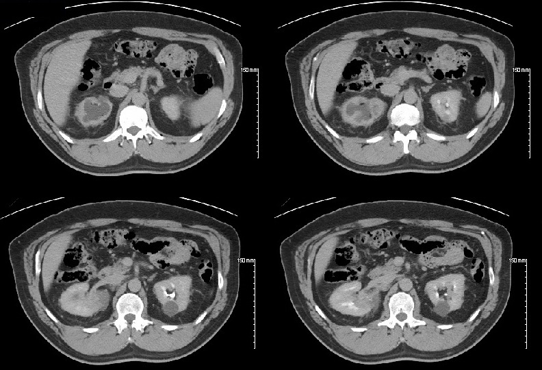
CT Bilateral Disease
CT with contrast, reconstructed in the coronal plane shows bilateral solid masses in the upper poles of the kidneys consistent with bilateral renal cell carcinoma (RCC?s). This patient has acquired cystic disease of Chronic renal failure
Additional key words: renal
Ashley Davidoff MD TheCommonVein.net
CT Solid Spontaneous Hemorrhage
Patient has acquired cystic disease of chronic failure and presents wit spontaneous subcapsular hemorrhage of the left kidney. A hemorrhage from a malignancy known to occur as a complication of this entity was suspected. At the inferior aspect of the hemorrhage there is extension of the process into the retroperitoneum giving the appearance of the ?spider web sign? or ?cobweb sign?
Ashley Davidoff MD TheCommonVein.net RnD
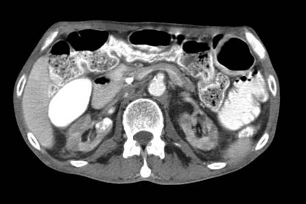
CT Focal Mass and Renal Vein Involvement
16736c.jpg
RCC Invasion into the Renal Vein and IVCRenal Cell Carcinoma of the right kidney shows extension of hypervascular tumor thrombus into the right renal vein and IVC
Ashley Davidoff MD
CT Diffuse Infiltration and Renal Vein Involvement
CT of the upper abdomen in axial projection shows an enlarged right kidney in cortical phase, effaced by an infiltrating renal cell carcinoma invading the renal parenchyma, intrarenal collecting system, the IVC and the left renal vein. The left kidney is in the nephrogram phase, so at this time of contrast handling there is evidence of delayed nephrographic phase of the right kidney. The right kidney also demonstrates the face less kidney sign which implies that the the normal appearance of the renal sinus on cross-sectional imaging is absent.
Ashley Davidoff MD TheCommonVein.net 19937
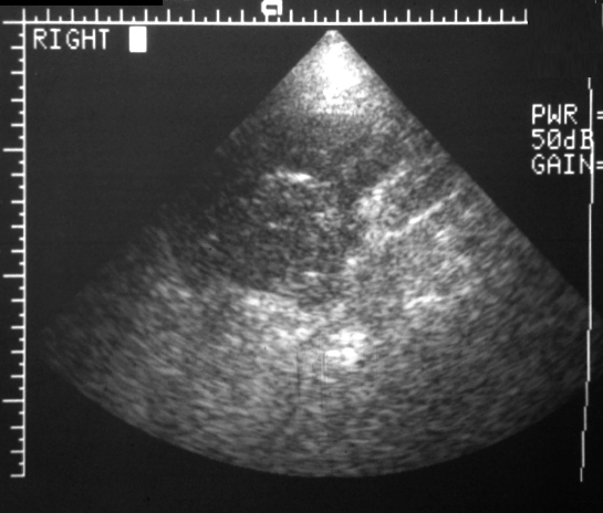
US Solid Enhancing
Ashley Davidoff MD
39056c
US Echogenic Mass Papillary Form
Ashley Davidoff MD
64 year old male
IVP at 20 seconds shows a mass in the right lower pole and 20 minute film conforms the finding.. CT shows a deforming mass and US shows mildly hypoechoic mass. Pathology reveals papillary cell RCC
Ashley Davidoff MD
RCC CT presenting with Hydronephrosis
Top left ? Hydronephrosis
Top right Right upper pole mass and hydronephrosis
Bottom 2 images ? Mass invading the Calyceal system
Ashley Davidoff MD
RCC MRI originating in the Ducts of Bellini and Presenting with Hydronephrosis
125380b.jpg
Renal Cell Carcinoma originating in the Cells of the Duct of Bellini with secondary Hydronephrosis of the Upper Pole CalycesMultiphase T2 Fat Sat Images
Top right and left ? Hydronephrosis right upper pole calyceal system, and mass with T2 bright component
Bottom 2 images ? Perinephric fluid collection around the mass
Ashley Davidoff MD
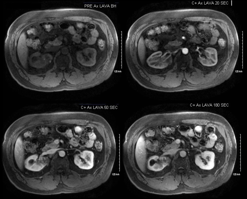
Multiphase Contrast Enhanced MRI
Top left ? Pre gadolinium shows mass like lesion
Top right Poorly visualized mass seen in the early arterial phase
Bottom 2 images ? heterogeneously enhancing mass in the right upper pole
Ashley Davidoff MD
Top left ? Hydronephrosis
Top right Right upper pole mass and hydronephrosis
Bottom 2 images ? Mass invading the Calyceal system
Ashley Davidoff MD

23 F
kidney
fx delayed nephrogram
dx renal cell carcinoma
dx probable renal vein thrombosis
imaging radiology CTscan C+
CT scan
Ashley Davidoff MD
Patient has acquired cystic disease of chronic failure and presents wit spontaneous subcapsular hemorrhage of the left kidney. A hemorrhage from a malignancy known to occur as a complication of this entity was suspected. At the inferior aspect of the hemorrhage there is extension of the process into the retroperitoneum giving the appearance of the ?spider web sign? or ?cobweb sign?
Ashley Davidoff MD TheCommonVein.net RnD
CT through the kidneys reveal a hyperdense nodule that measured 56.HU. Following contrast, the density of the nodule increased to 101.8 HU. An enhancing mass in the kidney is highly suspicious for a renal cell carcinoma
Ashley Davidoff MD TheCommonVein.net 135674
RCC hypervascular Mass Angiography

40437c Courtesy Ashley Davidoff MD code kidney renal artery fx hypervascular mass fx arteriovenous shunting fx neovascularity code IVC invasion “string and thread” sign fx filling defect code left kidney hypervascular mass code pancreas celiac axis pancreatic head fx hypervascular mass code dx primary RCC renal cell carcinoma complicated by metastases to the left kidney and pancreatic parenchyma imaging radiology angiogram venogram code neoplasm primary metastasis malignant tumor carcinoma cancer
RCC Angiography

IVP and Angiogram
a IVP in the nephrographic phase shows an enhancing mass off the upper pole of the right kidney.
b ? IVP in the pyelographic phase shows an a deformity of the upper and outer part of the right kidney
c ? Nephrographic phase of the arteriogram confirms a hyper vascular mass
d Renal angiogram with epinephrine confirms arterial enhancement of the mass with constriction of the normal vessels and failure of the abnormal response
Ashley Davidoff MD

On CT there is dystrophic calcification. On angiography there is hypervascularity, neovascularity, AV shunting. Excretory phase after angiography shows distorted calyces
Ashley Davidoff MD