|
The Common Vein Copyright 2008
Ashley Davidoff MD
Introduction
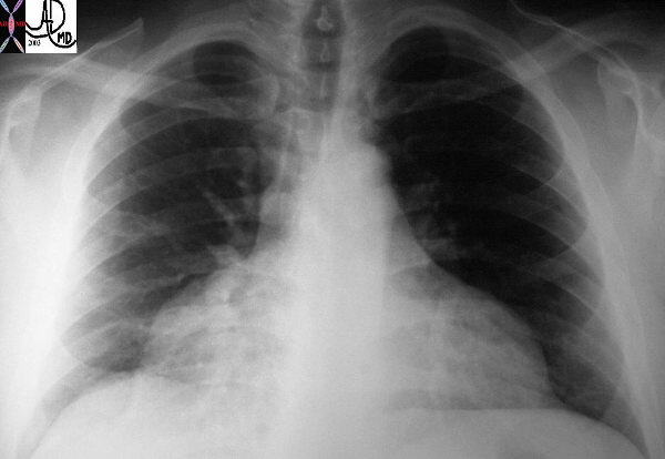
Unusual shape of the cardiac shadow characterised by a narrow nediastinum and a broad base caused by accumulation of fluid in the pericardial space

30095

Pericardial effusion
 
43704 43702
CAUSES
Infectious – viral, tuberculosis, HIV related Idiopathic
Virus – echovirus, influenza virus, and coxsackievirus group B.
Bacteria streptococci, staphylococci, and gram-negative bacilli. Haemophilus influenzae is a common cause in children
Inflammation
rheumatoid arthritis,
SLE
systemic sclerosis,
vasculitides
radiation
Neoplastic –
Malignant (20% of patients with malignancy have nmetastases to pericardium),
nonmalignant.
Not all cancer-associated effusions are malignant. Mediastinal lymphoma, Hodgkin disease, and metastatic breast cancer have been found to cause transient effusions, likely due to impaired lymphatic drainage, which do not cause long-term sequelae.
Metabolic
Myxedema
Uremia
Drugs
-
- Warfarin, heparin
- Penicillin
- Phenytoin
- Procainamide
- Hydralazine, minoxidil
- Cromolyn sodium
- Methysergide
- Doxorubicin
Mechanical
Postpericardiotomy syndrome
Trauma
penetrating or blunt
Circulatory –
Congestive heart failure, valvular disease
Idiopathic
PEARLS
Normal amount of fluid – 10-50ccs
The rapid accumulationamount of only 80ccs can lead to tamponade wheras a volume of up to 2 litres if slowly accumulated can be tolerated.
Malignant effusions
Up to 20 percent of autopsy series – lung breast and leukemias lymphomas
Infections HIV -40-80 PERCENT with or without AIDS –
Mycobacterium avium, M. tuberculosis, Nocardia, fungal infection, viral disease, lymphoma, or Kaposi’s sarcoma.
MI and pericardial effusions
early consequence of acute MI (10 to 15% of cases).
late post-MI syndrome (Dressler’s syndrome) 10 days to 2 mo post-MI (1 to 3%) -fever, pericarditis friction rub, pericardial effusion, pleurisy, pleural effusions, and joint pain.
rupture post-MI, causing hemopericardium. This usually occurs 1 to 10 days post-MI and is more common in women.
TB may not have pulmonary involvement |
DOMElement Object
(
    [schemaTypeInfo] =>
    [tagName] => table
    [firstElementChild] => (object value omitted)
    [lastElementChild] => (object value omitted)
    [childElementCount] => 1
    [previousElementSibling] =>
    [nextElementSibling] =>
    [nodeName] => table
    [nodeValue] =>


The Common Vein Copyright 2008
Ashley Davidoff MD

Introduction
Unusual shape of the cardiac shadow characterised by a narrow nediastinum and a broad base caused by accumulation of fluid in the pericardial space

30095

 Pericardial effusion


43704                                                                    43702
CAUSES
Infectious – viral,  tuberculosis, HIV related Idiopathic
Virus  – echovirus, influenza virus, and coxsackievirus group B.
Bacteria  streptococci, staphylococci, and gram-negative bacilli. Haemophilus influenzae is a common cause in children
Inflammation
rheumatoid arthritis,
SLE
systemic sclerosis,
vasculitides
radiation
Neoplastic –
Malignant  (20% of patients with malignancy have nmetastases to pericardium),
nonmalignant.
Not all cancer-associated effusions are malignant. Mediastinal lymphoma, Hodgkin disease, and metastatic breast cancer have been found to cause transient effusions, likely due to impaired lymphatic drainage, which do not cause long-term sequelae.
Metabolic
Myxedema
Uremia
Drugs


Warfarin, heparin
Penicillin
Phenytoin
Procainamide
Hydralazine, minoxidil
Cromolyn sodium
Methysergide
Doxorubicin


Mechanical
Postpericardiotomy syndrome
Trauma
penetrating or blunt
Circulatory –
Congestive heart failure, valvular disease

Chylopericardium

Idiopathic
PEARLS
Normal amount of fluid – 10-50ccs
The rapid accumulationamount of only 80ccs can lead to tamponade wheras a volume of up to 2 litres if slowly accumulated can be tolerated.
Malignant effusions
Up to 20 percent of autopsy series – lung breast and leukemias lymphomas
Infections   HIV -40-80 PERCENT with or without AIDS –
Mycobacterium avium, M. tuberculosis, Nocardia, fungal infection, viral disease, lymphoma, or Kaposi’s sarcoma.
MI and pericardial effusions
early consequence of acute MI (10 to 15% of cases).
late post-MI syndrome (Dressler’s syndrome) 10 days to 2 mo post-MI (1 to 3%) -fever, pericarditis friction rub, pericardial effusion, pleurisy, pleural effusions, and joint pain.
rupture post-MI, causing hemopericardium. This usually occurs 1 to 10 days post-MI and is more common in women.

TB may not have pulmonary involvement


    [nodeType] => 1
    [parentNode] => (object value omitted)
    [childNodes] => (object value omitted)
    [firstChild] => (object value omitted)
    [lastChild] => (object value omitted)
    [previousSibling] =>
    [nextSibling] => (object value omitted)
    [attributes] => (object value omitted)
    [ownerDocument] => (object value omitted)
    [namespaceURI] =>
    [prefix] =>
    [localName] => table
    [baseURI] =>
    [textContent] =>


The Common Vein Copyright 2008
Ashley Davidoff MD

Introduction
Unusual shape of the cardiac shadow characterised by a narrow nediastinum and a broad base caused by accumulation of fluid in the pericardial space

30095

 Pericardial effusion


43704                                                                    43702
CAUSES
Infectious – viral,  tuberculosis, HIV related Idiopathic
Virus  – echovirus, influenza virus, and coxsackievirus group B.
Bacteria  streptococci, staphylococci, and gram-negative bacilli. Haemophilus influenzae is a common cause in children
Inflammation
rheumatoid arthritis,
SLE
systemic sclerosis,
vasculitides
radiation
Neoplastic –
Malignant  (20% of patients with malignancy have nmetastases to pericardium),
nonmalignant.
Not all cancer-associated effusions are malignant. Mediastinal lymphoma, Hodgkin disease, and metastatic breast cancer have been found to cause transient effusions, likely due to impaired lymphatic drainage, which do not cause long-term sequelae.
Metabolic
Myxedema
Uremia
Drugs


Warfarin, heparin
Penicillin
Phenytoin
Procainamide
Hydralazine, minoxidil
Cromolyn sodium
Methysergide
Doxorubicin


Mechanical
Postpericardiotomy syndrome
Trauma
penetrating or blunt
Circulatory –
Congestive heart failure, valvular disease

Chylopericardium

Idiopathic
PEARLS
Normal amount of fluid – 10-50ccs
The rapid accumulationamount of only 80ccs can lead to tamponade wheras a volume of up to 2 litres if slowly accumulated can be tolerated.
Malignant effusions
Up to 20 percent of autopsy series – lung breast and leukemias lymphomas
Infections   HIV -40-80 PERCENT with or without AIDS –
Mycobacterium avium, M. tuberculosis, Nocardia, fungal infection, viral disease, lymphoma, or Kaposi’s sarcoma.
MI and pericardial effusions
early consequence of acute MI (10 to 15% of cases).
late post-MI syndrome (Dressler’s syndrome) 10 days to 2 mo post-MI (1 to 3%) -fever, pericarditis friction rub, pericardial effusion, pleurisy, pleural effusions, and joint pain.
rupture post-MI, causing hemopericardium. This usually occurs 1 to 10 days post-MI and is more common in women.

TB may not have pulmonary involvement


)
DOMElement Object
(
    [schemaTypeInfo] =>
    [tagName] => td
    [firstElementChild] => (object value omitted)
    [lastElementChild] => (object value omitted)
    [childElementCount] => 40
    [previousElementSibling] =>
    [nextElementSibling] =>
    [nodeName] => td
    [nodeValue] =>

The Common Vein Copyright 2008
Ashley Davidoff MD

Introduction
Unusual shape of the cardiac shadow characterised by a narrow nediastinum and a broad base caused by accumulation of fluid in the pericardial space

30095

 Pericardial effusion


43704                                                                    43702
CAUSES
Infectious – viral,  tuberculosis, HIV related Idiopathic
Virus  – echovirus, influenza virus, and coxsackievirus group B.
Bacteria  streptococci, staphylococci, and gram-negative bacilli. Haemophilus influenzae is a common cause in children
Inflammation
rheumatoid arthritis,
SLE
systemic sclerosis,
vasculitides
radiation
Neoplastic –
Malignant  (20% of patients with malignancy have nmetastases to pericardium),
nonmalignant.
Not all cancer-associated effusions are malignant. Mediastinal lymphoma, Hodgkin disease, and metastatic breast cancer have been found to cause transient effusions, likely due to impaired lymphatic drainage, which do not cause long-term sequelae.
Metabolic
Myxedema
Uremia
Drugs


Warfarin, heparin
Penicillin
Phenytoin
Procainamide
Hydralazine, minoxidil
Cromolyn sodium
Methysergide
Doxorubicin


Mechanical
Postpericardiotomy syndrome
Trauma
penetrating or blunt
Circulatory –
Congestive heart failure, valvular disease

Chylopericardium

Idiopathic
PEARLS
Normal amount of fluid – 10-50ccs
The rapid accumulationamount of only 80ccs can lead to tamponade wheras a volume of up to 2 litres if slowly accumulated can be tolerated.
Malignant effusions
Up to 20 percent of autopsy series – lung breast and leukemias lymphomas
Infections   HIV -40-80 PERCENT with or without AIDS –
Mycobacterium avium, M. tuberculosis, Nocardia, fungal infection, viral disease, lymphoma, or Kaposi’s sarcoma.
MI and pericardial effusions
early consequence of acute MI (10 to 15% of cases).
late post-MI syndrome (Dressler’s syndrome) 10 days to 2 mo post-MI (1 to 3%) -fever, pericarditis friction rub, pericardial effusion, pleurisy, pleural effusions, and joint pain.
rupture post-MI, causing hemopericardium. This usually occurs 1 to 10 days post-MI and is more common in women.

TB may not have pulmonary involvement
    [nodeType] => 1
    [parentNode] => (object value omitted)
    [childNodes] => (object value omitted)
    [firstChild] => (object value omitted)
    [lastChild] => (object value omitted)
    [previousSibling] => (object value omitted)
    [nextSibling] => (object value omitted)
    [attributes] => (object value omitted)
    [ownerDocument] => (object value omitted)
    [namespaceURI] =>
    [prefix] =>
    [localName] => td
    [baseURI] =>
    [textContent] =>

The Common Vein Copyright 2008
Ashley Davidoff MD

Introduction
Unusual shape of the cardiac shadow characterised by a narrow nediastinum and a broad base caused by accumulation of fluid in the pericardial space

30095

 Pericardial effusion


43704                                                                    43702
CAUSES
Infectious – viral,  tuberculosis, HIV related Idiopathic
Virus  – echovirus, influenza virus, and coxsackievirus group B.
Bacteria  streptococci, staphylococci, and gram-negative bacilli. Haemophilus influenzae is a common cause in children
Inflammation
rheumatoid arthritis,
SLE
systemic sclerosis,
vasculitides
radiation
Neoplastic –
Malignant  (20% of patients with malignancy have nmetastases to pericardium),
nonmalignant.
Not all cancer-associated effusions are malignant. Mediastinal lymphoma, Hodgkin disease, and metastatic breast cancer have been found to cause transient effusions, likely due to impaired lymphatic drainage, which do not cause long-term sequelae.
Metabolic
Myxedema
Uremia
Drugs


Warfarin, heparin
Penicillin
Phenytoin
Procainamide
Hydralazine, minoxidil
Cromolyn sodium
Methysergide
Doxorubicin


Mechanical
Postpericardiotomy syndrome
Trauma
penetrating or blunt
Circulatory –
Congestive heart failure, valvular disease

Chylopericardium

Idiopathic
PEARLS
Normal amount of fluid – 10-50ccs
The rapid accumulationamount of only 80ccs can lead to tamponade wheras a volume of up to 2 litres if slowly accumulated can be tolerated.
Malignant effusions
Up to 20 percent of autopsy series – lung breast and leukemias lymphomas
Infections   HIV -40-80 PERCENT with or without AIDS –
Mycobacterium avium, M. tuberculosis, Nocardia, fungal infection, viral disease, lymphoma, or Kaposi’s sarcoma.
MI and pericardial effusions
early consequence of acute MI (10 to 15% of cases).
late post-MI syndrome (Dressler’s syndrome) 10 days to 2 mo post-MI (1 to 3%) -fever, pericarditis friction rub, pericardial effusion, pleurisy, pleural effusions, and joint pain.
rupture post-MI, causing hemopericardium. This usually occurs 1 to 10 days post-MI and is more common in women.

TB may not have pulmonary involvement
)


