Apple Core Lesion
The Common Vein Copyright 2008
Definition
DOMElement Object
(
    [schemaTypeInfo] =>
    [tagName] => table
    [firstElementChild] => (object value omitted)
    [lastElementChild] => (object value omitted)
    [childElementCount] => 1
    [previousElementSibling] => (object value omitted)
    [nextElementSibling] =>
    [nodeName] => table
    [nodeValue] =>


Napkin Ring in the Antrum


55 year old female with vague right upper quadrant pain. Upper GI shows napkin ring lesion in the antrum aupper GI study barium double contrast
83306.8s  Courtesy Ashley Davidoff MD copyright 2009


    [nodeType] => 1
    [parentNode] => (object value omitted)
    [childNodes] => (object value omitted)
    [firstChild] => (object value omitted)
    [lastChild] => (object value omitted)
    [previousSibling] => (object value omitted)
    [nextSibling] => (object value omitted)
    [attributes] => (object value omitted)
    [ownerDocument] => (object value omitted)
    [namespaceURI] =>
    [prefix] =>
    [localName] => table
    [baseURI] =>
    [textContent] =>


Napkin Ring in the Antrum


55 year old female with vague right upper quadrant pain. Upper GI shows napkin ring lesion in the antrum aupper GI study barium double contrast
83306.8s  Courtesy Ashley Davidoff MD copyright 2009


)
DOMElement Object
(
    [schemaTypeInfo] =>
    [tagName] => td
    [firstElementChild] => (object value omitted)
    [lastElementChild] => (object value omitted)
    [childElementCount] => 1
    [previousElementSibling] =>
    [nextElementSibling] =>
    [nodeName] => td
    [nodeValue] => 55 year old female with vague right upper quadrant pain. Upper GI shows napkin ring lesion in the antrum aupper GI study barium double contrast
83306.8s  Courtesy Ashley Davidoff MD copyright 2009
    [nodeType] => 1
    [parentNode] => (object value omitted)
    [childNodes] => (object value omitted)
    [firstChild] => (object value omitted)
    [lastChild] => (object value omitted)
    [previousSibling] => (object value omitted)
    [nextSibling] => (object value omitted)
    [attributes] => (object value omitted)
    [ownerDocument] => (object value omitted)
    [namespaceURI] =>
    [prefix] =>
    [localName] => td
    [baseURI] =>
    [textContent] => 55 year old female with vague right upper quadrant pain. Upper GI shows napkin ring lesion in the antrum aupper GI study barium double contrast
83306.8s  Courtesy Ashley Davidoff MD copyright 2009
)
DOMElement Object
(
    [schemaTypeInfo] =>
    [tagName] => td
    [firstElementChild] => (object value omitted)
    [lastElementChild] => (object value omitted)
    [childElementCount] => 2
    [previousElementSibling] =>
    [nextElementSibling] =>
    [nodeName] => td
    [nodeValue] =>
Napkin Ring in the Antrum
    [nodeType] => 1
    [parentNode] => (object value omitted)
    [childNodes] => (object value omitted)
    [firstChild] => (object value omitted)
    [lastChild] => (object value omitted)
    [previousSibling] => (object value omitted)
    [nextSibling] => (object value omitted)
    [attributes] => (object value omitted)
    [ownerDocument] => (object value omitted)
    [namespaceURI] =>
    [prefix] =>
    [localName] => td
    [baseURI] =>
    [textContent] =>
Napkin Ring in the Antrum
)
DOMElement Object
(
    [schemaTypeInfo] =>
    [tagName] => table
    [firstElementChild] => (object value omitted)
    [lastElementChild] => (object value omitted)
    [childElementCount] => 1
    [previousElementSibling] => (object value omitted)
    [nextElementSibling] => (object value omitted)
    [nodeName] => table
    [nodeValue] =>


Napkin Ring Circunferential Tumor -aka Apple Core


Portion of left colon with a constricting carcinoma. The carcinoma has infiltrated the full thickness of the wall. This can be seen on the cut portion of the wall, where the wall is much paler than the surrounding mucosa.
colon large bowel fx apple core fx constriction fx narrowing fx wall thickening fx mass dx carcinoma malignant stricture grosspathology
Courtesy Barbara Banner MD aka applecore12313


    [nodeType] => 1
    [parentNode] => (object value omitted)
    [childNodes] => (object value omitted)
    [firstChild] => (object value omitted)
    [lastChild] => (object value omitted)
    [previousSibling] => (object value omitted)
    [nextSibling] => (object value omitted)
    [attributes] => (object value omitted)
    [ownerDocument] => (object value omitted)
    [namespaceURI] =>
    [prefix] =>
    [localName] => table
    [baseURI] =>
    [textContent] =>


Napkin Ring Circunferential Tumor -aka Apple Core


Portion of left colon with a constricting carcinoma. The carcinoma has infiltrated the full thickness of the wall. This can be seen on the cut portion of the wall, where the wall is much paler than the surrounding mucosa.
colon large bowel fx apple core fx constriction fx narrowing fx wall thickening fx mass dx carcinoma malignant stricture grosspathology
Courtesy Barbara Banner MD aka applecore12313


)
DOMElement Object
(
    [schemaTypeInfo] =>
    [tagName] => td
    [firstElementChild] => (object value omitted)
    [lastElementChild] => (object value omitted)
    [childElementCount] => 2
    [previousElementSibling] =>
    [nextElementSibling] =>
    [nodeName] => td
    [nodeValue] => Portion of left colon with a constricting carcinoma. The carcinoma has infiltrated the full thickness of the wall. This can be seen on the cut portion of the wall, where the wall is much paler than the surrounding mucosa.
colon large bowel fx apple core fx constriction fx narrowing fx wall thickening fx mass dx carcinoma malignant stricture grosspathology
Courtesy Barbara Banner MD aka applecore12313
    [nodeType] => 1
    [parentNode] => (object value omitted)
    [childNodes] => (object value omitted)
    [firstChild] => (object value omitted)
    [lastChild] => (object value omitted)
    [previousSibling] => (object value omitted)
    [nextSibling] => (object value omitted)
    [attributes] => (object value omitted)
    [ownerDocument] => (object value omitted)
    [namespaceURI] =>
    [prefix] =>
    [localName] => td
    [baseURI] =>
    [textContent] => Portion of left colon with a constricting carcinoma. The carcinoma has infiltrated the full thickness of the wall. This can be seen on the cut portion of the wall, where the wall is much paler than the surrounding mucosa.
colon large bowel fx apple core fx constriction fx narrowing fx wall thickening fx mass dx carcinoma malignant stricture grosspathology
Courtesy Barbara Banner MD aka applecore12313
)
DOMElement Object
(
    [schemaTypeInfo] =>
    [tagName] => td
    [firstElementChild] => (object value omitted)
    [lastElementChild] => (object value omitted)
    [childElementCount] => 1
    [previousElementSibling] =>
    [nextElementSibling] =>
    [nodeName] => td
    [nodeValue] => Napkin Ring Circunferential Tumor -aka Apple Core
    [nodeType] => 1
    [parentNode] => (object value omitted)
    [childNodes] => (object value omitted)
    [firstChild] => (object value omitted)
    [lastChild] => (object value omitted)
    [previousSibling] => (object value omitted)
    [nextSibling] => (object value omitted)
    [attributes] => (object value omitted)
    [ownerDocument] => (object value omitted)
    [namespaceURI] =>
    [prefix] =>
    [localName] => td
    [baseURI] =>
    [textContent] => Napkin Ring Circunferential Tumor -aka Apple Core
)
DOMElement Object
(
    [schemaTypeInfo] =>
    [tagName] => table
    [firstElementChild] => (object value omitted)
    [lastElementChild] => (object value omitted)
    [childElementCount] => 1
    [previousElementSibling] => (object value omitted)
    [nextElementSibling] => (object value omitted)
    [nodeName] => table
    [nodeValue] =>


Apple core lesion of the sigmoid colon


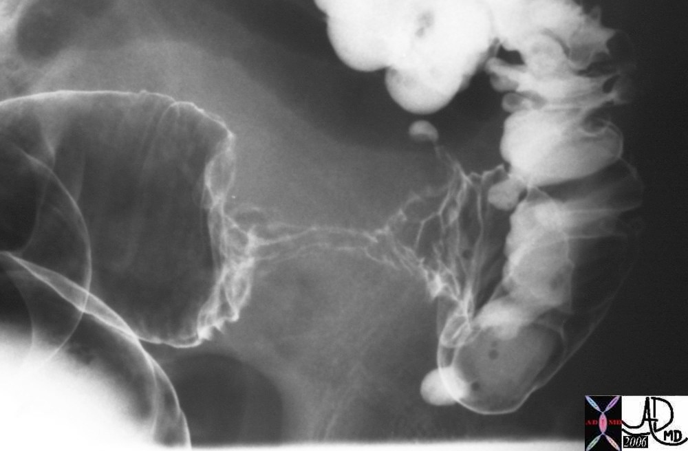
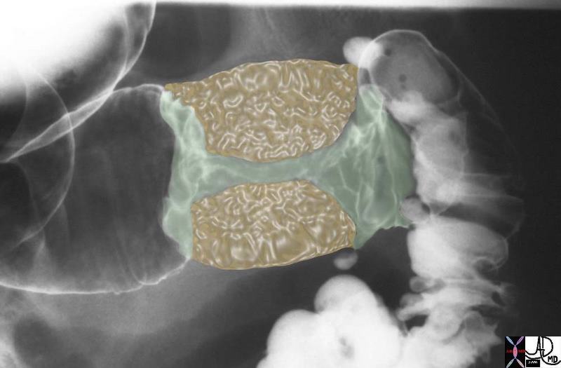
The double contrast barium enema shows an apple core lesion caused by circumferential growth of a malignant tumor of the colon in ?napkin-ring? fashion.  The second image shows the apple core-shape of the lumen.  The third image shows the yellow mass of tumor that has surrounded the lumen in napkin ring fashion essentially strangling the lumen.  Note how the distal colon which is receiving air and barium via the enema tip is relatively dilated when compared to the descending colon which is relatively decompressed suggesting an obstructive process.  Note also the presence of contrast filled diverticuli at the rectosigmoid junction.  The two entities of carcinoma and diverticulosis often coexist since diverticulosis is very common in Westernized nations.
Courtesy Ashley Davidoff MD
28655


    [nodeType] => 1
    [parentNode] => (object value omitted)
    [childNodes] => (object value omitted)
    [firstChild] => (object value omitted)
    [lastChild] => (object value omitted)
    [previousSibling] => (object value omitted)
    [nextSibling] => (object value omitted)
    [attributes] => (object value omitted)
    [ownerDocument] => (object value omitted)
    [namespaceURI] =>
    [prefix] =>
    [localName] => table
    [baseURI] =>
    [textContent] =>


Apple core lesion of the sigmoid colon


The double contrast barium enema shows an apple core lesion caused by circumferential growth of a malignant tumor of the colon in ?napkin-ring? fashion.  The second image shows the apple core-shape of the lumen.  The third image shows the yellow mass of tumor that has surrounded the lumen in napkin ring fashion essentially strangling the lumen.  Note how the distal colon which is receiving air and barium via the enema tip is relatively dilated when compared to the descending colon which is relatively decompressed suggesting an obstructive process.  Note also the presence of contrast filled diverticuli at the rectosigmoid junction.  The two entities of carcinoma and diverticulosis often coexist since diverticulosis is very common in Westernized nations.
Courtesy Ashley Davidoff MD
28655


)
DOMElement Object
(
    [schemaTypeInfo] =>
    [tagName] => td
    [firstElementChild] => (object value omitted)
    [lastElementChild] => (object value omitted)
    [childElementCount] => 2
    [previousElementSibling] =>
    [nextElementSibling] =>
    [nodeName] => td
    [nodeValue] => The double contrast barium enema shows an apple core lesion caused by circumferential growth of a malignant tumor of the colon in ?napkin-ring? fashion.  The second image shows the apple core-shape of the lumen.  The third image shows the yellow mass of tumor that has surrounded the lumen in napkin ring fashion essentially strangling the lumen.  Note how the distal colon which is receiving air and barium via the enema tip is relatively dilated when compared to the descending colon which is relatively decompressed suggesting an obstructive process.  Note also the presence of contrast filled diverticuli at the rectosigmoid junction.  The two entities of carcinoma and diverticulosis often coexist since diverticulosis is very common in Westernized nations.
Courtesy Ashley Davidoff MD
28655
    [nodeType] => 1
    [parentNode] => (object value omitted)
    [childNodes] => (object value omitted)
    [firstChild] => (object value omitted)
    [lastChild] => (object value omitted)
    [previousSibling] => (object value omitted)
    [nextSibling] => (object value omitted)
    [attributes] => (object value omitted)
    [ownerDocument] => (object value omitted)
    [namespaceURI] =>
    [prefix] =>
    [localName] => td
    [baseURI] =>
    [textContent] => The double contrast barium enema shows an apple core lesion caused by circumferential growth of a malignant tumor of the colon in ?napkin-ring? fashion.  The second image shows the apple core-shape of the lumen.  The third image shows the yellow mass of tumor that has surrounded the lumen in napkin ring fashion essentially strangling the lumen.  Note how the distal colon which is receiving air and barium via the enema tip is relatively dilated when compared to the descending colon which is relatively decompressed suggesting an obstructive process.  Note also the presence of contrast filled diverticuli at the rectosigmoid junction.  The two entities of carcinoma and diverticulosis often coexist since diverticulosis is very common in Westernized nations.
Courtesy Ashley Davidoff MD
28655
)
DOMElement Object
(
    [schemaTypeInfo] =>
    [tagName] => td
    [firstElementChild] => (object value omitted)
    [lastElementChild] => (object value omitted)
    [childElementCount] => 1
    [previousElementSibling] =>
    [nextElementSibling] =>
    [nodeName] => td
    [nodeValue] => Apple core lesion of the sigmoid colon
    [nodeType] => 1
    [parentNode] => (object value omitted)
    [childNodes] => (object value omitted)
    [firstChild] => (object value omitted)
    [lastChild] => (object value omitted)
    [previousSibling] => (object value omitted)
    [nextSibling] => (object value omitted)
    [attributes] => (object value omitted)
    [ownerDocument] => (object value omitted)
    [namespaceURI] =>
    [prefix] =>
    [localName] => td
    [baseURI] =>
    [textContent] => Apple core lesion of the sigmoid colon
)


