Differential consideration is an angiomyolipoma
Ashley Davidoff MD
Ashley Davidoff MD
64 year old male
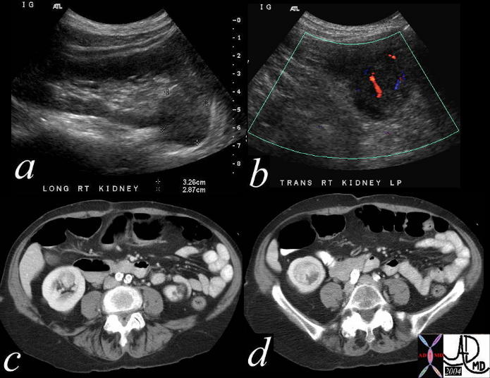
IVP at 20 seconds shows a mass in the right lower pole and 20 minute film conforms the finding.CT shows a deforming mass and US shows mildly hypoechoic mass. Pathology reveals papillary cell RCC
Ashley Davidoff MD
Ashley Davidoff MD

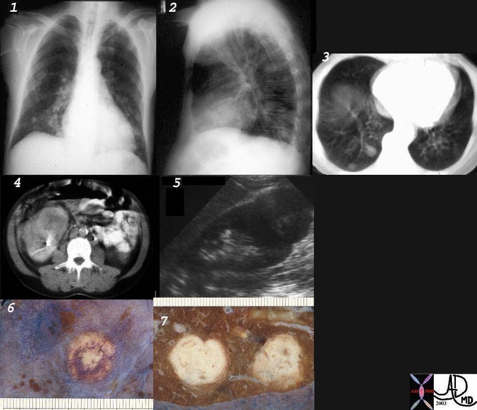
32303c01 code lungs pulmonary pleura nodules neoplasm malignant metastases metastasis primary renal kidney RCC imaging plain film CXR CTscan USscan grosspathology
Ashley Davidoff MD

CT’ scans show two CT scans performed 2 years apart showing no significant growth
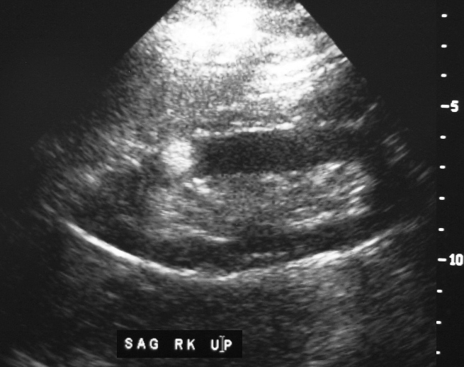
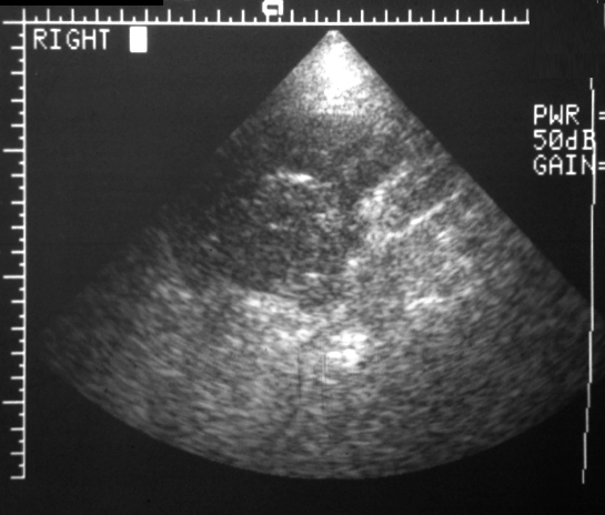
The ultrasounds below show mildly hyperechoic character of RCC
# renal cell cardcinoma#time
Ashley Davidoff MD
