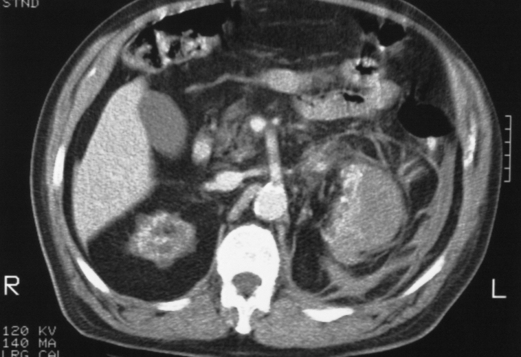
Patient has acquired cystic disease of chronic failure and presents wit spontaneous subcapsular hemorrhage of the left kidney. A hemorrhage from a malignancy known to occur as a complication of this entity was suspected. At the inferior aspect of the hemorrhage there is extension of the process into the retroperitoneum giving the appearance of the ?spider web sign? or ?cobweb sign?
Ashley Davidoff MD TheCommonVein.net RnD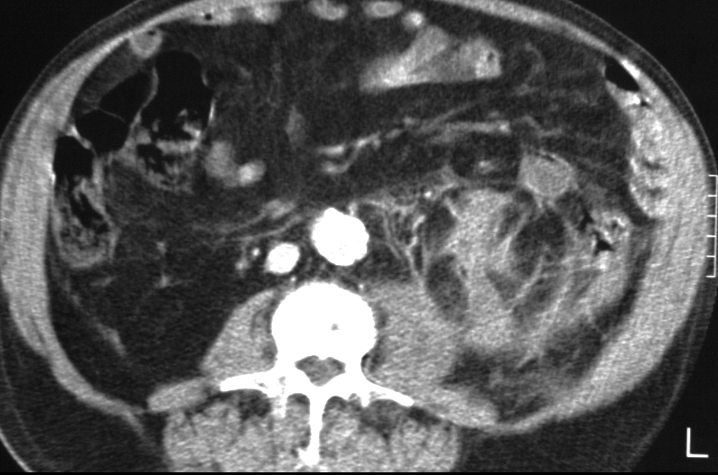
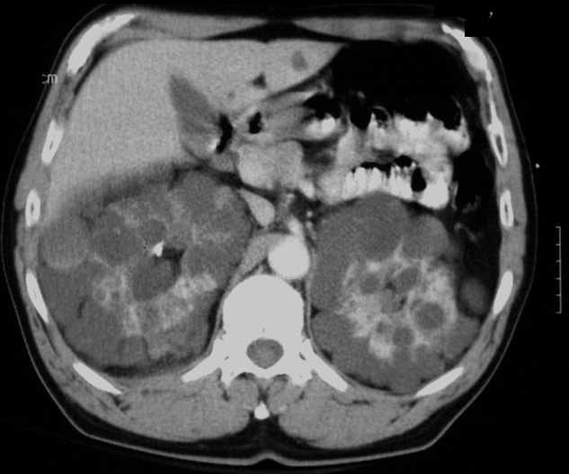
CT scan with contrast of a 54 year old male with polycystic kidney disease. In this case, the cysts are distributed in a relatively orderly and homogeneous fashion in the periphery of the both kidneys with an unusual symmetry. There is a reasonable amount of functioning parenchyma. One of the cysts in the periphery of the right kidney has a higher density and high density sediment suggesting hemorrhage
Ashley Davidoff MD TheCommonVein.net
CT scan with contrast of a 54 year old male with polycystic kidney disease. In this case, the cysts are distributed in a relatively orderly and homogeneous fashion in the periphery of the both kidneys with an unusual symmetry. There is a reasonable amount of functioning parenchyma. One of the cysts in the periphery of the right kidney (yellow arrowhead) has a higher density and high density sediment (red arrowhead lower image suggesting hemorrhage
Ashley Davidoff MD TheCommonVein.net
Hemorrhagic or Proteinaceous Cysts CT Non Contrast
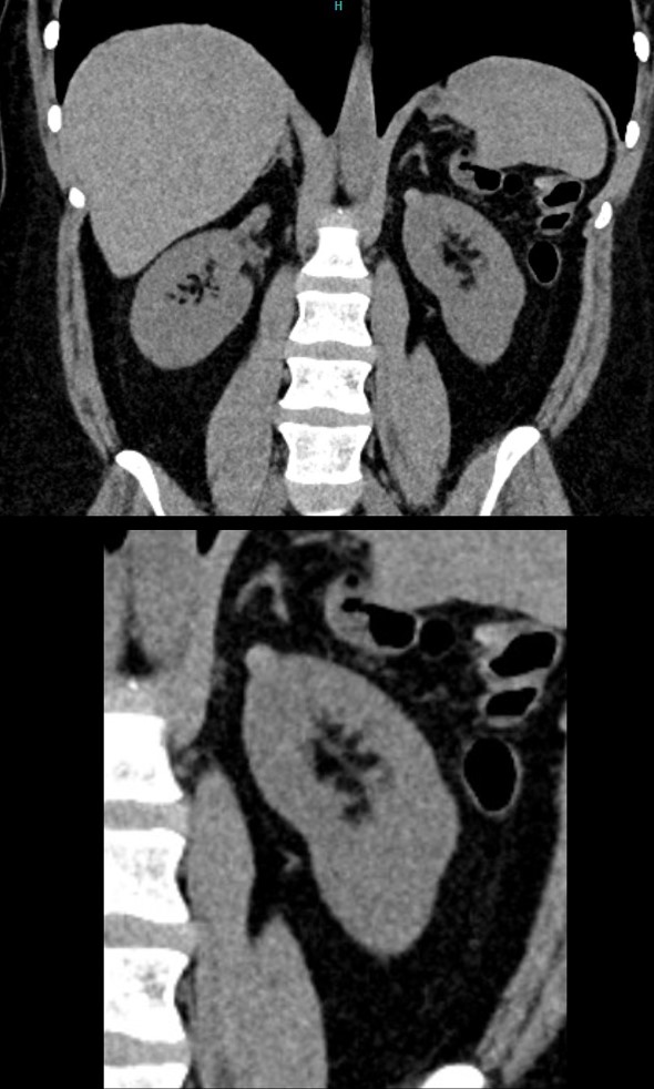
Non-Contrast CT in the coronal plane shows an 8mm homogeneous hyperdense lesion, exophytic off the upper pole of the left kidney. A hyperdense cyst as a result of hemorrhage or accumulation of proteinaceous material are most likely. The right kidney has a deformity of the upper pole secondary to atrophy of the upper pole moiety of a duplicated collecting system
Ashley Davidoff MD TheCommonVein.net TCV 24K 135915c
Hemorrhagic or Proteinaceous Cysts CT with Contrast

High Density Hemorrhagic or Proteinaceous Cyst of the Upper Pole of the Left Kidney
CT in the coronal plane shows an 8mm homogeneous that remains relatively hypodense, exophytic off the upper pole of the left kidney. .A hemorrhagic or proteinaceous cyst is most likely. MRI subsequently confirmed the diagnosis
The right kidney shows contrast in a mildly dilated collecting system in an upper pole moiety with secondary atrophy of the upper pole moiety of a duplicated collecting system
Ashley Davidoff MD TheCommonVein.net TCV 24K 135920c
T1 Bright Hemorrhagic or Proteinaceous Cyst
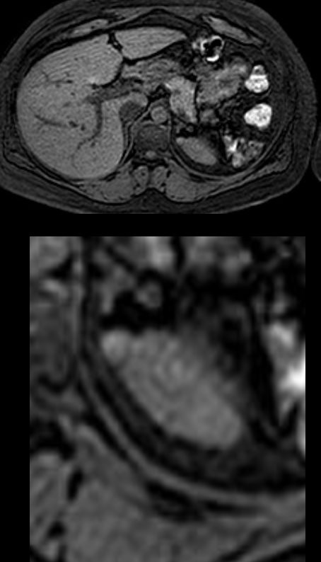
T1 weighted MRI in the axial plane shows a T1 bright 8mm homogeneous lesion, exophytic off the upper pole of the left kidney.
This finding is compatible with a hemorrhagic or proteinaceous cyst
Ashley Davidoff MD TheCommonVein.net TCV 24K 135921c
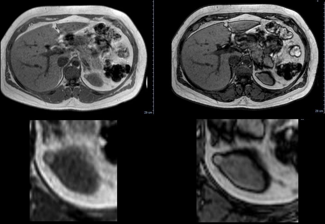
T1 weighted MRI in the axial plane shows a T1 bright 8mm homogeneous lesion, exophytic off the upper pole of the left kidney.
This finding is compatible with a hemorrhagic or proteinaceous cyst
Ashley Davidoff MD TheCommonVein.net TCV 24K 135923c
T2 Dark Hemorrhagic or Proteinaceous Cyst
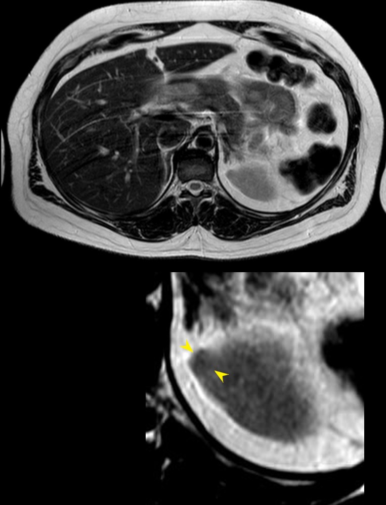
T2 weighted MRI in the axial plane shows a T1 dark 8mm homogeneous lesion, (yellow arrowheads lower image) exophytic off the upper pole of the left kidney confirming that the cyst contains protein or blood.
This finding is compatible with a hemorrhagic or proteinaceous cyst
Ashley Davidoff MD TheCommonVein.net TCV 24K 135925cL
