Barton’s Fracture of the Wrist
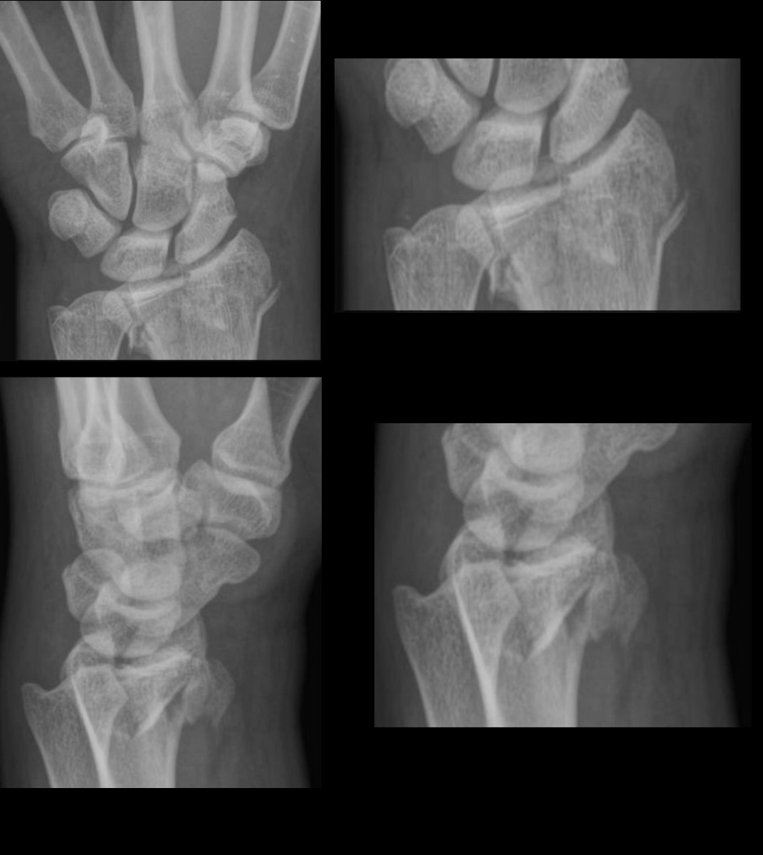
Upper AP radiographs from a left wrist show a comminuted fracture of the distal radial metadiaphysis with intraarticular extension.
The lower two images are a lateral projection, and show a comminuted fracture of the distal radial metadiaphysis with volar angulation consistent with a diagnosis of volar Barton?s fracture. Soft tissue edema is noted.
Ashley Davidoff MD TheCommonVein.net Barton’s fracture 003
Barton’s fracture is a
type of wrist fracture that involves the
distal radius bone.
occurs near the wrist jo
named after John Rhea Barton, an American surgeon who first described the injury in 1838.
Caused by
fall on an outstretched hand or a
direct blow to the wrist.
more common in young adults
often associated with
sports injuries or
motor vehicle accidents.
Two types
dorsal
fracture line extends from the back (dorsal) of the radius bone into the wrist joint
volar
fracture line extends from the front (volar) of the radius bone
into the wrist joint.
Treatment of Barton’s fracture
depends on the severity and
type of the
Mild
a cast or splint may be used to immobilize the wrist while the bone heals.
In more severe cases,
surgery may be necessary to
realign the bone and
stabilize the joint.
Physical therapy may also be recommended to help regain strength and flexibility in the wrist.
 Barton’s Fracture of the Wrist