The Common Vein Copyright 2011
Definition
Fractures of the scaphoid are a common carpal injury are usually caused by a fall onto the outstretched hand and are often characterized by poor healing ability.
Scaphoid fractures can be classified by the fracture pattern, level of displacement, or location. The fracture patterns are horizontal oblique, transverse, or vertical oblique. The displacement can be stable or unstable. Unstable is when there is more than 1 mm of step-off or increased angulation. The locations for scaphoid fractures can be at the tuberosity, distal pole, waist, or proximal pole. Most scaphoid fractures occur at the waist.
The fracture may be complicated in the acute phase by neurovascular injury, or in the subacute or chronic phases by nonunion, malunion, infection, osteonecrosis, or osteoarthritis. More specific to scaphoid injuries is poor healing. Nonunion and avascular necrosis can lead to a SLAC or SNAC wrist..
The diagnosis of this injury is usually made by a combination of physical examination and x-ray imaging. Patients may have ?snuff box? tenderness.
Imaging includes the use of plain x-rays, and if indicated CT-scan, or MRI. Initial x-rays commonly are not diagnostic for scaphoid fractures prompting follow-up x-rays and CT-scans.
All suspected scaphoid fractures are initially treated with a thumb spica cast, splint or a brace. These methods of immobilization should be left on for 1 to 2 weeks until follow-up x-rays or CT either show a fracture or show no fracture.
Indications for surgery include a displacement of fragments greater than 1 mm, a radiolunate angle greater than 15 degrees, a scapholunate angle greater than 60 degress, a ?Humpback? deformity, or a nonunion. Surgery usually involves screw insertion. Nonunions can be treated with bone grafting or vascularized bone grafts.

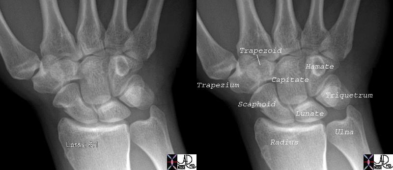
The AP examination of the normal wrist shows the normal scaphoid
Courtesy Ashley Davidoff MD 45738c01

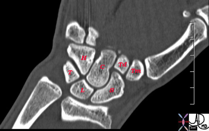
The coronal reconstruction of the wrist shows the normal scaphoid (s)
Courtesy Ashley Davidoff MD Copyright 2011 46616b01

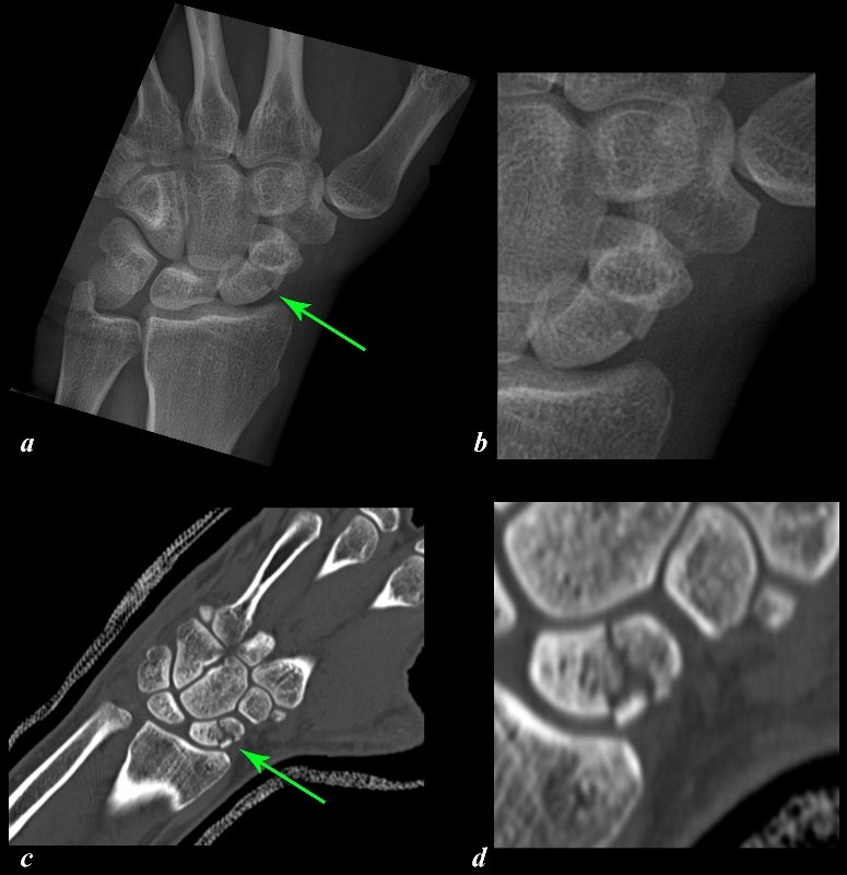
The X-Ray (a, and magnified in b) and CT scan (c and magnified in d) reveal a transverse fracture (green arrows) through the middle of the left scaphoid of a 29 year old patient after a fall on his outstretched hand. This is the most common site for fracture of the scaphoid and in this location the injury carries a small risk of avascular necrosis. A fracture through the proximal 1/3 would carry a higher risk of AVN approximating 30%.
Courtesy Ashley Davidoff Copyright 2011 107550c01L.8

CT scan Reconstruction of a Normal Scaphoid |
| Courtsy of Ashley Davidoff MD 46616b01 |
References
Davis MF, Davis PF, Ross DS. Expert Guide to Sports Medicine. ACP Series, 2005.
Elstrom J, Virkus W, Pankovich (eds), Handbook of Fractures (3rd edition), McGraw Hill, New York, NY, 2006.
Koval K, Zuckerman J (eds), Handbook of Fractures (3rd edition), Lippincott Williams & Wilkins, Philadelphia, PA, 2006.
Lieberman J (ed), AAOS Comprehensive Orthopaedic Review, American Academy of Orthopaedic Surgeons, 2008.
Moore K, Dalley A (eds), Clinically Oriented Anatomy (5th edition), Lippincott Williams & Wilkins, Philadelphia, PA, 2006.
Wheeless?s Textbook of Orthopaedics: Scaphoid/ Scaphoid Fracture (http://www.wheelessonline.com/ortho/scaphoid_scaphoid_fracture)
