Ashley Davidoff MD Gregory Waryasz MD
The Common Vein Copyright 2011
Definition
A fracture is defined as the disruption of the integrity of living bone. A fracture is caused when the force applied to the bone is greater than the integrity of the bone. Sudden unexpected injury is the most common cause of a fracture. Less commonly fractures are caused when underlying conditions such as osteoporosis or metastatic disease result in weakened bones. These fractures are called pathological fractures. The force required to fracture this type of bone may be minimal. Lastly fractures may be caused by recurrent and repeated small forces on the bone. These fractures are called stress fractures or fatigue fractures and are commonly seen in athletes and dancers. Fractures result in pain and loss of function provided by the specific bone.
Structural change includes disruption of the compact and spongy bone, bone marrow and periosteum, associated with injury and hematoma of the surrounding soft tissues.
Functional changes depend on the specific bone involved. Long bones of the limbs for example are usually involved primarily in support and locomotion, and this functionality becomes limited when the bone is fractured.
Fractures may be complicated by damage to nearby blood vessels, nerves, muscles, tendons, ligaments, and joints.
The diagnosis of a fracture is first suspected clinically when the patient presents with an appropriate history of a severe force and on examination shows deformity of the limb associated with loss of function.
Imaging by plain X-ray is the gold standard of diagnosis, and enables confirmation of the fracture and assessment of the size shape, and position – factors necessary for treatment planning.
Treatment usually requires anatomical alignment, support and protection of the injury to enable optimal healing and return to function.

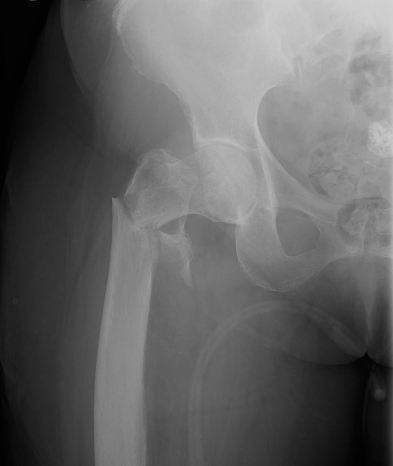
Fracture of the Hip through the Neck of the Femur
|
|
This A-P examination of the proximal left femur shows a comminuted (more than two pieces) transverse fracture of the intertrochanteric region of the right femur with deformity. A fragment of the lesser trochanter is seen medially. This fracture requires intraoperative repair with hardware.
Courtesy Ashley Davidoff Copyright 2011 101640b01.8
|
Hip fractures involving the proximal femur are one of the most common fractures in the adult population. They are usually closed injuries and may be simple or comminuted but almost all require intraoperative intervention. They are seldom life threatening in themselves.
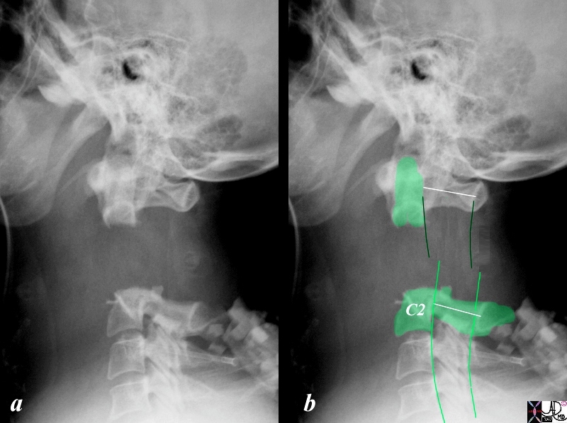
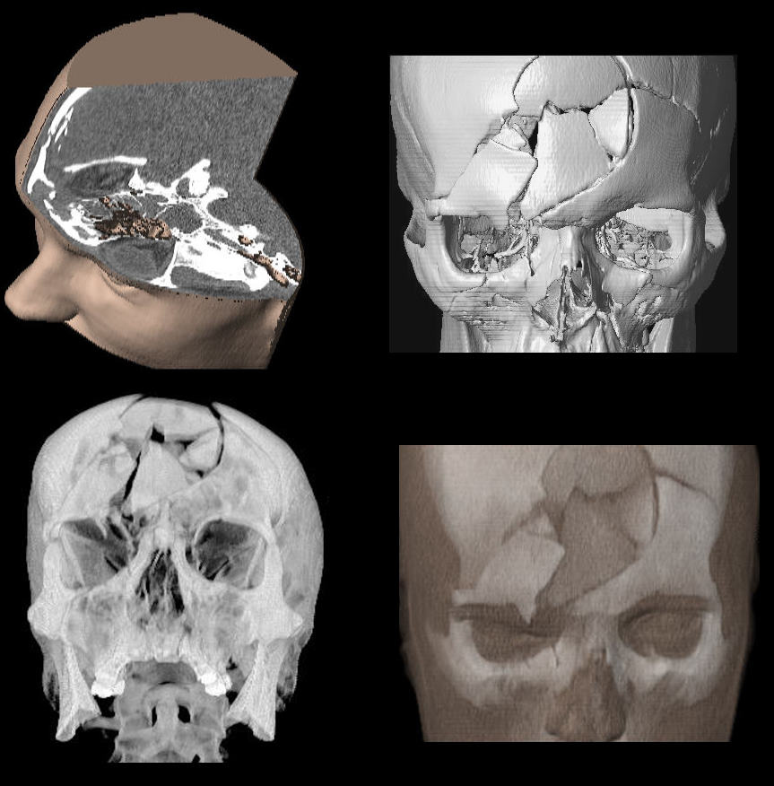
The life threatening fractures include cervical spine fractures and skull fractures, though any fracture that may cause excessive hemorrhage can be life threatening if the bleeding is not recognized and controlled. Fractures of the cervical spine are dangerous because of the close anatomical relationship to the spinal cord. Jefferson’s fracture, (C1 fracture) and hangman’s fracture (C2 fracture) are unstable fractures. Skull fractures can also be life threatening if an associated space occupying subdural or extradural bleed is present. Since the skull has extremely limited reserve for any new space occupying abnormality, bleeding can cause the brain to herniate resulting in coning and subsequent arrest and death through interruption of vital cardiopulmonary functions that are impeded by pressure on the brainstem.

Fracture Dislocation of the Odontoid of C2
|
|
This lateral examination of the cervical spine shows a type II transverse fracture of the odontoid of C2 with upward and anterior dislocation of the proximal fragment. The parallel dark green lines project the position of the spinal cord superiorly and the light green lines project the expected location of the spinal cord inferiorly with an almost 10-15% discrepancy in position. This difference infers pressure on the spinal cord and a life threatening situation. The anterior translation of more than 3.5 mm of one vertebrae relative to the adjacent vertebra is indicative of an unstable situation.
Courtesy Ashley Davidoff Copyright 2011 15915c04
|

Severe Skull Fracture – Life Threatening
|
|
This series of volume rendered 3D CT images reveals a severe comminuted fracture of the frontal bone. The loss of integrity of the skull is obvious. It is shattered in multiple pieces inferring a comminuted fracture.
Courtesy Philips Medical Systems 88462c
|
—
DOMElement Object
(
    [schemaTypeInfo] =>
    [tagName] => table
    [firstElementChild] => (object value omitted)
    [lastElementChild] => (object value omitted)
    [childElementCount] => 1
    [previousElementSibling] => (object value omitted)
    [nextElementSibling] => (object value omitted)
    [nodeName] => table
    [nodeValue] =>


Severe Skull Fracture – Life Threatening


This series of volume rendered 3D CT images reveals a severe comminuted fracture of the frontal bone. The loss of integrity of the skull is obvious. It is shattered in multiple pieces inferring a comminuted fracture.
Courtesy Philips Medical Systems 88462c


    [nodeType] => 1
    [parentNode] => (object value omitted)
    [childNodes] => (object value omitted)
    [firstChild] => (object value omitted)
    [lastChild] => (object value omitted)
    [previousSibling] => (object value omitted)
    [nextSibling] => (object value omitted)
    [attributes] => (object value omitted)
    [ownerDocument] => (object value omitted)
    [namespaceURI] =>
    [prefix] =>
    [localName] => table
    [baseURI] =>
    [textContent] =>


Severe Skull Fracture – Life Threatening


This series of volume rendered 3D CT images reveals a severe comminuted fracture of the frontal bone. The loss of integrity of the skull is obvious. It is shattered in multiple pieces inferring a comminuted fracture.
Courtesy Philips Medical Systems 88462c


)
DOMElement Object
(
    [schemaTypeInfo] =>
    [tagName] => td
    [firstElementChild] => (object value omitted)
    [lastElementChild] => (object value omitted)
    [childElementCount] => 2
    [previousElementSibling] =>
    [nextElementSibling] =>
    [nodeName] => td
    [nodeValue] =>
This series of volume rendered 3D CT images reveals a severe comminuted fracture of the frontal bone. The loss of integrity of the skull is obvious. It is shattered in multiple pieces inferring a comminuted fracture.
Courtesy Philips Medical Systems 88462c

    [nodeType] => 1
    [parentNode] => (object value omitted)
    [childNodes] => (object value omitted)
    [firstChild] => (object value omitted)
    [lastChild] => (object value omitted)
    [previousSibling] => (object value omitted)
    [nextSibling] => (object value omitted)
    [attributes] => (object value omitted)
    [ownerDocument] => (object value omitted)
    [namespaceURI] =>
    [prefix] =>
    [localName] => td
    [baseURI] =>
    [textContent] =>
This series of volume rendered 3D CT images reveals a severe comminuted fracture of the frontal bone. The loss of integrity of the skull is obvious. It is shattered in multiple pieces inferring a comminuted fracture.
Courtesy Philips Medical Systems 88462c

)
DOMElement Object
(
    [schemaTypeInfo] =>
    [tagName] => td
    [firstElementChild] => (object value omitted)
    [lastElementChild] => (object value omitted)
    [childElementCount] => 2
    [previousElementSibling] =>
    [nextElementSibling] =>
    [nodeName] => td
    [nodeValue] =>
Severe Skull Fracture – Life Threatening

    [nodeType] => 1
    [parentNode] => (object value omitted)
    [childNodes] => (object value omitted)
    [firstChild] => (object value omitted)
    [lastChild] => (object value omitted)
    [previousSibling] => (object value omitted)
    [nextSibling] => (object value omitted)
    [attributes] => (object value omitted)
    [ownerDocument] => (object value omitted)
    [namespaceURI] =>
    [prefix] =>
    [localName] => td
    [baseURI] =>
    [textContent] =>
Severe Skull Fracture – Life Threatening

)
DOMElement Object
(
    [schemaTypeInfo] =>
    [tagName] => table
    [firstElementChild] => (object value omitted)
    [lastElementChild] => (object value omitted)
    [childElementCount] => 1
    [previousElementSibling] => (object value omitted)
    [nextElementSibling] => (object value omitted)
    [nodeName] => table
    [nodeValue] =>


Fracture Dislocation of the Odontoid of C2


This lateral examination of the cervical spine shows a type II transverse fracture of the odontoid of C2 with upward and anterior dislocation of the proximal fragment. The parallel dark green lines project the position of the spinal cord superiorly and the light green lines project the expected location of the spinal cord inferiorly with an almost 10-15% discrepancy in position. This difference infers pressure on the spinal cord and a life threatening situation.  The anterior translation of more than 3.5 mm of one vertebrae relative to the adjacent vertebra is indicative of an unstable situation.
Courtesy Ashley Davidoff Copyright 2011 15915c04


    [nodeType] => 1
    [parentNode] => (object value omitted)
    [childNodes] => (object value omitted)
    [firstChild] => (object value omitted)
    [lastChild] => (object value omitted)
    [previousSibling] => (object value omitted)
    [nextSibling] => (object value omitted)
    [attributes] => (object value omitted)
    [ownerDocument] => (object value omitted)
    [namespaceURI] =>
    [prefix] =>
    [localName] => table
    [baseURI] =>
    [textContent] =>


Fracture Dislocation of the Odontoid of C2


This lateral examination of the cervical spine shows a type II transverse fracture of the odontoid of C2 with upward and anterior dislocation of the proximal fragment. The parallel dark green lines project the position of the spinal cord superiorly and the light green lines project the expected location of the spinal cord inferiorly with an almost 10-15% discrepancy in position. This difference infers pressure on the spinal cord and a life threatening situation.  The anterior translation of more than 3.5 mm of one vertebrae relative to the adjacent vertebra is indicative of an unstable situation.
Courtesy Ashley Davidoff Copyright 2011 15915c04


)
DOMElement Object
(
    [schemaTypeInfo] =>
    [tagName] => td
    [firstElementChild] => (object value omitted)
    [lastElementChild] => (object value omitted)
    [childElementCount] => 2
    [previousElementSibling] =>
    [nextElementSibling] =>
    [nodeName] => td
    [nodeValue] =>
This lateral examination of the cervical spine shows a type II transverse fracture of the odontoid of C2 with upward and anterior dislocation of the proximal fragment. The parallel dark green lines project the position of the spinal cord superiorly and the light green lines project the expected location of the spinal cord inferiorly with an almost 10-15% discrepancy in position. This difference infers pressure on the spinal cord and a life threatening situation.  The anterior translation of more than 3.5 mm of one vertebrae relative to the adjacent vertebra is indicative of an unstable situation.
Courtesy Ashley Davidoff Copyright 2011 15915c04

    [nodeType] => 1
    [parentNode] => (object value omitted)
    [childNodes] => (object value omitted)
    [firstChild] => (object value omitted)
    [lastChild] => (object value omitted)
    [previousSibling] => (object value omitted)
    [nextSibling] => (object value omitted)
    [attributes] => (object value omitted)
    [ownerDocument] => (object value omitted)
    [namespaceURI] =>
    [prefix] =>
    [localName] => td
    [baseURI] =>
    [textContent] =>
This lateral examination of the cervical spine shows a type II transverse fracture of the odontoid of C2 with upward and anterior dislocation of the proximal fragment. The parallel dark green lines project the position of the spinal cord superiorly and the light green lines project the expected location of the spinal cord inferiorly with an almost 10-15% discrepancy in position. This difference infers pressure on the spinal cord and a life threatening situation.  The anterior translation of more than 3.5 mm of one vertebrae relative to the adjacent vertebra is indicative of an unstable situation.
Courtesy Ashley Davidoff Copyright 2011 15915c04

)
DOMElement Object
(
    [schemaTypeInfo] =>
    [tagName] => td
    [firstElementChild] => (object value omitted)
    [lastElementChild] => (object value omitted)
    [childElementCount] => 2
    [previousElementSibling] =>
    [nextElementSibling] =>
    [nodeName] => td
    [nodeValue] =>
Fracture Dislocation of the Odontoid of C2

    [nodeType] => 1
    [parentNode] => (object value omitted)
    [childNodes] => (object value omitted)
    [firstChild] => (object value omitted)
    [lastChild] => (object value omitted)
    [previousSibling] => (object value omitted)
    [nextSibling] => (object value omitted)
    [attributes] => (object value omitted)
    [ownerDocument] => (object value omitted)
    [namespaceURI] =>
    [prefix] =>
    [localName] => td
    [baseURI] =>
    [textContent] =>
Fracture Dislocation of the Odontoid of C2

)
DOMElement Object
(
    [schemaTypeInfo] =>
    [tagName] => table
    [firstElementChild] => (object value omitted)
    [lastElementChild] => (object value omitted)
    [childElementCount] => 1
    [previousElementSibling] => (object value omitted)
    [nextElementSibling] => (object value omitted)
    [nodeName] => table
    [nodeValue] =>


Fracture of the Hip through the Neck of the Femur


This A-P examination of the proximal left femur shows a comminuted (more than two pieces) transverse fracture of the intertrochanteric region of the right femur with deformity. A fragment of the lesser trochanter is seen medially.  This fracture requires intraoperative repair with hardware.
Courtesy Ashley Davidoff Copyright 2011 101640b01.8


    [nodeType] => 1
    [parentNode] => (object value omitted)
    [childNodes] => (object value omitted)
    [firstChild] => (object value omitted)
    [lastChild] => (object value omitted)
    [previousSibling] => (object value omitted)
    [nextSibling] => (object value omitted)
    [attributes] => (object value omitted)
    [ownerDocument] => (object value omitted)
    [namespaceURI] =>
    [prefix] =>
    [localName] => table
    [baseURI] =>
    [textContent] =>


Fracture of the Hip through the Neck of the Femur


This A-P examination of the proximal left femur shows a comminuted (more than two pieces) transverse fracture of the intertrochanteric region of the right femur with deformity. A fragment of the lesser trochanter is seen medially.  This fracture requires intraoperative repair with hardware.
Courtesy Ashley Davidoff Copyright 2011 101640b01.8


)
DOMElement Object
(
    [schemaTypeInfo] =>
    [tagName] => td
    [firstElementChild] => (object value omitted)
    [lastElementChild] => (object value omitted)
    [childElementCount] => 2
    [previousElementSibling] =>
    [nextElementSibling] =>
    [nodeName] => td
    [nodeValue] =>
This A-P examination of the proximal left femur shows a comminuted (more than two pieces) transverse fracture of the intertrochanteric region of the right femur with deformity. A fragment of the lesser trochanter is seen medially.  This fracture requires intraoperative repair with hardware.
Courtesy Ashley Davidoff Copyright 2011 101640b01.8

    [nodeType] => 1
    [parentNode] => (object value omitted)
    [childNodes] => (object value omitted)
    [firstChild] => (object value omitted)
    [lastChild] => (object value omitted)
    [previousSibling] => (object value omitted)
    [nextSibling] => (object value omitted)
    [attributes] => (object value omitted)
    [ownerDocument] => (object value omitted)
    [namespaceURI] =>
    [prefix] =>
    [localName] => td
    [baseURI] =>
    [textContent] =>
This A-P examination of the proximal left femur shows a comminuted (more than two pieces) transverse fracture of the intertrochanteric region of the right femur with deformity. A fragment of the lesser trochanter is seen medially.  This fracture requires intraoperative repair with hardware.
Courtesy Ashley Davidoff Copyright 2011 101640b01.8

)
DOMElement Object
(
    [schemaTypeInfo] =>
    [tagName] => td
    [firstElementChild] => (object value omitted)
    [lastElementChild] => (object value omitted)
    [childElementCount] => 2
    [previousElementSibling] =>
    [nextElementSibling] =>
    [nodeName] => td
    [nodeValue] =>
Fracture of the Hip through the Neck of the Femur

    [nodeType] => 1
    [parentNode] => (object value omitted)
    [childNodes] => (object value omitted)
    [firstChild] => (object value omitted)
    [lastChild] => (object value omitted)
    [previousSibling] => (object value omitted)
    [nextSibling] => (object value omitted)
    [attributes] => (object value omitted)
    [ownerDocument] => (object value omitted)
    [namespaceURI] =>
    [prefix] =>
    [localName] => td
    [baseURI] =>
    [textContent] =>
Fracture of the Hip through the Neck of the Femur

)


