Copyright 2011
Introduction
The position of the fracture and the position of the component fragments are the most important elements that will determine the management of a fracture.
Position of the Fracture Line
In long bones fractures may occur in the shaft (diaphysis), the neck (metaphysis), the growth plate itself (epiphyseal plate), the epiphysis but often affects two or more of these sites.
Diaphyseal fractures are described as being in the proximal, mid or distal shaft. Since compact bone is mostly involved in diaphyseal fractures, healing is slower.

The A-P examinations of these long bones demonstrate an oblique fracture through the proximal portion of the shaft of the femur (a), transverse fractures through the midshafts of the radius and ulna, and a transverse fracture through the distal shaft of the middle phalanx of the index finger (c).
Courtesy Ashley Davidoff Copyright 2011 102037cL.8
The metaphysis is relatively short and therefore fractures often involve the diaphysis and or the epiphysis. Since the metaphysis is mostly made up from cancellous bone healing is more rapid in this region.
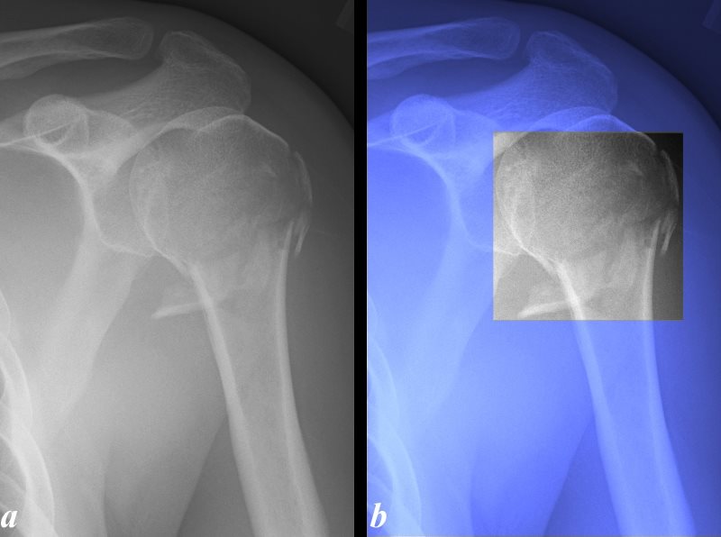
The X-ray of the left humerus in A-P projection is from a 56 year old male and shows comminuted transverse fracture of the corticocancellous junction just as the cortical bone thins in transition to the metaphysis (a) with the fracture highlighted in black and white in (b). The corticocancellous region is well known for its vulnerability. The fracture extends into the superolateral portion of the metaphysis of the head of the humerus.
Courtesy Ashley Davidoff Copyright 2011 100028c.8L
Epiphyseal plate fractures occur in immature individuals when the growth plate is still active. Thus fractures in this region may be complicated by aberrant and therefore discrepant growth of the affected side.
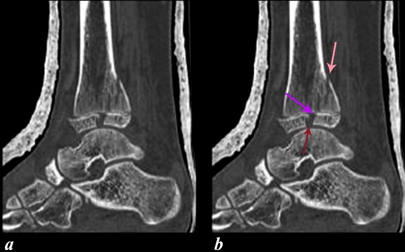
The sagittal reconstruction of the CT scan of the ankle of a youth shows a fracture through the tibia involving the posterior metaphysis (salmon arrow), epiphyseal plate (purple arrow), and epiphysis (maroon arrow). Since the growth plate is not calcified and is also a thin structure, fractures are characteristically difficult to diagnose. The fracture of the epiphyseal plate in this instance can be inferred by interpolating the vector line of the metaphyseal fracture (salmon arrow) to the epiphyseal fracture (maroon arrow). In addition the subluxation of the anterior portion of the epiphysis confirms significant injury to the growth plate. Involvement of the growth plate raises concern about necrosis and consequent failure to grow. This complication occurs in about 15% of injuries.
Courtesy of Philips Medical Systems 2011 91309c02L.8
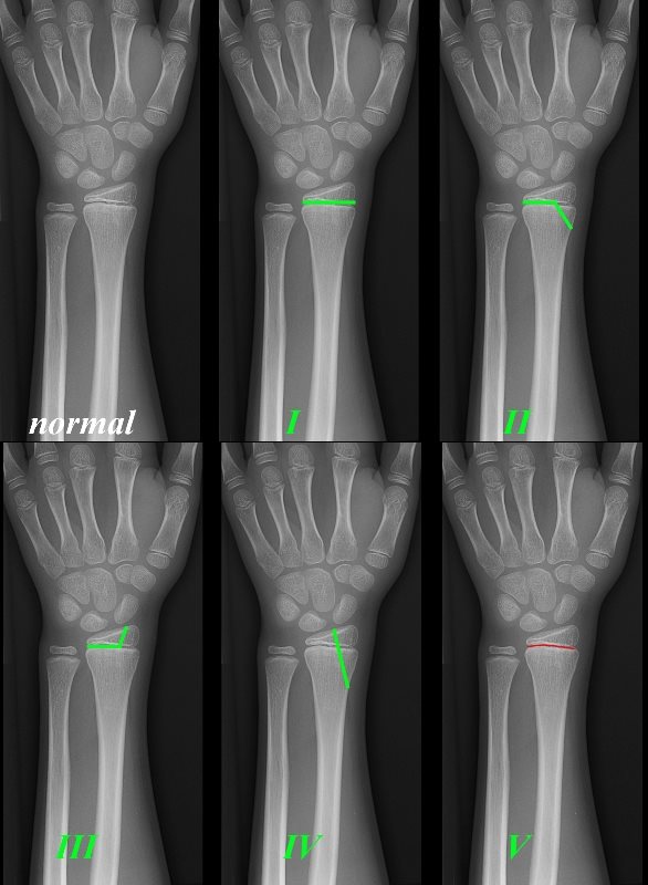
The series of A-P X-Rays are from a 10 year old who had pain in the wrist after sustaining an injury.
Salter Harris
I = epiphyseal plate only
II= epiphyseal plate + metaphysis
III = epiphyseal plate and epiphysis
IV = epiphyseal plate + metaphysis + epiphysis
V = compression of epiphyseal plate
Courtesy Ashley Davidoff Copyright 2011 99794bc01L.8
Salter Harris Classification
Type I – Epiphyseal separation: there is displacement of the epiphysis from the metaphysis at the growth plate. This fracture is most common in newborns and infants and the prognosis is excellent.
Type II – A small corner of metaphyseal bone fractures and displaces, with the epiphysis displaced from the metaphysis at the growth plate. This is the most common fracture and carries an excellent prognosis as well.
Type III-Fracture is through the epiphysis and part of the growth plate, but the metaphysis is unaffected. This is an uncommon fracture and open reduction and internal fixation (ORIF) is usually necessary.
Type IV-Fracture is through the epiphysis, growth plate, and metaphysis. Several fracture lines may be seen. The prognosis is poor since blood supply is stripped and requires perfect reduction.
Type V-Impaction of the epiphyseal plate occurs, with the metaphysis driven into the epiphysis. This is a rare fracture and carries a poor prognosis. The early X-ray may be negative.
With each progressive type, the fracture described becomes increasingly difficult to treat and carries a poorer prognosis for return to normal function.
Epiphyseal fractures in adults may be isolated or may involve the joint. Fractures involving the joint infer injury to the articulating cartilage. These fractures require perfect alignment for healing to take place so that joint function can return with prevention of degenerative arthritis. Hence it is usual for the fracture involving joint surfaces to require orthopedic attention and unless perfect closed reduction can be attained and maintained, open reduction is required.
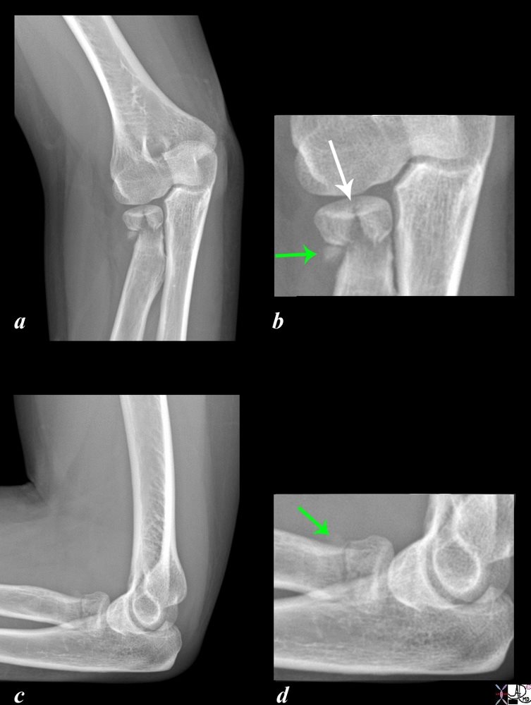
Two views of the proximal radius of a 38 year female following blunt trauma in oblique and lateral projection show a comminuted fracture of the head of the radius with involvement of the joint surface (white arrow). A transverse fracture at the junction of the diaphysis with the metaphysic is better seen on the lateral examination An avulsed fragment is shown by the green arrow, easily seen in b, but barely visible in d.
Courtesy Ashley Davidoff MD 103258c02.8s
Position of the Component Parts of the Fracture
Displacement of the fracture fragments may lead to abnormal alignment. There are three major aspects that encompass displacement including length discrepancy, angulation, and rotation.
Alignment
Alignment of the component fracture fragments is a key component in the evaluation of fractures. Alignment describes the relationship of the longitudinal axis of one fragment to another.
When a fracture has anatomical alignment it means that almost 100% of the surface of the two components of the fracture are appropriately aligned in the longitudinal axis. Anatomical alignment means that continuity of the fragments is maintained. Since the fracture needs bone on bone contact for optimal reparative process it is most important that optimal contact is achieved.
Partial continuity implies that there is some contact between the components.
Fractures that have some continuity of osseous fragments may, in general, be treated by closed reduction, while fractures that have lost continuity usually require open reduction.
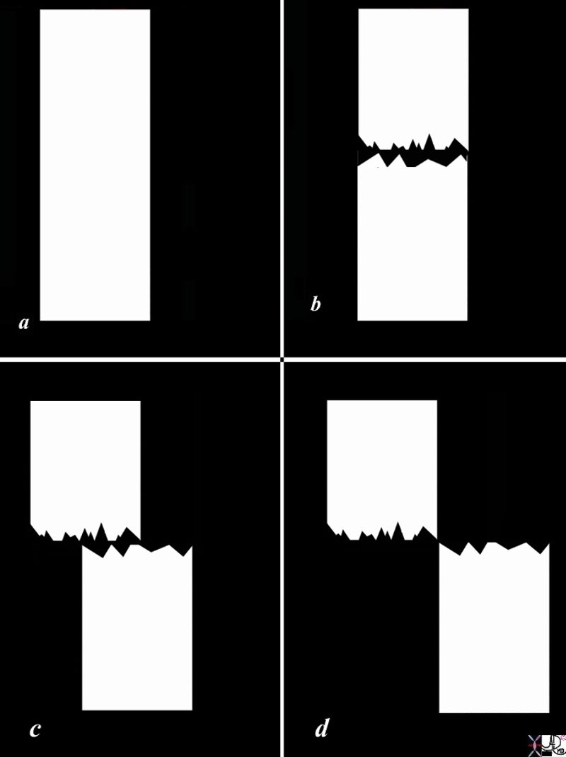
The diagram shows a diagram of a bone without fracture (a), an example of an anatomically aligned fracture with no length change, a fracture that is displaced by about 50% (c), and a fracture in which the component parts are totally displaced (d) without any bone on bone contact. As long as the finding on the A-P examination is in agreement with the lateral examination regarding alignment, the fracture exemplified in b, will likely not require reduction, the fracture in d probably require reduction and the fracture in c, may or may not require reduction.
Courtesy Ashley Davidoff Copyright 2011 103498l01bc02.8s
Displacement
Displacement is the term used to describe the malalignment of two fracture fragments where there is loss of cortical continuity. There are many adjectives used to describe the displacement, since the fracture fragments can be displaced anteriorly posteriorly medially, laterally, superiorly inferiorly or any combination of these. Displacement is the amount of translation of the distal fragment in relation to the proximal fragment in either the anterior/posterior or the medial/lateral planes. Displacement is the opposite of apposition.
No “Bone on Bone” Alignment
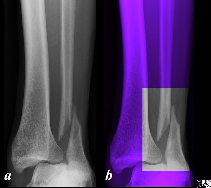
The X-ray is taken in the antero-posterior (A-P) projection and shows a simple displaced oblique fracture of the distal shaft of the fibula in a 36 year old male following blunt trauma. In image b the ankle is overlaid in purple and the area of interest is highlighted in black and white. There is significant, medial displacement of the proximal fragment so there is virtually no bone on bone alignment. This morphology is not conducive to satisfactory healing and likely would require open reduction. In addition to the fracture there has been significant damage to soft tissues and the mortise is not intact with medial displacement and dislocation of the tibia in relation to the talus suggesting rupture of at least the deltoid ligament. The mechanism of this injury is likely due to a direct hit on the lateral aspect of the fibula causing the oblique fracture and dislocation of the tibia off the talus.
Courtesy Ashley Davidoff Copyright 2011 99854.6b01c001
There are three major aspects that encompass displacement including length discrepancy, angulation, and rotation.
This diagnosis of displacement requires two views on an X -ray since a foreshortened appearance may for example also be due to the one component being pushed down and posterior and the other being pushed up and anteriorly.
Displacement – Length Discrepancy
Length discrepancy occurs when the two major fragments become overlapped or distracted.
Overlap occurs as a result of the downward movement of the one component and or the upward movement of the second fragment. This is also called apposition (or bayonet apposition) which infers fragment overlap. As a result there is shortening of the bone.
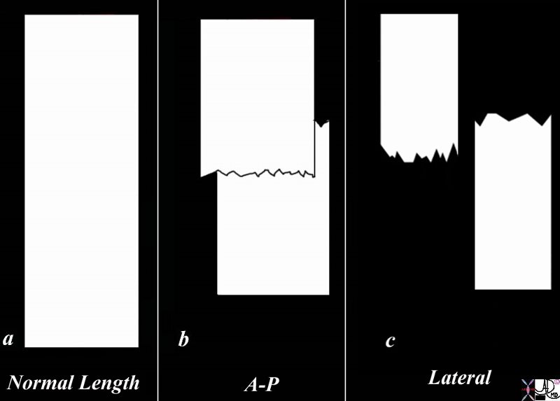
The diagram shows a normal length bone represented by a white rectangle without fracture (a), and (b) an example of overlap fracture in the A-P projection with overall shortening of the bone, while the lateral examination (c) shows posterior and superior displacement of the distal fragment accounting for the foreshortening. Sometimes this fracture in the one projection may look anatomically aligned but in the orthogonal projection, no bone on bone contact is seen. Two views are essential in the evaluation of all fractures.
Courtesy Ashley Davidoff Copyright 2011 103498k04.8s

The A-P (a,b) examination suggests an almost anatomically aligned transverse fracture. Image a, is the A-P projection of the right femur and image b is the same image overlaid in green with the fracture highlighted in black and white. The lateral examination (c,d) is far more revealing and shows a foreshortened, malaligned fracture with the distal segment posteriorly and superiorly positioned with resultant posterior angulation. Image c, is the lateral projection of the right femur and image d is the same image overlaid in green with the fracture highlighted in black and white. The most remarkable aspects of the fracture is that only in the lateral examination do we appreciate the most relevant therapeutic implications of the fracture; the foreshortening of the femur as a result of the superior malposition of the distal fragment and the absence of bone on bone contact. Since there is no ?bone on bone? contact hence healing would be near to impossible and open reduction therefore necessary. Lastly on close inspection multiple lytic elements in the bone represent metastatic disease. This is therefore an example of a transverse pathological fracture caused by metastatic malignant disease with misalignment with foreshortening and overlap.
Courtesy Ashley Davidoff Copyright 2011 101121c03b.6L
Impaction fractures occur when one component of a fracture is driven into, or telescoped into the second part causing foreshortening of the bone.
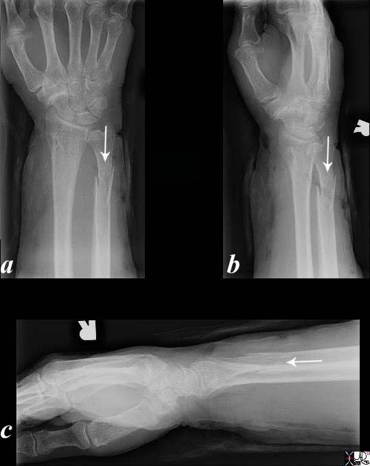
Three views of the distal radius and ulna in the anteroposterior (a), oblique (b), and lateral (c), projection show an acute simple transverse and impacted (arrow) fracture of the shaft of the distal ulna with near anatomic alignment.
Courtesy Ashley Davidoff MD 76722c.81L
Distraction infers the longitudinal separation of the fragments, and overall lengthening of the bone.
Displacement – Angulation
Angulation infers an angular relationship of the longitudinal axis of the fracture fragments, and usually describes the distal fragment. Angulation may be due to medial lateral anterior or posterior displacement of the distal fragment. Angulation has been discussed in detail in the section on the shape of fractures.
Certain alternate terms have been used that are synonyms sometimes specifically related to the part of the body being described and sometimes as a matter of preference. Anterior and posterior are sometimes described as ventral, and dorsal, palmar when in reference to the hand, plantar when in reference to the foot, volar, varus and valgus angulation as a matter of preference.
Anterior angulation (ventral angulation, palmar angulation)
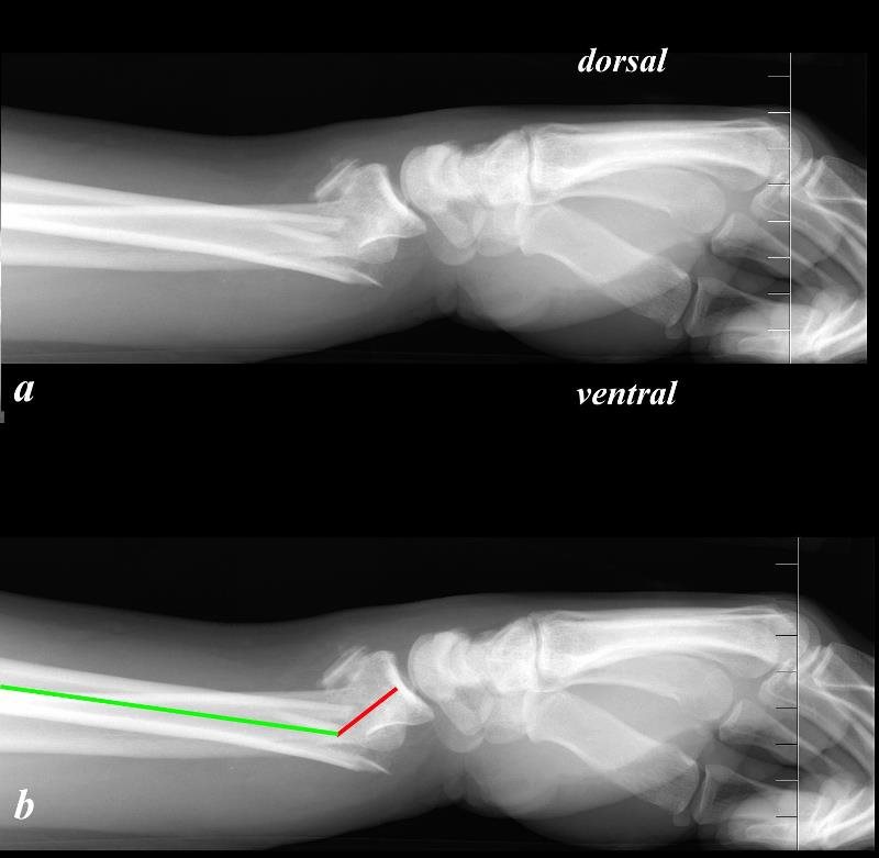
aka Distal Displacement
The image demonstrates the lateral projection of an X-ray from a 40 year old man who fell on an outstretched hand and sustained a fracture of the distal radius. This fracture is called a Colle?s fracture and is characterized by its location being about 1.5 inches from the distal end of the radius, at the weak portion of the distal radius where the diaphysis meets the metaphysis called the at the cortico-cancellous junction. Additionally it is characterized by dorsal displacement of the distal fracture fragment but with ventral angulation of the fracture. The shape of the combined appearance of the proximal fragment, distal fragment and the carpals and metacarpals on clinical and lateral radiological examination is reminiscent of a dinner fork and hence the deformity is called “dinner fork deformity”.
The anatomical position of the forearm is by convention with the palm of the hand facing forward (anteriorly), and so the palm of the hand is the ventral surface. The back of the hand is called the dorsal or posterior surface.
The direction of the forces and weight pushes on the wrist so that the radius gets pushed to the anatomically named ventral portion of the forearm and the distal fragment attached to the wrist gets pushed dorsally.
Courtesy Ashley Davidoff Copyright 2011 101288c02.8
Posterior angulation (dorsal angulation, volar angulation, plantar angulation)

This X-ray shows a simple transverse fracture of the middle of the shaft of the left second metacarpal with the hand held in the antero-posterior (A-P) projection. Image b shows the fracture overlaid in black and the green line is almost straight suggesting near anatomical alignment. The presence of minimal foreshortening suggests angulation which is better appreciated on the oblique projection, (c and d). In this view there is dorsal or posterior angulation and some impaction accounting for the foreshortening seen on the A-P projection.
Courtesy Ashley Davidoff Copyright 2011. 101198.8cL03
Medial angulation (valgus)
Lateral angulation (varus)
Some of these terms we have already used such as impaction fractures
The terms varus and valgus are very similar and a variety of mnemonics have been used to try and remember which one is which. The best mnemonic relates to the story of a Russian woman walking down the street when a loose pig runs between her legs. As it does so, she looks down opens her legs and shouts “Varus the pig?” This scene is so comical and unforgettable that it has stuck in my mind for 30 years and I have never forgotten the difference between the two types of angulation. An image relating to this scene has been depicted in the section on shapes of fractures.

Courtesy Ashley Davidoff 103519cL.8
This diagnosis of displacement requires two views on an X -ray since a foreshortened appearance may for example also be due to the one component being pushed down and posterior and the other being pushed up and anteriorly.
The Relevance of the Position of Fractures in Other Bones
The position of the fracture line or the position of the fragments has relevance in the other types of bones as well. For example the position of the fracture line in a scaphoid injury has relevance to involvement of its blood supply such that a fracture involving the medial third has the highest risk of avascular necrosis (AVN up to 30%). Fractures in the middle third are the most common site for scaphoid fracture but are less commonly complicated by AVN, while distal third fractures are rarely complicated by AVN.
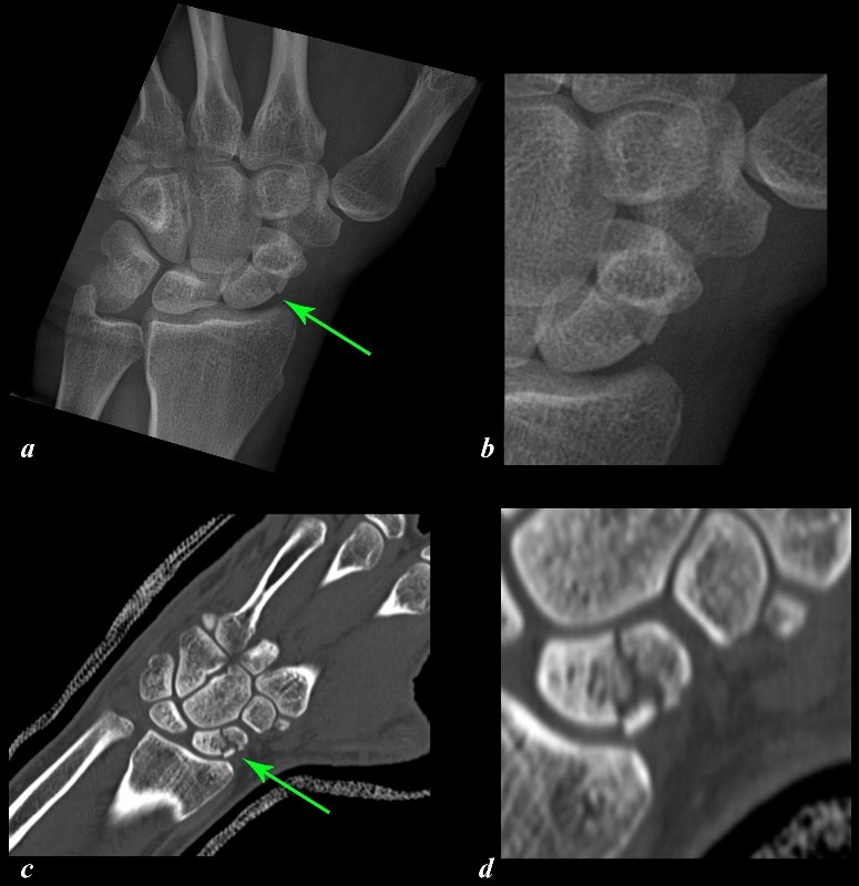
The X-Ray (a, and magnified in b) and CT scan (c and magnified in d) reveal a transverse fracture (green arrows) through the middle of the left scaphoid of a 29 year old patient after a fall on his outstretched hand. This is the most common site for fracture of the scaphoid and in this location the injury carries a small risk of avascular necrosis. A fracture through the proximal 1/3 would carry a higher risk of AVN approximating 30%.
Courtesy Ashley Davidoff Copyright 2011 107550c01L.8
Fractures of the vertebral bodies may result in retropulsion of the fragments into the spinal cord with potentially devastating functional results as quadriplegia and even death.
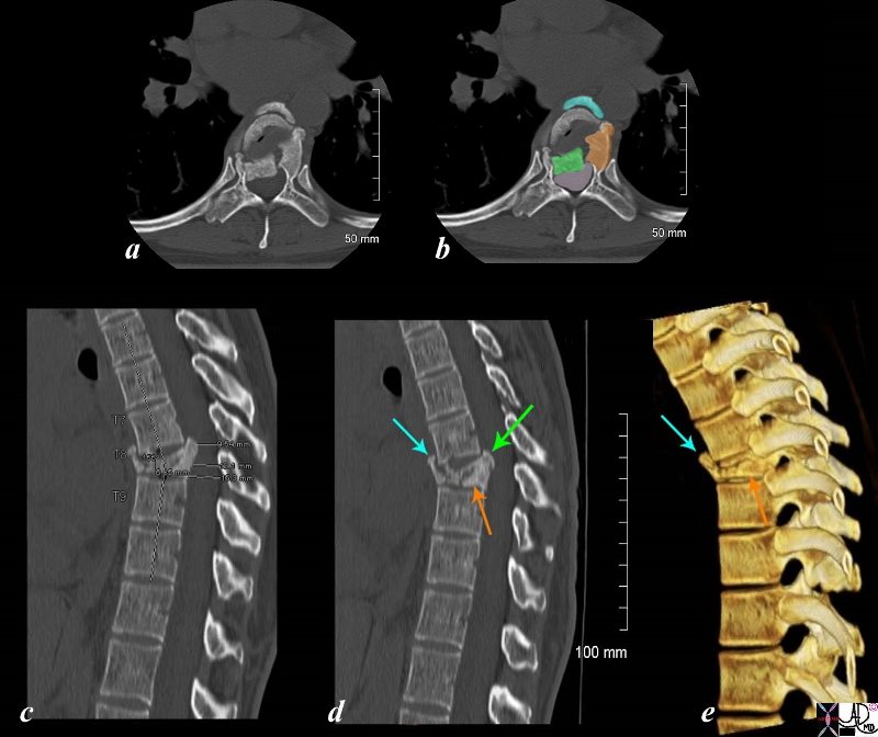
The panel of images using CT technology revealing a compression fracture of T8 in a 43 year old female who sustained compressive trauma to the thoracic spine resulting in a kyphosis at that level. Images a and b are transverse images showing a right sided retropulsed fragment (green) impinging on the thecal sac (light pink in b) The fragment that has ruptured anteriorly is overlaid in teal blue. The middle portion of the fracture is overlaid in orange. Image c is a mid sagittal reconstruction of the thoracic spine and shows the compression fracture with a 9.5mm retropulsed fragment with reduction of the thecal sac diameter to 11mm. The overall bony density of the fracture is increased suggesting a non pathological compression fracture. Image d is also a midsagittal reconstruction showing the retropulsed fragment (green arrow), the anteriorly displaced fragment (teal arrow) and the fragment in the middle indicated by the orange arrow. Image e is a 3D reconstruction of the T8 fracture enabling the visualization of the anterior fragment (teal arrow) and the middle portion of the fracture (orange arrow).
Courtesy Ashley Davidoff Copyright 2011 102190c05L.8s
The position of the fracture line and the fracture fragments in relation to each other is the most important determinant of how fractures should be managed. When the fracture in the pediatric population involves the growth plate, the potential for growth disturbances becomes a concern particularly in the Salter Harris type III,IV, and V. When a fracture involves the joint surface special attention to optimal alignment is even more important because of the involvement of the joint cartilage and failure to align exactly can be complicated by degenerative disease in later life.
When there is near anatomical alignment of the fracture fragments, with bone on bone contact, and this can be maintained for the duration of healing, then there is no need for reduction. If there is displacement, angulation distraction, shortening or rotation reduction is necessary. If this cannot be attained by a closed method, then open reduction and internal fixation is required (ORIF).
References
McGuigan Francis X. Skeletal Trauma www.springer.com/cda/content/
Rang, M, Pring, M.E., Wenger, D R. , Rang’s Children’s Fractures 3rd Edition 2005 Lippincott Williams and Wilkins Philadelphia
—
