The Common Vein Copyright 2011
Introduction
Bone is a specialised form of connective tissue that serves both as a connective tissue as well as an organ.
As a connective tissue it is composed of the three basic elements that comprise all connective tissues; cells, fibers and an extracellular matrix. In this realm its function is to connect body tissues with each other, provide a framework for the body, protect the organs, and store energy.
It is also considered an organ since it is made up of a collection of tissues joined together in a structural unit to perform a function. These functions include hematopoeietic function, protection, mobility, and mineral homeostasis. It stores minerals such as calcium and phosphorus and participates in acid base metabolism by absorbing or releasing alkaline salts to maintain and sustain a stable PH environment.
Its dominant structural characteristics are that it is hard and lightweight. Its character has been described as being similar to a tree trunk – strong and sponge like.
The cellular component is made of osteoblasts, (generate bone) osteoclasts (resorb bone), and osteocytes (bone-maintaining cells). Osteocytes are inactive osteoblasts trapped in the extracellular matrix.
The extracellular matrix is responsible for the strength of the bone. It contains organic elements and minerals. The organic component is made of proteins mostly of type I collagen providing resilience and tensile strength. The mineral component is composed of calcium, phosphate, and hydroxyl ions. Together the minerals form a compound called hydroxyapatite (Ca5(PO4)3(OH)). The hydroxyapatite provides hardness, strength and rigidity for the bone.
The mineral (hydroxyapatite) represents 70%, the proteins (type 1 collagen) 22% and water 8% by weight
The mineral component resists compression but has poor ability to withstand tensile loads. In essence it is hard but brittle and acts like glass when subjected to a force – ie it cracks. In contrast the collagen arranged at oblique angles in the lamellae affords tensile strength. This provides a rubbery and to some degree elastic nature. Overall however bone acts more like glass than like rubber.
Stiffness of bone therefore depends on mineral content while toughness depends on collagen (Augat). The combination of the hydroxyapatite and collagen provides bone with a high compressive strength, but poor tensile strength and very low shear stress strength. This infers that it resists pushing forces but not pulling or torsional forces.
There are 206 bones in the adult human body grouped as being either part of the axial skeleton (80) or the appendicular skeleton (126). The component bones of the skeleton include the following:
skull (28):
vertebral column (26),
hyoid bone, (1)
sternum and ribs (25),
upper extremities (64),
lower extremities (62)
Of the sesamoid bones only the patellae are included in the above evaluation, but the smaller sesamoid bones are not
Compact Bone and Spongy Bone
There are two types of bone tissue; compact bone and spongy bone. They are very different entities and have different functions bur are interdependent and collaborative. Compact bone is hard and dense while spongy bone is softer and porous.

The image reflects a coronally reconstructed CT scan of the knee of a 23 year old male and shows the basic anatomy of a long bone noting the distal end of the femur and the proximal portion of the tibia. Two types of bone are identified; the compact bone is easily identified on the surface of the bone (notated in b, white). It is white and thick around the shaft of the femur and thins as it progresses to its distal end. At the proximal end of the tibia the compact bone is thin and becomes thicker as it progresses to the shaft. Compact bone is the hard stiff outer shell of all bone providing support, protection and acting as a lever for movement and site of attachment for ligaments tendons and muscle. In addition it stores and releases calcium that is needed in the rest of the body for muscle contraction and signal transduction. It is much denser than spongy bone (cancellous bone) The cancellous bone is overlaid in purple and has a lacy trabeculated appearance better appreciated on image a. Red marrow is usually found in the spongy bone and yellow marrow in the diaphysis within the medullary cavity.
Courtesy Ashley Davidoff Copyright 2011 104676c09L01L.8sCompact Bone
Compact bone (aka cortical bone) is the hard outer shell of bone. A cortical layer surrounds all bone. The shafts of bone have thicker layers of compact bone. It is called cortical bone because it surrounds the bone as a hard external frame and cover. The word cortex derives from the Latin word “cortex” which means bark of a tree, and it is therefore used in describing an outer shell or husk of a structure. The cortex of the kidney is the outer part of the kidney for example.
Structurally compact bone is hard strong and stiff and represents 80% of the weight of the skeletal system. The long bones contain most of the compact bone in the body. Compact bone forms an intricate system around vessels known as Haversian systems or osteons. Volkmann?s canals help to connect the Haversian systems. At a microscopic level the osteon is the unit (brick equivalent) of compact bone. It has a lamellar structure, which implies that it is made of thin disc like layers of tissue.
Functionally compact bone facilitates support of the body in general, provides anchorage for muscles and ligaments, functions as lever for movement, stores calcium and other chemicals that are used in neural transmission and muscles contraction.
Compact bone and spongy bone are so diverse in structure and function that they should really be considered two distinct entities. For example when their turnover/year is considered compact bone has about a 2% change whereas spongy bone has a turnover of 25%.
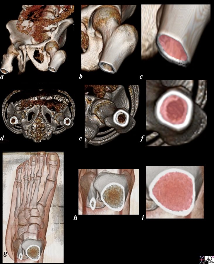
The 3D reconstructions of a normal examination of the hips and proximal femur progressively magnified (a-f) and the foot with distal tibia progressively magnified (g,h, i). The compact bone is the white rim or ring of bone that takes up little volume and is relatively thin compared to the spongy bone which is more voluminous and is depicted in pink in the magnified views (c,f,i).
In images a-c, the hip is turned in left anterior oblique projection with left hip forward. In these images we are looking obliquely down the barrel of the pelvis, while in images d,e,and f we are viewing it directly down the barrel as if we were standing at the feet.
The images also depict the inner spongy bone (pink in c, f, and i) surrounded by the ring of compact bone (white ring around the pink spongy bone in c, f,and i).
Courtesy Ashley Davidoff Copyright 2011 106241c03L.8s
Osteon – Haversian System
The osteon (aka Haversian system) is the morphological unit that makes up compact bone.
Structurally it is described as a cylindrical structure that is made up from plates of tissue called lamellae. The osteon has a central canal that contains the lifelines for the bone including the arteries veins and nerves. The osteon measures about 200 micrometers in diameter (.2mm) and may be up to 10mms tall. A group of osteons form a series of columns along the long axis of bone.
The word lamella is the diminutive of the Latin word lamina which means “thin plate”, and lamella therefore means a small thin plate. The lamellae consist of concentric layers of collagen, bone cells and the hydroxyapatite matrix. Most of the lamellae are arranged around the central canal as plates of tissue shaped like thick old fashioned records. Each lamella is between 3 and 7 µm in width, and each osteon has between 4 and 20 lamellae. (Wojnar)
The collagen in the lamellae is organized so that for a given lamella the collagen project in one direction but the next layer has the collagen vector in the opposite direction creating a zig-zag pattern.
The lacunae are spaces in the osteon that contain the osteocytes which communicate with each other via the canaliculi. The canaliculi (small canals) are small communicating channels carved out in the hard bone. The osteocytes are the cells which are housed in the lacunae.
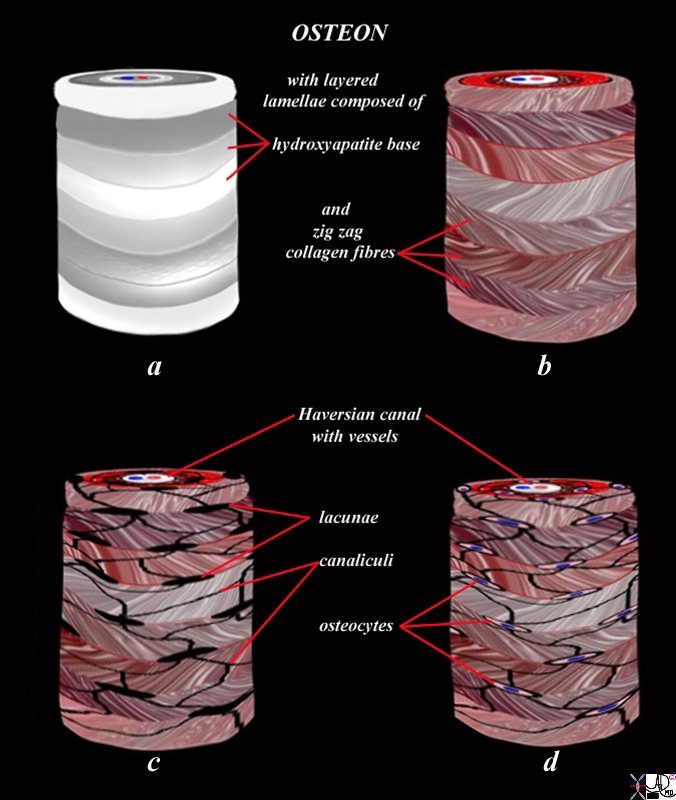
The diagram illustrates the appearance of an osteon (Haversian system) in 3D. The osteon is the unit or building brick of compact bone and consists of a column of bone with layered subunits called lamellae (flat plate like structures). The lamellae consist of calcium, phosphate, and hydroxyl ions which form a compound called hydroxyapatite (Ca5(PO4)3(OH) – seen in (a) as shades of white and gray overlay. The hydroxyapatite gives bone its hardness. The lamella also contains type I collagen fibers. (color overlay of reds and pinks) that are arranged in different directions on adjacent lamellae so that a zig zag pattern of the collagen is created allowing for strengthening of the tissue. The collagen provides elasticity. Lacunae (c) are spaces in the lamellae that house the osteocytes (d). The canaliculi (c) are a network of fine tubular channels that allow the osteocytes to communicate with each other. The Haversian canal (c,d) are spaces in the middle of the osteon that contain an artery, vein and nerve (c)
Courtesy Ashley Davidoff Copyright 2011 106275c03L11.43kce02L.81s

The diagram shows a microscopic cross section of compact bone One of the osteons is overlaid in red.
The osteon (aka Haversian system) is about .2 mms. in diameter and up to 10mms high, and consists of 4-20 concentric layers or lamellae that surround the Haversian canal that contains blood vessels (red and blue ) and nerves The arteries (red) and veins (blue) are shown in the Haversian canal and the concentric layers of lamellae are shown as well. When viewed from this projection the shape with concentric rings and the “hole” in the middle are reminiscent of an old fashioned LP record.
106272b06b01.8 Artistic rendering Ashley Davidoff
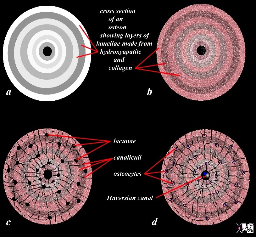
This diagram serves to illustrate the component parts of the osteon in cross section. The extracellular matrix of calcium, phosphate, and hydroxyl ions (hydroxyapatite (Ca5(PO4)3(OH)) is represented in diagram in (a) as shades of white and gray overlay. The hydroxyapatite gives bone its hardness. The type I collagen fibers shown in an overlay of reds and pinks and are reflected in images b, c, and d.
Lacunae are shown as black holes in c. The canaliculi are shown as fine communicating channels in image c as well.
The osteocytes with pink cytoplasm and blue nuclii are shown in the lacunae in d. The Haversian canal (c,d) that contain artery, vein and nerve is shown in image d.
Courtesy Ashley Davidoff Copyright 2011 106275c03L08b09cLd02.81s
Applied Anatomy
Compact bone is hard and brittle and can fracture, but will usually fracture at its thinnest and weakest point unless the force is directly imposed on the thickest portion. Since the middle of the shaft of long bones contains the thickest compact bone fractures are not as commonly seen in this region as they are at the distal ends of the long bones near the metaphyses.
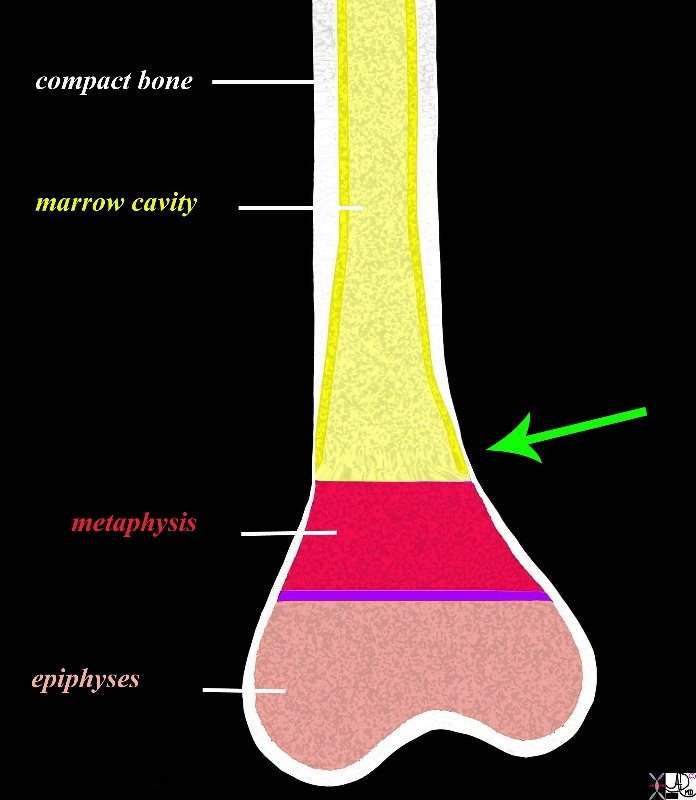
The diagram shows the basic anatomy of the distal end of a long bone such as the humerus or femur which consists of a shaft (diaphysis) and an expanded end each containing an epiphyses (pink), the growth plate in children (purple line) or epiphyseal line in adults, and the metaphysis (red). The shaft (diaphysis) consists of a thick outer layer of compact bone (white) which thins significantly as it progresses to the metaphysis (green arrow). The metaphysis and epiphyses contain spongy bone (trabecular or cancellous bone) which is more porous and softer. The fracture of the Colle?s fractures for example, occurs at this juncture where the compact bone thins out.
Courtesy Ashley Davidoff Copyright 2011 104007b06c19L.8s

Spongy Bone (aka Cancellous Bone Trabecular Bone)
Spongy bone (aka trabecular bone and cancellous bone) is the porous and softer trabeculated bone that has a spiculated appearance. It is found in the epiphyses and metatphysis of long bones and forms the matrix of the vertebra, short bones, flat bones of the ribs and the pelvis.
Structurally it is characterized by its spongy appearance (aka trabecular and cancellous appearance). The word trabecular derives from the Latin trab?cula, which is the diminutive of trabs meaning a small beam, rod or strut since the bone is marked with small vertical bars and horizontal cross bars. The word cancellous originates from the Latin word cancellus that means a lattice which also describes its morphological appearance. The structure is not random. Rather it is organized with bars of bone oriented along the lines of physiological loads. The morphology is dependent on the loads exerted on the bone, and this depends on its position in the body, and the unique load imposed on the bone. The morphology serves to dissipate the force as well as to provide support.

The anatomical specimen of a vertebral body between two discs exemplifies the trabecular bone highlighted in black and white in the second image. The spongy, trabeculated or cancellous nature is readily apparent with the vertical and horizontal cross bars easily visualized
Courtesy Ashley Davidoff Copyright 2011 05521b02c.8
Functionally spongy bone creates a dynamic infrastructure within the relatively adynamic compact bone and responds rapidly to changing forces on the bone. It also houses hematopoietic tissue – aka the red marrow.
There is a significant difference in the activity of spongy bone and compact bone. The latter is slow and steady and is not subject to day to day change whilst spongy bone is a dynamic tissue always responding to the day to day stresses on the bone.

3 Different Regions in the Metaphysis
A Story of the Forces
The image reflects a coronally reconstructed CT scan of the right knee of a normal 23 year old male and shows the dynamic nature of trabecular bone by reflecting the heterogeneous trabecular pattern of the spongy bone at the distal femur with very different morphologies in each of three adjacent metaphyseal regions. In the lateral part of the metaphysis (b) the density of the bone is greatest (whitest) while the middle portion is the least dense suggesting that the lateral aspect of the femur is subject to the most stress of the three regions while the middle receives the least. Note also the net vertical vector of the medial and lateral trabeculae with most the trabeculae organized in a craniocaudal direction with fewer horizontal bars. In the middle section there are many more horizontal bars and the bone has the characteristic spongy appearance. The morphology of the compact bone on CT is restricted to a white dense region on the outside of the metaphysis. The difference in the slow steady compact bone and the dynamic nature of spongy bone is exemplified in this image.
Courtesy Ashley Davidoff Copyright 2011 104672ce01L.8

Note the Vector of the Trabeculae in A-P Direction
The image reflects axial cut from CT scan of the knee of a normal person and shows the dynamic nature of trabecular bone by reflecting the heterogeneous but organized trabecular pattern of the spongy bone at the distal femur with very different morphologies in each of three adjacent metaphyseal regions. As opposed to the image above where the dominant vector is vertical, the dominant vector is in an anteroposterior direction thus serving as a support for the weight rather than the transmission and dissipation of the weight. The area between the red lines (b) is slightly denser than the area between the green lines (d) and much less dense than the middle region (c) inferring that area between the red lines (b) supports the most weight. The morphology of the compact bone on CT is restricted to a white dense region on the outside of the metaphysis and in this region is relatively thin.
Courtesy Ashley Davidoff Copyright 2011 106585c05L.8
Applied Anatomy of the Spongy Bone
Since spongy bone is softer than compact bone it will fracture more easily
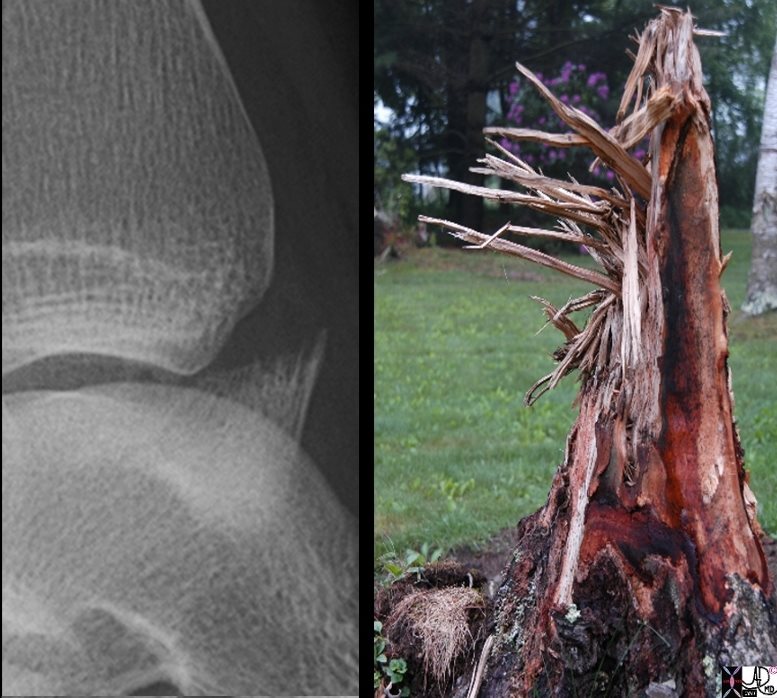
These images afford us the opportunity to get a sense of the shape and pathogenesis of the fracture at the fracture line because the resolution of the fracture is so clear in the X-ray image. The X-ray is of the ankle in a 15 year old male in the anteroposterior projection (A-P) and shows an avulsion fracture of the anterior portion of the distal tibia. Note the spikes of broken bone at the edge of the distal fragment, reminiscent of the spikes of splintered tree from the weather induced fracture of this tree. The photograph was taken in the Berkshires Connecticut.
Courtesy Ashley Davidoff Copyright 2011 99940c02.8s
The normal turnover of cancellous bone is far quicker than compact bone and therefore by implication is far more relevant in the healing process than is compact bone. When a patient fractures a bone attention is paid to the amount of ?bone on bone? contact that is retained. This “bone on bone” contact is directed to the evaluation of the more voluminous spongy bone. When the fragments are anatomically aligned and there is good “bone on bone” contact then the bone will heal well without intervention. On the other hand when the fragments are significantly distracted, angulated or rotated then the patient requires surgical intervention.
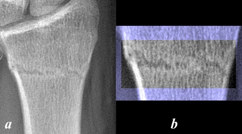
The A-P examination of the distal radius of a young adult male shows a non displaced two week old transverse fracture of the cortico-cancellous junction of the distal radius (a) with excellent ?bone on bone? contact and with excellent anatomical alignment (highlighted in black and white in b). This fracture does not require any hardware intervention and will continue to heal well. Note the alignment of the vertical trabeculae is hardly disturbed at the site of the fracture. The subacute fracture reveals early resorption of the dead bone along the fracture line accounting for the minimal widening of the fracture line and early increased density on either side.
Courtesy Ashley Davidoff Copyright 2011 106764b03L
Poor Bone on Bone Contact
Courtesy Ashley Davidoff Copyright 2011 99816c031L.81s

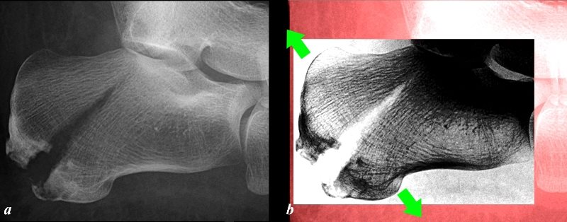
The lateral examination of the calcaneus of a 64 year old female (4 years later) shows two screws traversing the previously identified distracted fracture, with not a trace of the former fracture line (region highlighted in black and white in b). Open reduction and internal fixation (ORIF) was required to bring the fragments together which allowed bridging of the trabeculae to take place.
Courtesy Ashley Davidoff Copyright 2011 99818c.81L
Interaction of Spongy and Compact Bone
The interaction of spongy and compact bone is a dynamic relationship that evolves to strengthen the bone in response to forces imposed on them by body weight and the forces of movement that they are subjected to on a day to day basis.
The head and neck of the femur for example are exposed to the primary vertical force of the body transferred via the spinal column and pelvis, and in response develop spongy bone called primary trabeculae that have a vertical orientation (see diagram below). The primary vertical trabeculae in turn act as a bridge to dissipate the force to the compact bone on the medial border of the shaft of the femur.
The secondary horizontal trabeculae respond to the horizontal forces imposed on the femur and transfer these forces to the compact bone on the lateral aspect of the shaft of the femur.
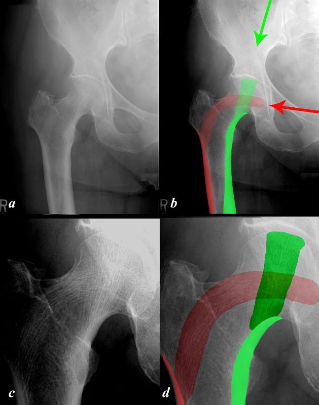
The X-ray in A-P projection of a normal right hip is from a 62 year old female and displays the two major trabecular groups of the head of the femur. The vertical group in green is called the primary trabecular group which has developed in response to vertical pressures of the body (green arrow). These vertical forces are transferred via the primary group to the medial compact bone (solid green) in the diaphysis
The secondary horizontal trabeculae (overlaid in red) in the head of the femur respond to transferred forces from the pelvis (red arrow) and the secondary trabeculae in turn transfer the force to the lateral compact bone in the diaphysis.
Courtesy Ashley Davidoff Copyright 2011 106282c05L.8s Hall
As the force of gravity with the weight of the body progresses down the leg and through the knee, it needs to be transferred to the ankle and foot which are at right angles to the force.
Examination of the trabeculae of the foot reveals to some extent the manner with which the body handles the dissipation of the full body weight to the foot.

The image reflects a lateral projection of the left ankle of a normal adult and reveals the almost microscopic anatomy of the trabecular patterns in the component bones. The arrows represent at least two of the major vectors of the force, one (green arrow) toward the calcaneus (c), and the second (red arrows) toward the tarsals and metatarsals of the foot.
Image a is an inverted image so that bone that usually shows up as white is black and that which is usually black is white. This technique allows improved visualization of the trabeculae.
Two basic forces are shown that get divided and dissipated to the foot. The green arrows reflect a vertical force created by the weight of the person which is transmitted relatively posteriorly through the anterior part of the tibia via the talus (t) and then to the posterior part of the calcaneus (c). The trabecular pattern runs parallel to the force thus reinforcing the bone. The posterior and inferior aspect of the calcaneus is the final destination (“buck ends here!”) and this region is reinforced with compact bone. This compact bone is the thickest cortical bone of the foot and reflects the final destination of the vector of the (green arrow) force.
Some of posterior trabeculations of the tibia and the trabeculations of fibula are directed inferiorly and anteriorly along the other vector (red arrows). The anterior aspect of the talus and the anterior aspect of the calcaneus redirect this force (red arrows) toward the other tarsal bones and then toward the metatarsals and phalanges. The pattern of the trabeculations in horizontal direction therefore reflects the response of the bone to the forces imposed on them.
Courtesy Ashley Davidoff Copyright 2011 72326c12Lb01.8s
The Periosteum
The periosteum is a tough double layer of “skin” that surrounds bone except at the level of the joint surfaces.
The periosteum consists of an outer fibrous layer and an inner cellular layer that has the capacity to produce bone. Tendons that attach to bone traverse the periosteum and insert into the compact bone via Sharpey’s fibers.
It is structurally characterized by its component two layers. The outer layer consists of dense fibrous tissue containing fibroblasts and the inner layer is an osteogenic layer that contains progenitor cells that can develop into osteoblasts. There are blood vessels in the periosteum which act as a source of blood supply and venous drainage for the outer parts of the bone but also carry the nutrient artery that penetrates the bone. There are pain sensitive fibers in the periosteum.
Functionally the periosteum is responsible for providing access to the compact bone for the attachment of muscles, tendons and ligaments with the assistance of Sharpey’s fibers. The inner layer is responsible for circumferential growth. The osteogenic nature and prominent vascularity make the periosteum an important structure in providing assistance in the healing of fractures.
In children the periosteum is thicker, metabolically more active, more vascular and not as firmly attached to the periosteum. In an acute fracture subperiosteal hematoma is usually more prominent in the children. All these factors enable more rapid repair and accelerated healing of pediatric fractures.

The X-ray of the left knee is from a 13 year old male who was complaining of pain. The most significant finding is the presence of periosteal reaction on the medial aspect of the tibia (highlighted in b and magnified in c), at the junction of the diaphysis with the metaphysis. Although no fracture is identified the presence of periosteal reaction and its location ( a common location for fractures) the possibility of a healing occult fracture is entertained.
Courtesy Ashley Davidoff Copyright 2011 106588c02.81L
Endosteum
The endosteum is considered a layer of “resting” bone marrow that abuts the inner surface of the bone.
Blood Supply
Bone has a rich blood supply and receives 10-20% of the cardiac output. The nutrient artery enters the diaphysis of the long bone and divides into an ascending and descending branch and supplies the inner 2/3 of the cortex and the medullary cavity. The metaphysis and epiphyses are supplied by vessels that supply the joints. The nutrient arteries and arteries around the joints anastomose with each other. The periosteum has its own blood supply and these vessels also supply the outer 1/3 of the cortical compact bone. (Bonakdarpour)
Bone Marrow
Bone marrow is a soft tissue that acts as a packing within the bone, but more importantly has hematopoietic function. There are two types of marrow; yellow marrow and red marrow. The yellow marrow is composed mostly of fat and found in the medullary cavity of the diaphysis of long bones in adults. The red marrow which has hematopoietic function is found in the cancellous bone of the metaphysis and epiphyses of long bones as well as in the flat bones. The bone marrow produces 2.5 billion red cells per kg/day, 2.5billion platelets per kg/day and 1 billion white cells per kg/day. For a 70kg man that is more than 400 billion blood cells ( Bakitas, Erslev). There are in comparison 7 billion people on earth. Imagine the enormity of the function of the bone marrow!
Thus bone is thus intimately associated with the hematopoietic system and they share cells and local factors that regulate both of them.
Classification of the Bones Based on Size and Shape
The classification that revolves around the size and shape of bones is the most common method of classifying bones. It has proved to be practical, since the classification allows one to predict the compact /spongy bone content, the type of function the bone fulfils, and the type of fractures they may sustain.
There are basically 5 types of bones in this classification
long bones eg femur tibia and phalanges
short bones eg carpals and tarsals of the wrist and ankle
flat bones eg skull sternum and ribs
irregular bones eg vertebra and pelvis
sesamoid bones– eg patella
Long Bones
The long bones will be discussed in detail at this point since they demonstrate examples of both compact and spongy bone and also demonstrate the physiological changes of growth exemplifying the role of the metaphysis growth plate, and epiphysis.
The long bones such as the femur, humerus and phalanges are characterized by having a shaft and two expanded extremities which usually function as two articulating surfaces.
The shaft has the thickest compact bone of all bones in the body while the proximal and distal ends have spongy bone surrounded by a thin layer of compact bone.
The shaft is also called the diaphysis. The word diaphysis originates from the Greek words ?dia? meaning across or through, and physis meaning growth and it is therefore the part of the long bone that grows between or across the two growth areas. It is the first part of the bone to ossify during growth. It consists of an outer cortical layer and an inner medullary cavity that contains yellow marrow in the adult.
The ends of the long bone house the metaphysis, growth plate (subsequently becoming the growth line) and the epiphysis.
The metaphysis and epiphysis contain spongy bone that usually contains red marrow.
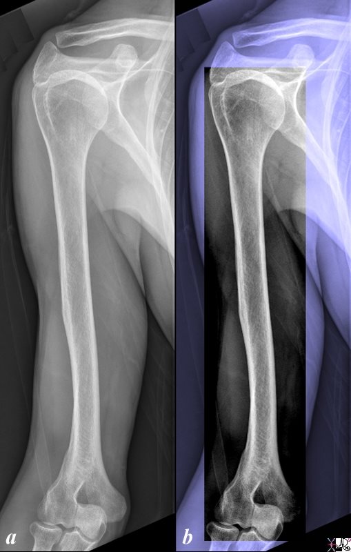
An Example of a Long Bone
The plain X-ray of the right humerus is from an 56 year old male and is normal.. The humerus is an example of a long bone consisting of a shaft and two articulating ends. The shaft is longer than it is wide and the two ends are usually wider and more globular than the shaft
Courtesy Ashley Davidoff Copyright 2011 104579b.8cLs

Long bones consist of a shaft called the diaphysis and two expanded ends. The expanded ends consist of the metaphysis and epiphysis Between the metaphysis and epiphysis is the growth plate or epiphyseal plate, consisting of hyaline cartilage. The cells of the growth plate are called chondrocytes and are under constant division. The new cells are projected forward toward the epiphysis and the older cells are pushed toward the metaphysis and shaft. As these cells age the osteoblasts- cells that generate bone form bone from the degenerating cartilage cells. The epiphysis contains red marrow and is covered with cartilage.
Courtesy Ashley Davidoff Copyright 2011 104007b04.9s
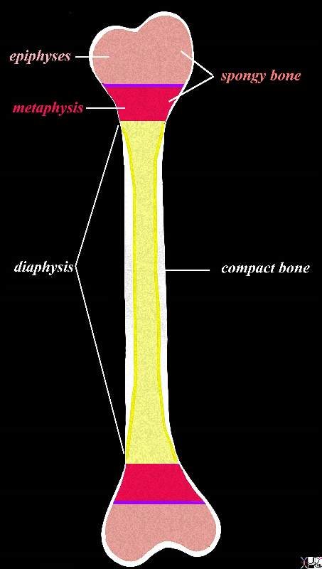
The diagram shows the basic anatomy of a long bone such as the humerus or femur which consists of a shaft (diaphysis) and two ends each containing an epiphyses (pink)covered by cartilage on the outside and subtended by the growth plate in children (epiphyseal plate purple) and the metaphysis (red) The shaft (diaphysis) consists of an outer thick cortical bone (white) (aka compact bone) which is hard with an inner cavity called the medullary cavity (yellow) containing yellow marrow in the adult. The metaphysis and epiphyses contain spongy bone. The compact bone thins significantly at the junction of the diaphysis and metaphysis and is a weak part of long bones. This weakness is exemplified in Colle?s fractures for example in which the fracture of the distal radius occurs at this junction.
Courtesy Ashley Davidoff MD 104007b06c18b.8s
From Fetus to Adult – From Cartilage to Bone
In the 3-5 week fetus the long bones are made entirely from cartilage. By 6 weeks a primary ossification center appears in the shaft and is surrounded by a collar of compact bone. At birth the shaft is fully ossified. Between 2-5 years a marrow cavity is formed in the shaft and secondary ossification centers appear at the ends of the shafts consisting of a core of spongy bone and an outer rim of cartilage. By adulthood (approximately 18-25 years) the cartilage in the epiphysis and metaphysis disappear and the secondary ossification center merges with the diaphysis.
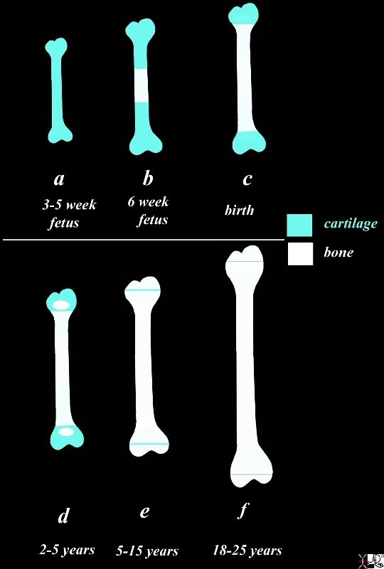
Cartilage to Bone
The diagram reflects the growth of long bone from a purely cartilagenous structure (teal blue) to a purely bony structure (white) except for the cartilage protecting the bone at the joint surfaces. In the 3-5 week fetus (a) the long bones are made entirely from cartilage (teal blue). By 6 weeks a primary ossification center appears in the shaft (b, white)and is surrounded by a collar of compact bone. At birth the shaft is fully ossified (c) but the ends remain as cartilage. Between 2-5 years (d) secondary ossification centers appear at the ends of the shafts consisting of a core of spongy bone and an outer rim of cartilage. Between 5-15 years the growth plate made of cartilage is a linear structure (e) By adulthood (f) the cartilage in the epiphysis and metaphysis disappear and the secondary ossification center merges with the diaphysis. The epiphyseal line remains as a thin white line on the X-ray image.
Courtesy Ashley Davidoff Copyright 2011 104007b03b04c05L.8b
The first phase of ossification is the appearance of the primary ossification center which usually occurs in the prenatal period and for long bones is the diaphysis and for irregular bones is in the main body. There is usually only one primary ossification center per bone.
The secondary ossification centers represent the second site in bone that starts to ossify. At birth in general there is a paucity of secondary ossification centers and often none are present. Those that may be present at birth include the center at the distal femur, proximal tibia, calcaneus, talus, and cuboid.
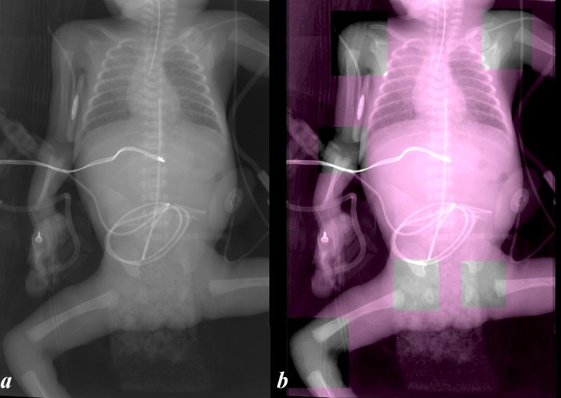
The image reflects an A-P projection of the normal X-ray of a 1 day old infant and shows the basic appearance of the skeletal system with lack of any secondary ossification centers in any of the visualized joints highlighted in black and white in image b. What is visible and dense on the radiographs are the calcified and ossified portions of the primary ossification centers. The shafts of the bones are ossified and these are easily recognized. As the shaft flares at its extremities they transition into the metaphysis and assume an early and characteristic trumpet shape.
Courtesy Ashley Davidoff Copyright 2011 106590cL.81
Secondary Ossification Centers Not Calcified and Not Visible
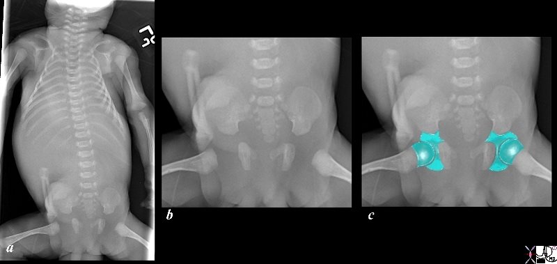
The image reflects an A-P projection of a full term still birth revealing the basic appearance of part of the skeletal system of the neonate. Image b is a magnified view of the pelvis and hip bones showing the absence of calcified secondary ossification centers in the pelvic bones, hip joints and proximal femurs. The absent spaces around the hip joints consist of cartilage (blue in c) which will evolve into the growth plate, epiphyses and articulating cartilage What is visible and dense on the radiographs are the calcified and ossified portions of the bone at this stage
Courtesy Ashley Davidoff Copyright 2011 106109bc05.81s
Over the next few years all the other secondary ossification centers develop progressively, each bone evolving at a different time. There is usually more than one secondary ossification center per bone. These centers of secondary ossification occur in the epiphyses at both ends of long bones.
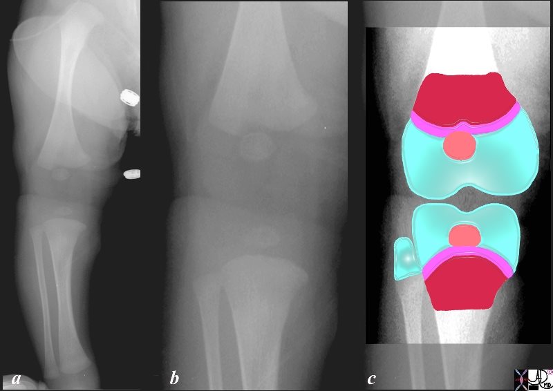
The image reflects an A-P projection of the normal right leg of a 26day old infant and shows the basic appearance of the femur, knee joint, tibia and fibula (a). Image b is the magnified portion of the knee joint and image c reflects the projected components in color. What is visible and dense on the radiographs are the calcified and ossified portions of the bone at this stage The shafts of the bones are ossified and these are easily recognized. As the shaft flares at its extremities they transition into the metaphysis (maroon in c) and assume a characteristic trumpet shape. The growth plates (purple in c) at this stage are not calcified and are shown as an extrapolated ridge at the edge of the metaphysis. The rounded calcified epiphyses (salmon pink in c) aka secondary ossification center is only part of the larger epiphysis at this stage and it lies in a sea of cartilage (pale blue in c) The secondary ossification center of the fibula is not yet formed and so its proximal end consists purely of cartilage.
Courtesy Ashley Davidoff Copyright 2011 104235b07b04L.8s
The proximal femoral secondary ossification center appears at about 6 months. The ossification center for the capitellum appears by about 8 months, the radial head at about age 3-4, the medial epicondyle at about age 5, the trochlea at about age 7, the olecranon at about age 9, and the lateral epicondyle at about age 11.
With time the cartilage progressively ossifies and the growth centre – epiphyseal plate – remain as a linear stripe between the metaphysis and epiphysis.

The image reflects an oblique projection of the normal left ankle of a 15 year old male and shows the basic anatomy of the distal aspect of the tibia and fibula. Specifically it exemplifies the appearance of the epiphyseal (growth plate) which is still open at this age since the patient is still growing. The epiphyseal plate is now visible as a discrete structure since the metaphysis (maroon c) superiorly and the epiphysis salmon pink inferiorly now are dense because their cartilage has calcified into spongy bone while the growth plate remains as cartilage and is thus a weak part of the bone and more susceptible to fracture. The edge of the metaphysis is marked by a sclerotic line, followed by a lucent space of the growth plate, and then a sclerotic line of the upper end of the epiphysis. These findings are noted in both the distal tibia and fibula.
Courtesy Ashley Davidoff Copyright 2011 103479c01Lb06.81
The Epiphyseal Plate – Active Child – Sports and Fractures
As the child grows stronger and more active, physical activity becomes a large part of the child’s day. Many of the bones will have lost almost all the cartilage except for the growth plate. The physical activities put the weakest part of the bone at risk. A fracture in the epiphyseal plate may have significant implications on the child’s growth.
The focus of the Salter Harris classification of pediatric fractures is the method used to predict the implications of the fracture on the epiphyseal plate and on growth.
.
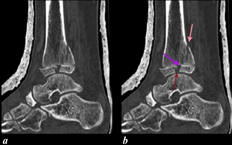
The sagittal reconstruction of the CT scan of the ankle of a youth shows a fracture through the tibia involving the posterior metaphysis (salmon arrow), epiphyseal plate (purple arrow), and epiphysis (maroon arrow). Since the growth plate is not calcified and is also a thin structure, fractures are characteristically difficult to diagnose. The fracture of the epiphyseal plate in this instance can be inferred by interpolating the vector line of the metaphyseal fracture (salmon arrow) to the epiphyseal fracture (maroon arrow). In addition the subluxation of the anterior portion of the epiphysis confirms significant injury to the growth plate. Involvement of the growth plate raises concern about necrosis and consequent failure to grow. This complication occurs in about 15% of injuries.
Courtesy of Philips Medical Systems 2011 91309c02L.8
Once the growth plate closes the only remaining cartilage in the bone is at the articular surface. The epiphyseal plate becomes a non active sclerotic line and the concerns for injury of the plate specifically are no longer viable.
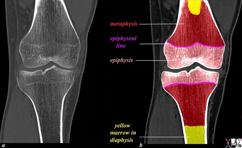
The image reflects a coronally reconstructed CT scan of the knee of a 23 year old male and shows the basic anatomy of a long bone noting the distal end of the femur and the proximal portion of the tibia. This image reflects the growth line (purple in b) which separates the metaphysis from the diaphysis. In this 23 year old patient no further growth will occur and so the active growth plate of childhood has become a line. In addition in the adult the marrow of the medullary canal is mostly made of yellow marrow whereas the red marrow of the adult is mostly in the epiphysis and metaphysis.
Courtesy Ashley Davidoff Copyright 2011 104676c09L02L.8s
Short Bones
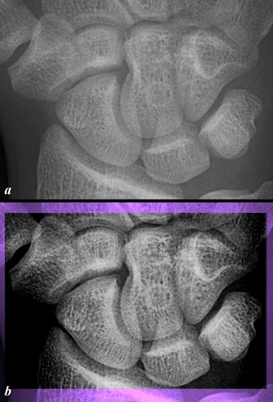
Short bones are roughly cube shaped, consisting primarily of spongy bone covered by a thin layer of compact bone. The carpals and tarsal bones are examples of short bones.
An Example of Short Bones
The plain X-ray of the right wrist is from an 16 year old male and is normal. The carpal bones are an example of short bones characterized by their cube or cuboid shape with a thin cortical layer and trabeculated bone in the interior. Image b has been enhanced to increase the contrast so as to exaggerate the trabeculated pattern.
Courtesy Ashley Davidoff Copyright 2011 104171cL.81
Flat Bones
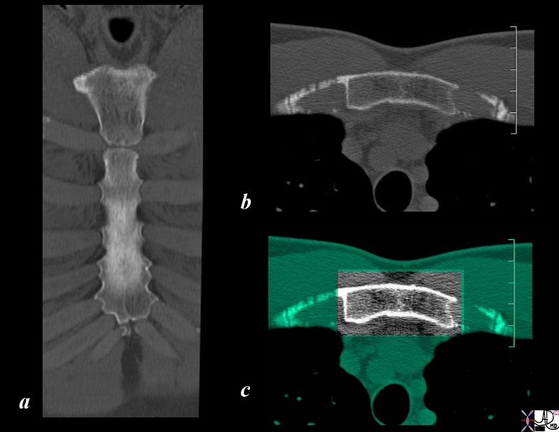
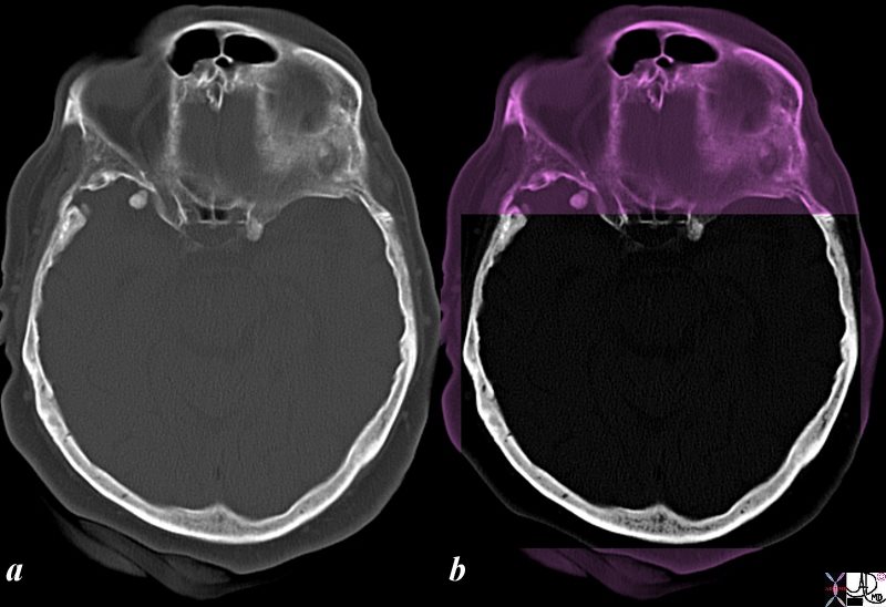
Flat bones such as the skull, ribs and sternum are thin, flattened and curved with the cortical have the two cortical margins running closely parallel to each other.
An Example of a Flat Bone
The CT scan of the sternum reconstructed in coronal projection (a) and in transverse projection (b). They exemplify a flat bone which is thin with parallel layers of cortex (compact bone) surrounding trabeculated bone. Image c amplifies the cortical margin surrounding the trabeculated bone.
Courtesy Ashley Davidoff Copyright 2011 106103c.8sL
Example of a Flat Bone
The CT scan of the skull in the axial plane (a,b) is normal. They exemplify a flat bone which is thin with parallel layers of cortex (compact bone) surrounding trabeculated bone. Image b amplifies the cortical margin surrounding the trabeculated bone.
Courtesy Ashley Davidoff Copyright 2011 105533cL
Irregular Bones
Irregular bones that have an unusual shape such as the vertebra and some of the bones in the cranium such as the sphenoid bones and bones of the middle ear. They have a thin cortical margin and are mostly composed of spongy bone.

Example of Irregular Bones
The CT scan of the lumbosacral spine and pelvis (a,b) is a volume rendered and 3D reconstructed CT scan from a 30 year old male and is normal. The lumbar vertebra and the bones that make up the pelvis have such an unique and unusual shape that they are classified as irregular bones. The thin cortical layer and trabeculated interior is exemplified in the transverse image (c) and enhanced by increasing contrast (d) where the trabeculations are best appreciated in the vertebral body.
Courtesy Ashley Davidoff Copyright 2011 81843.8c01.81L.9
Sesamoid Bones
Sesamoid bones are characterized by their location within a tendon. A classical example is the patella which is found between the quadriceps femoris tendon, the tendons of the vastus muscles and the ligament of the patella which runs inferiorly.
Character of Bone and Aging
In the pediatric population the bone is less brittle and more elastic. Hence bending fractures or greenstick fractures are more common and exclusive to the pediatric population. Additionally the periosteum is not as adherent to the bone in children and hence stripping of the periosteum by the hematoma is more common.
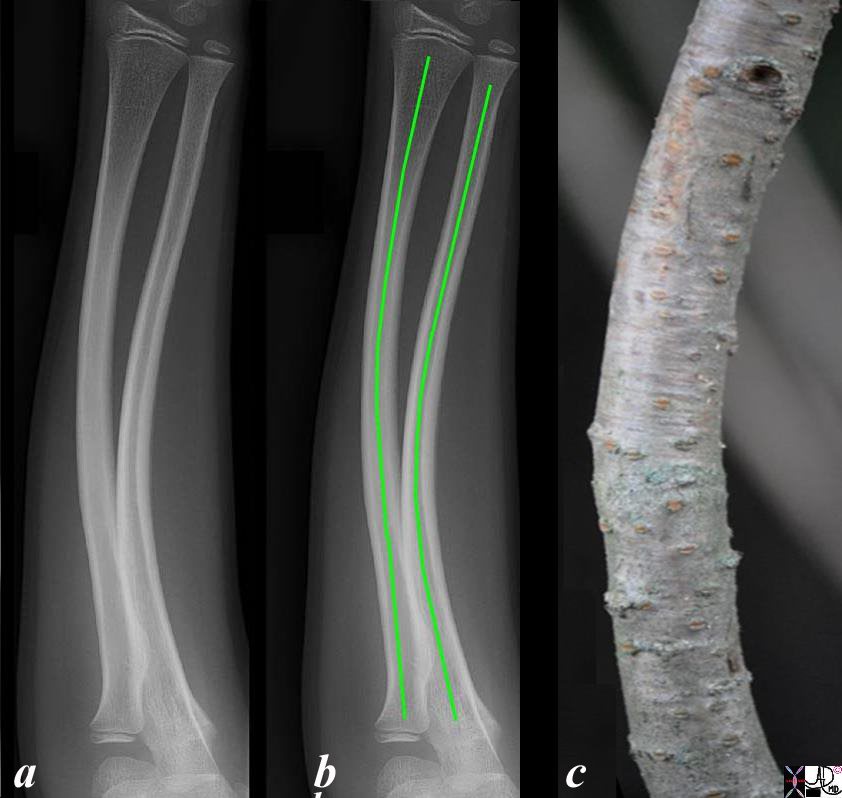
This X-ray shows a bowing deformity fracture of the shaft of both the radius and ulna in a young child. Image b shows the deformity overlaid in green. Bowing fractures usually occur in the forearm and is a bending deformity without a grossly visible fracture or macroscopic break of the cortex. Microfractures are visible on microscopic evaluation. Periosteal reaction may not occur on subsequent imaging
Ashley Davidoff Copyright 2011 99790c.8L
At the other end of the spectrum the bones become more brittle with age as osteopenia and osteoporosis evolve.
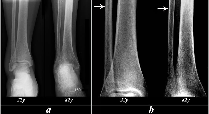
The images reflect the A-P examinations of the distal tibia and fibula of a normal 21 year old female (mild soft swelling lateral malleolus) and the tibia and fibula of an 82 year old osteopenic female (a). The images have been equally modified by increasing the contrast and magnification to enhance the difference in the thickness of compact bone (white arrows), and the overall loss of density as seen on the metaphyseal and epiphyseal regions. The trabeculae of the 82 year old are easily distinguished because of the overall loss of density of the bone. Osteoporosis is an entity where the bone becomes weakened by loss of component elements. The trabeculae become prominent and the overall density of the bone is decreased. The osteoporotic bone is at risk for pathological fracture. Normal forces of daily living are sufficient to cause these pathological compression fractures
Courtesy Ashley Davidoff Copyright 2011 103356c05L.8
Thus this section is a preamble to the section on fractures. It is important at this stage to understand that all bone is not created equal. Most important is the distinction between compact bone and spongy bone. Age and time has significant implications as the bone starts out as a purely cartilagenous tissue and slowly ossifies as it matures by about age 16-18years. Each bone and individual has a time line that should fit within a bell curve for maturation. In the context of fractures, cartilage cannot normally be visualized by plain film examination and thus some fractures can be invisible. Health care givers should pursue the diagnosis of a fracture in patients where there is a high index of clinical suspicion despite a negative radiology report. At the other extreme the same vigilance has to be paid to the elderly in whom fractures may be invisible because of the loss of density of bone.
References
Augat P, Schorlemmer S The Role of Cortical Bone in Bone Strength Age and Ageing 35 S-2 ii 27-ii312006
Bakitas M and Wujcik D Blood and Marrow Stem Cell Transplanation : Principles, Practice and Nursing Insights Jones and Bartlett Learning Second Edition 1997 Sudbury Massachusetts and London
Bonakdarpour Akbar, Reinus WWilliam R, Khurana Javsvir S Diagnostic Imaging of Musculoskeletal Diseases First Edition 2010 Springer New York Dordrecht Heidelberg London
Erslev, A.J., Weiss, L. Structure and Function of Marrow in Williams W Beutler E, Erslev AJ Eds Hematology 1983 New York Mc Graw-Hill
Fleisch H Bisphosphonates in Bone Disease: from the laboratory to the patient 2000 4th Edition Academic Press London
Hall M Trabecular Patterns of the head of the Femur Canad. M A. J. Nov 1961 Vol 85 pages 1141-1143.
Wojnar R Bone and Cartilage – Its Structure and Physical Properties From
Biomechanics of Hard Tissues: Modeling, Testing, and Materials. Edited by Andreas Ochsner and Waqar Ahmed ¨ Copyright 2010 WILEY-VCH Verlag GmbH & Co. KGaA, Weinheim
Links
Bone Development and Growth SEER Training Modules
University of Michigan School of Engineering
