The Common Vein Copyright 2011
Definition
Fractures of the femur are usually caused either by low-energy injuries that lead to individual bone fractures or high-energy fractures that may disrupt the pelvic ring. Crush injuries may cause a variety of fracture types. and are often characterized by
The fractures may be stable or an unstable . Stable injuries do not deform with normal physiologic forces. Unstable injuries are associated with a rotational or vertical displacement.
Femoral head fractures occur in 6 to 16 percent of patients who have a dislocation of the hip posteriorly. There are four types of femoral head fractures, according to the Pipkin classification system: intrafoveal, suprafoveal, associated femoral neck and associated acetabulum. The intrafoveal fracture refers to when there is disruption of the ligamentum teres from the head fragment. The ligamentum teres is a ligament that connects the head of the femur to the acetabulum. Suprafoveal refers to when the ligamentum teres remains attached to the head fragment. Either the suprafoveal or the infrafoveal can be associated with a fracture of the acetabulum or the femoral neck.
There are three main types of femoral neck fractures according to the AO/OTA classification system: subcapital, transcervical and basicervical. These fracture types refer to where the fracture line is. The subcapital is the most proximal, while the basicervical is the most distal (and closest to the intertrochanteric line of the femur).
The intertrochanteric fracture occurs when there is a fracture along the line of the femur that connects the greater and lesser trochanter. There have been no universally accepted classification systems to date; however most categorize fractures based on the level of destruction to the bony anatomy (comminution) and stability.
Isolated fracture of the greater trochanter is rare but can occur in older patients due to direct trauma or due to activity of the gluteus medius and minimus muscles.
Isolated fracture of the lesser trochanter is more common in adolescent patients. It is typically due to forceful contraction of the iliopsoas muscle leading to an avulsion type of injury. In the elderly, lesser trochanter fractures are usually a sign of a benign or malignant lesion in the proximal femur.
The subtrochanteric fracture refers to when the fracture occurs distal to the intertrochanteric line in the proximal femoral shaft. The fractures can create two or more fragments.
Femoral shaft fractures are described in the Winquist and Hansen classification by the level of fracture comminution. A type 1 has minimal or no comminution. A type 2 has at least 50% of both cortices of the bone fragments intact. A type 3 has 50 to 100% comminution of the cortex. A type 4 has circumferential comminution with no cortical contact. Other ways to describe the shaft fractures are as open vs closed, by location (proximal, middle, or distal 1/3), by angulation, by displacement, or by pattern (spiral, oblique, or transverse).
Distal femur fractures are approximately 7% of all femur fractures. They tend to occur in either young males from trauma or in elderly osteopenic females. The distal femur includes the supracondylar and condylar areas. The OTA classification system is used to describe these fractures. Type A fractures are extra-articular. Type B fractures are partially articulate and involve one condyle. Type C fractures are intercondylar or bicondylar and intra-articular. There can be varying amounts of comminution.
The fracture may be complicated in the acute phase by bleeding, neurovascular injury, fat emboli, or in the subacute or chronic phases by nonunion, malunion, infection, osteonecrosis, or osteoarthritis.
The diagnosis of this injury is usually made by a combination of physical examination and x-ray imaging.
Imaging includes the use of plain x-rays, and if indicated CT-scan, or MRI.
Femoral head fractures are treated with surgical or non-surgical treatments depending on the level of displacement of the fragments and the overall stability of the joint. When closed reduction is successful, patients often do not require surgery if there is less than 1mm of displacement.
Femoral neck fractures are treated differently depending upon the fracture location. The subcapital and transcervical get treated the same way with a compression screw fixation or an arthroplasty (replacement component of the joint). Basicervical fractures are treated like intertrochanteric fractures by fixation with a sliding hip compression screw to pull the pieces together.
The treatment of intertrochanteric fractures are surgical unless the patient is nonambulatory at baseline or is at risk for surgery. Surgery is either with a sliding hip screw/side plate or an intramedullary hip screw.
Lesser trochanter fractures are usually treated surgically. When associated with a bone tumor, the tumor also needs to be treated with medical and surgical treatments.
Greater trochanter fractures are usually treated non-operatively with bedrest that progresses to using crutches once symptoms improve. Surgery may be indication for patients who have greater than 1cm displacement of fracture fragments.
Subtrochanteric fractures are treated to attain an anatomic restoration of the femur. Treatment methods depend on the level of displacement of the fragment pieces and overall instability of the joint. Common treatments include intramedullary nailing or plate fixation.
Femoral shaft fractures are treated usually with surgery. Skeletal traction is the placement of a pin in either the distal femur or the proximal tibia to apply a traction force using weights to help keep the femur at length. Skeletal traction is only definitive management for patients with significant medical comorbidities. Surgery is the preferred treatment for most patients within 24 hours of the injury. Surgery can consist of antegrade intramedullary nailing, retrograde intramedullary nailing, or plate fixation. Open fractures require antibiotics.
Distal femur fractures are treated either non-operatively or operatively. Non-operative treatment is reserved for nondisplaced or incomplete fracture or for patients who cannot have surgery for medical reasons. Surgery consists of placement of screws and a plate, intramedullary nails, or with application of an external fixation device.
Complications of femur fracture surgery include infection, thromboembolism, neurovascular injury, pain, malunion, and nonunion.

This A-P examination of the proximal left femur shows a simple oblique fracture of the proximal shaft with mild varus deformity. This relatively simple fracture results in pain and loss of mobility.
Courtesy Ashley Davidoff Copyright 2011 102037.8
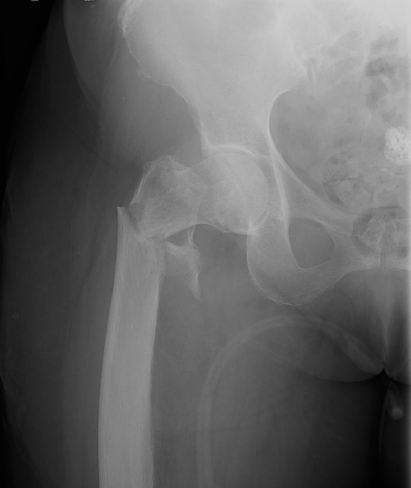
This A-P examination of the proximal left femur of a 91 year old female shows a comminuted transverse fracture of the intertrochanteric region of the right femur with mild varus deformity. A fragment of the lesser trochanter is seen medially
Courtesy Ashley Davidoff Copyright 2011 101640b01.8

The plain film of the pelvis in this elderly patient who presents with hip pain following fall. The examination shows an acute intercondylar fracture characterized by distinct borders of acute an fracture. There is also a subacute fracture of the left pubic ramus characterized by fuzzy borders of the fractures.
Courtesy Ashley Davidoff MD 75714c01
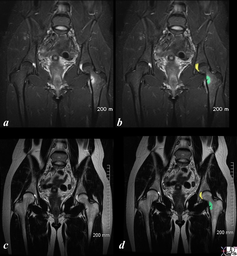
These MRI images demonstrate a stress fracture in the neck of the left femur. Images a and b are STIR images in the coronal plane while images c and d show a T2 sequence in the same plane. The fracture is characterized by a small medial positioned fracture (black) surrounded by a rim of edema (green in b and d). A small effusion is noted in the left hip (yellow)
Courtesy Ashley Davidoff Copyright 2011 82987cL01s.8
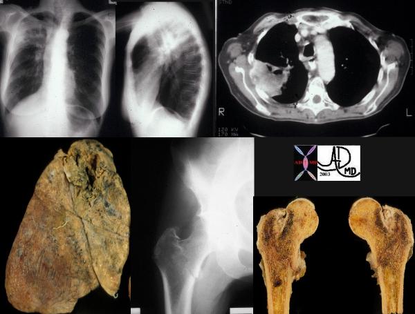
This combination represents a patient with stage IV cavitating primary squamous carcinoma of the RUL (a,b,c,d – white arrows) in this patient with COPD. A metastatic lesion to the right femur is complicated by a pathological fracture. (e,f black arrows).
Courtesy Ashley Davidoff MD 2011 32202c02
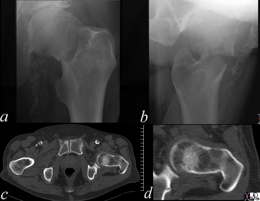
The plain x-ray in A-P and lateral projection (a,b) and CT scan in axial (c) and magnified axial (d) show a fracture through the neck of the left femur and lytic metastases. The patient has a history of metastatic lung carcinoma
Courtesy Ashley Davidoff MD 49471c03
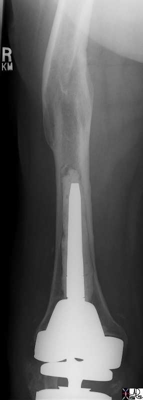
Courtesy Ashley Davidoff MD 49464 49465
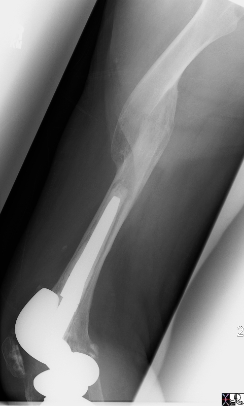
Courtesy Ashley Davidoff MD 49464 49465
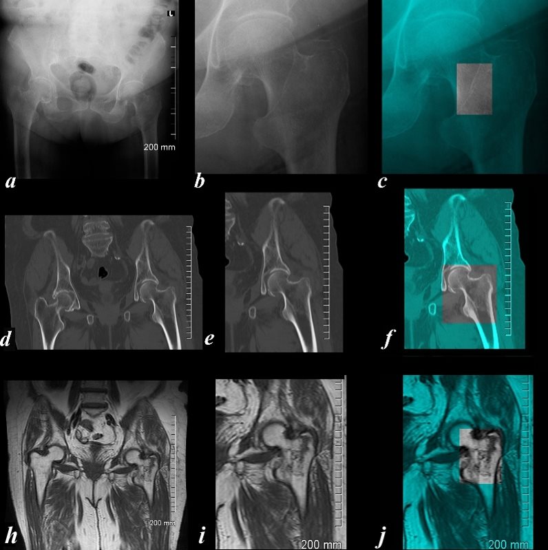
Plain Film, CTscan, and T1 Weighted MRI
The A-P examination of the proximal left femur of an 86 year old female shows the three major technologies in a difficult to diagnose fracture. Images a,b and c show a subtle change(?break) in the intertrochanteric line best seen in image c which is overlaid in teal green, and the area of interest shown in black and white. Image d,e,and f show a sagittally reformatted CT scan where there is a failure to visualize any hint of a fracture and repeated review of the axial images were never convincing for a fracture. Because the pain was severe and incapacitating an MRI was performed and the T1 component of that examination is shown in images (g,h,i) revealing an obvious non displaced intertrochanteric fracture of the left femur best visualized in i by a ragged intertrochanteric black line. CT is usually a quick and highly sensitive study for fractures but in rare instances such as this case, when the patient?s history creates ongoing concern, MRI proved to be highly sensitive to the injury.
Courtesy Ashley Davidoff Copyright 2011 103778c03L.8
References
Davis MF, Davis PF, Ross DS. Expert Guide to Sports Medicine. ACP Series, 2005.
Elstrom J, Virkus W, Pankovich (eds), Handbook of Fractures (3rd edition), McGraw Hill, New York, NY, 2006.
Koval K, Zuckerman J (eds), Handbook of Fractures (3rd edition), Lippincott Williams & Wilkins, Philadelphia, PA, 2006.
Lieberman J (ed), AAOS Comprehensive Orthopaedic Review, American Academy of Orthopaedic Surgeons, 2008.
Moore K, Dalley A (eds), Clinically Oriented Anatomy (5th edition), Lippincott Williams & Wilkins, Philadelphia, PA, 2006.
Wheeless?s Textbook of Orthopaedics: Femoral Shaft Fracture Menu (http://www.wheelessonline.com/ortho/femoral_shaft_fracture)
