
bonds-0012-catalogue-signed.jpg
atom-0002
An electrically neutral group of two or more atoms held together by chemical bonds. Molecules do not have an electrical charge.
bonds- 0012

size-0006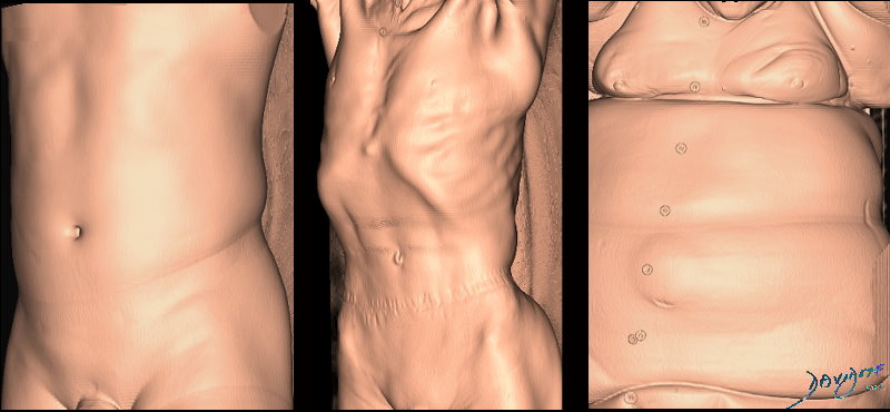
Image on the left shows a normal chest and abdomen. The middle image shows an emaciated body. The right image shows an overweight body
size-0001
Adrenal Gland

In this image we have been able to identify each of the four limbs that make up the two adrenal glands. Each has a medial and a lateral limb joined together at the apex. The left adrenal vein is noted as the blue overlay.
Courtesy of: Ashley Davidoff, M.D.

This diagram reflects the large left lobe of the liver in cirrhosis and the small right lobe. The caudate lob is not depicted.
Davidoff art
liver-00012c
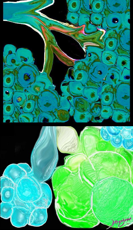
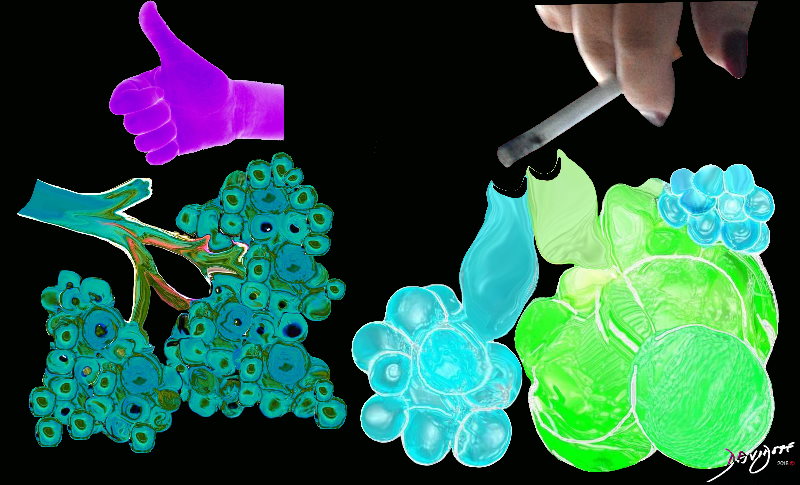
This diagram above illustrates the branching pattern of the tracheobronchial tree that extends from the bronchi to the terminal bronchioles transitioning into the alveoli via the alveolar sacs.
The diagram below shows alveoli and respiratory bronchioles that are too large due to loss of elasticity, so that air cannot be moved efficiently through them This is a diagram of emphysema causing hyperinflated lungs lung volumes
Davidoff art
size-0002
The CT scan on the left shows normal lung parenchyma while the CT scan on the right shows many black holes in the lung (“swiss cheese appearance”) characteristic of emphysema
Courtesy Ashley Davidoff MD
size-0004

The CT scan on the left shows normal lung parenchyma while the CT scan on the right shows many black holes in the lung (“swiss cheese appearance”) characteristic of emphysema
Courtesy Ashley Davidoff MD
size-0004
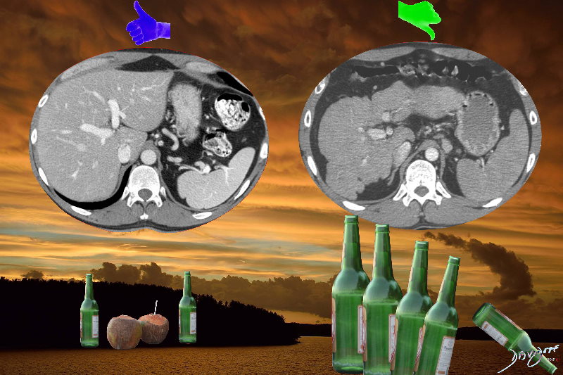
The CT scan on the left shows a normal sized liver in the context of a romantic evening with a couple sharing a beer at sunset by the sea .
The CT scan on the right shows a shrunken cirrhotic liver in the context of a person drinking alone and excesssively iwith raging clouds in the sky.
Courtesy Ashley Davidoff MD
size-0004
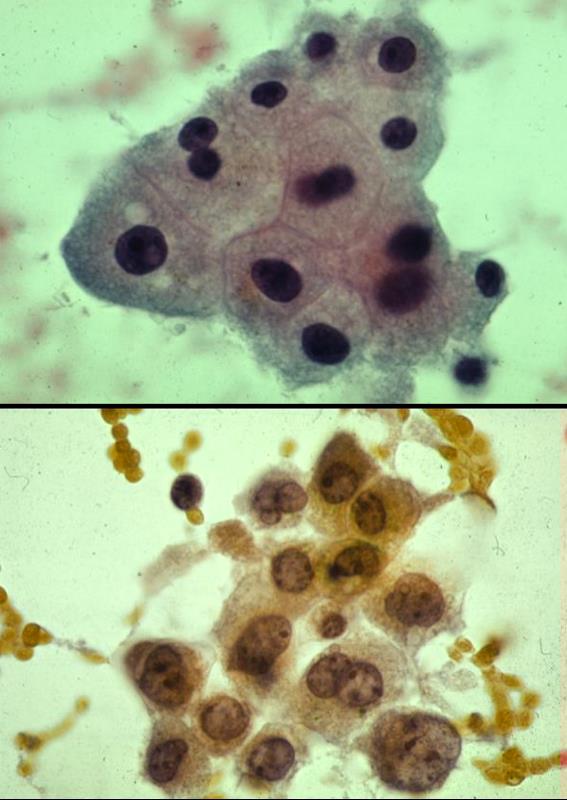
The normal (upper) and the abnormally large nucleus (lower)
Two histological sections above show a normal nuclear cytoplasmic ratio of some liver cells in the upper panel and cells with an increased nuclear cytoplasmic ratio on the lower panel indicating malignant change. The experienced eye and mind of the pathologist develops a geshtalt of what the normal ratio. This is a difficult measurement to make objectively.
Courtesy Barbara Banner MD
liver-0033