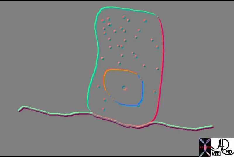
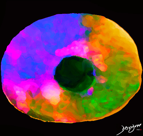
?The-Liver Cell? shows an artistically rendered liver cell.
The liver cell is cuboidal and measures about 20-30 µm.
The liver cell has many functions. It is involved with protein synthesis, protein storage, metabolism of carbohydrates, formation of cholesterol, bile salts and phospholipids, and participates in detoxification and excretion of both exogenous and endogenous products.
The style is reminiscent of surrealism . The complexity of brain function is reduced to its 3 major functions.
by Ashley Davidoff MD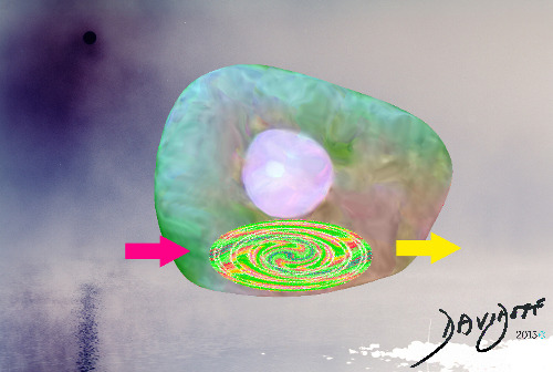
?Anatomy of Cell Function;-receive, process/produce, and export ? shows the basic process involved in human liver cellular function
The cell receives, processes, and exports.
The complexity of these three basic functions will unfold not only in the cell but in all functional systems.
The style is reminiscent of surrealism.
by Ashley Davidoff MD
by Ashley Davidoff MD
by Ashley Davidoff MD
by Ashley Davidoff MD
?Red Cell in the Blood Stream? shows a single red cell floating down the river of plasma. The deformability and biconcave shape of the red cell is essential to its function. The red cell is about 7.5 to 8.5?m in diameter and 1.5 to 2.0 ?m in thickness. They lack a nucleus. There are about 20?30 trillion red cells in the body representing 25% of cells in the body. In contrast there are 7.4 billion people in the world
Hemoglobin is the functional marvel in the red cell. The hemoglobin in the red cells take up oxygen in the lungs and transport it to the cells via the capillaries. The approximately 8?m cells have to fit into the approximately 5?m capillary. The red cells deform and release oxygen into the tissues .
by Ashley Davidoff MD
by Ashley Davidoff MD
by Ashley Davidoff MD
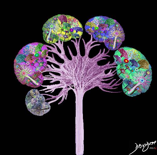
Tree cells in combination give life to the tree in accordance with the principles of the constitution.
by Ashley Davidoff MD
by Ashley Davidoff MD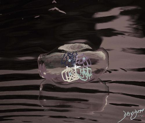
by Ashley Davidoff MD
by Ashley Davidoff MD
by Ashley Davidoff MD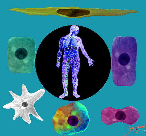
by Ashley Davidoff MD
by Ashley Davidoff MD
by Ashley Davidoff MD
by Ashley Davidoff MD
Ashley Davidoff MD
Adam and Eve ventured into the Garden of Eden Cell Shop with their shopping list of the functional needs for their society.
by Ashley Davidoff MD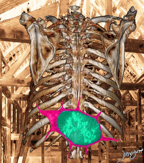
by Ashley Davidoff MD
by Ashley Davidoff MD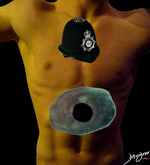
by Ashley Davidoff MD
by Ashley Davidoff MD
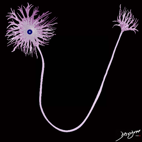
They chose the brain cell or neuron for its leadership, and they playfully called it ?Il Presidente.? Crucially, the neuron could connect not only with other brain cells, but also (directly or indirectly) with every cell in the body. The neuron could also react to the outside environment.
by Ashley Davidoff MD
by Ashley Davidoff MD
by Ashley Davidoff MD
by Ashley Davidoff MD
by Ashley Davidoff MD
by Ashley Davidoff MD
by Ashley Davidoff MD
by Ashley Davidoff MD
by Ashley Davidoff MD
by Ashley Davidoff MD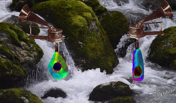
by Ashley Davidoff MD
by Ashley Davidoff MD
by Ashley Davidoff MD
by Ashley Davidoff MD

by Ashley Davidoff MD

by Ashley Davidoff MD

by Ashley Davidoff MD
by Ashley Davidoff MD

by Ashley Davidoff MD
by Ashley Davidoff MD

by Ashley Davidoff MD

by Ashley Davidoff MD

by Ashley Davidoff MD
by Ashley Davidoff MD

Adam tried on the brain cell and sought Eve?s approval. It was tailored for both the internal and external environments.
Modified public domain work of Henri Rousseau ?The Dream? 1910 (MoMA)
by Ashley Davidoff MD
?Il presidente? made his first speech referencing Hillel?s sage advice: ?If I am not for the body of my person who will be? And if I am only for the body of my person, then what am I? If not now when then??
Adam and Eve nodded at each other, satisfied with their choice.
by Ashley Davidoff MD
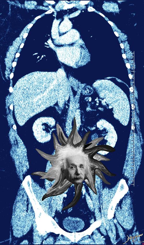
Enemy of the Common Good will be taken down by the Macrophage with Great Conviction for the sake of the peace of humanity
by Ashley Davidoff MD

by Ashley Davidoff MD

by Ashley Davidoff MD

by Ashley Davidoff MD

Adam and Eve realized that the cell was the key ?person? in their society
by Ashley Davidoff MD
by Ashley Davidoff MD

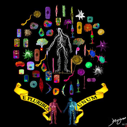
The cells knew they would be around for a long time; their grand scheme was to pursue Oneness. They understood the consequences of bullying, selfishness, greed, and jealousy. They also knew the power of goodness, integrity, humility, and sharing. The cells resolved to fight the evil aspects of humanity. 56 cells from all walks of life discussed the issue of collaborative community living, and drafted a constitution they all signed in Philadelphia. The cells submitted this proposal to Adam and Eve. It read:
?We the Cells of the Body, in Order to form a more perfect Union, establish Justice, insure domestic Tranquility, provide for the common defence, promote the general Welfare, and secure the Blessings of Liberty to ourselves and our Posterity, do ordain and establish this Constitution for the United Cells of the Body.?
by Ashley Davidoff MD
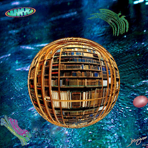
Courtesy of photograph provided by Ralf Roletschek / fahrradmonteur.de subsequently modified
by Ashley Davidoff MD

by Ashley Davidoff MD

by Ashley Davidoff MD

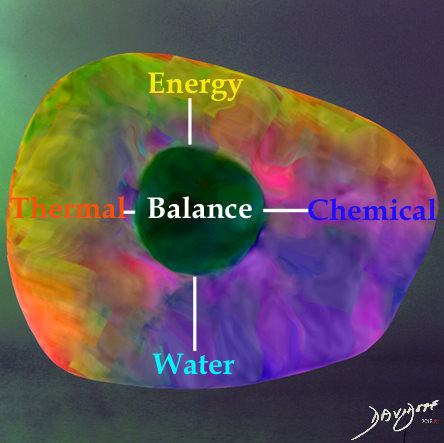
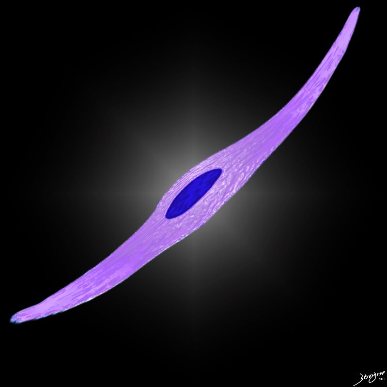
The 7 cells chosen by Adam and Eve to to advise the design of the structure and function of the body.
From left to right; Liver cell (manufacture), Macrophage (defense and protection) Ovum (reproduction) Neuron (government) Sperm (reproduction) Stem cell (maintenance and repair) and Red cell (transport)
by Ashley Davidoff MD
by Ashley Davidoff MD
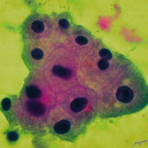
by Ashley Davidoff MD
by Ashley Davidoff MD
by Ashley Davidoff MD
by Ashley Davidoff MD
by Ashley Davidoff MD
by Ashley Davidoff MD

by Ashley Davidoff MD
by Ashley Davidoff MD

by Ashley Davidoff MD

by Ashley Davidoff MD

by Ashley Davidoff MD
Ashley DAvidoff MD
Ashley Davidoff MD
by Ashley Davidoff MD
Ashley Davidoff MD
Ashley Davidoff MD

Ashley Davidoff MD
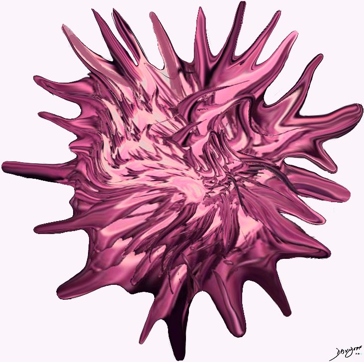
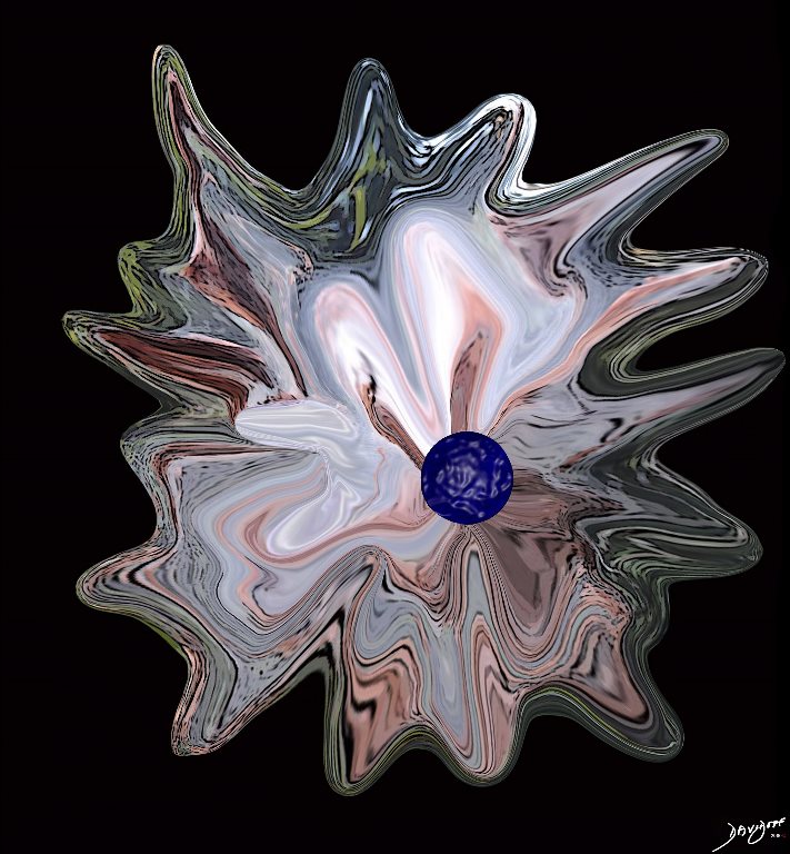
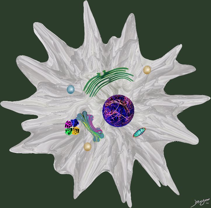
Macrophages

Ashley Davidoff MD

Ashley Davidoff MD
Ashley Davidoff MD

Ashley Davidoff MD
Ashley Davidoff MD

Ashley Davidoff MD
Ashley Davidoff MD
DOMElement Object
(
    [schemaTypeInfo] =>
    [tagName] => table
    [firstElementChild] => (object value omitted)
    [lastElementChild] => (object value omitted)
    [childElementCount] => 1
    [previousElementSibling] => (object value omitted)
    [nextElementSibling] =>
    [nodeName] => table
    [nodeValue] =>


Cells in a factory line


32347b01 colon large bowel columnar cell colonic mucosa epithelium simple columnar epithelium rectangle shape Manhattan apartments Davidoff art Courtesy Ashley Davidoff MD


    [nodeType] => 1
    [parentNode] => (object value omitted)
    [childNodes] => (object value omitted)
    [firstChild] => (object value omitted)
    [lastChild] => (object value omitted)
    [previousSibling] => (object value omitted)
    [nextSibling] => (object value omitted)
    [attributes] => (object value omitted)
    [ownerDocument] => (object value omitted)
    [namespaceURI] =>
    [prefix] =>
    [localName] => table
    [baseURI] =>
    [textContent] =>


Cells in a factory line


32347b01 colon large bowel columnar cell colonic mucosa epithelium simple columnar epithelium rectangle shape Manhattan apartments Davidoff art Courtesy Ashley Davidoff MD


)
DOMElement Object
(
    [schemaTypeInfo] =>
    [tagName] => td
    [firstElementChild] => (object value omitted)
    [lastElementChild] => (object value omitted)
    [childElementCount] => 1
    [previousElementSibling] =>
    [nextElementSibling] =>
    [nodeName] => td
    [nodeValue] => 32347b01 colon large bowel columnar cell colonic mucosa epithelium simple columnar epithelium rectangle shape Manhattan apartments Davidoff art Courtesy Ashley Davidoff MD
    [nodeType] => 1
    [parentNode] => (object value omitted)
    [childNodes] => (object value omitted)
    [firstChild] => (object value omitted)
    [lastChild] => (object value omitted)
    [previousSibling] => (object value omitted)
    [nextSibling] => (object value omitted)
    [attributes] => (object value omitted)
    [ownerDocument] => (object value omitted)
    [namespaceURI] =>
    [prefix] =>
    [localName] => td
    [baseURI] =>
    [textContent] => 32347b01 colon large bowel columnar cell colonic mucosa epithelium simple columnar epithelium rectangle shape Manhattan apartments Davidoff art Courtesy Ashley Davidoff MD
)
DOMElement Object
(
    [schemaTypeInfo] =>
    [tagName] => td
    [firstElementChild] => (object value omitted)
    [lastElementChild] => (object value omitted)
    [childElementCount] => 2
    [previousElementSibling] =>
    [nextElementSibling] =>
    [nodeName] => td
    [nodeValue] =>
Cells in a factory line
    [nodeType] => 1
    [parentNode] => (object value omitted)
    [childNodes] => (object value omitted)
    [firstChild] => (object value omitted)
    [lastChild] => (object value omitted)
    [previousSibling] => (object value omitted)
    [nextSibling] => (object value omitted)
    [attributes] => (object value omitted)
    [ownerDocument] => (object value omitted)
    [namespaceURI] =>
    [prefix] =>
    [localName] => td
    [baseURI] =>
    [textContent] =>
Cells in a factory line
)
DOMElement Object
(
    [schemaTypeInfo] =>
    [tagName] => table
    [firstElementChild] => (object value omitted)
    [lastElementChild] => (object value omitted)
    [childElementCount] => 1
    [previousElementSibling] => (object value omitted)
    [nextElementSibling] => (object value omitted)
    [nodeName] => table
    [nodeValue] =>


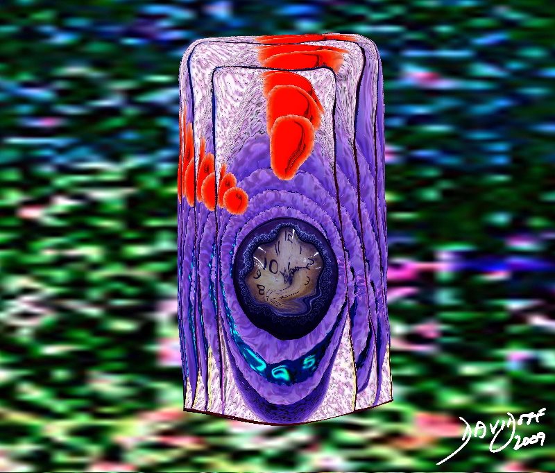
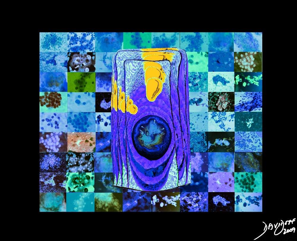
A Cancer Cell – Aberrant Sense of Time


The image represents the life of a single set of columnar cells showing a progressioon of generations as the cell lives dies and is regenerated. The orange secretions of the cell are seen in the background of the pink cytoplasm and the purple nucleus. The nucleus of the newesest generation and cell is seen as a clock that has become distorted and time has become disordered. This process is abnormal and is a forerunner of a malignant process.  The background is a collage of cytopatholagical specimens obtained from the liver representing a variety of metastases from different primary sites but all matked by large nuclear to cytoplasmic ratio.
03183c05.8s cell hyperchromatic increased nuclear cytoplasmic ratio disordered time cytopathology blue malignant cancer Davidoff art Copyright 2009


    [nodeType] => 1
    [parentNode] => (object value omitted)
    [childNodes] => (object value omitted)
    [firstChild] => (object value omitted)
    [lastChild] => (object value omitted)
    [previousSibling] => (object value omitted)
    [nextSibling] => (object value omitted)
    [attributes] => (object value omitted)
    [ownerDocument] => (object value omitted)
    [namespaceURI] =>
    [prefix] =>
    [localName] => table
    [baseURI] =>
    [textContent] =>


A Cancer Cell – Aberrant Sense of Time


The image represents the life of a single set of columnar cells showing a progressioon of generations as the cell lives dies and is regenerated. The orange secretions of the cell are seen in the background of the pink cytoplasm and the purple nucleus. The nucleus of the newesest generation and cell is seen as a clock that has become distorted and time has become disordered. This process is abnormal and is a forerunner of a malignant process.  The background is a collage of cytopatholagical specimens obtained from the liver representing a variety of metastases from different primary sites but all matked by large nuclear to cytoplasmic ratio.
03183c05.8s cell hyperchromatic increased nuclear cytoplasmic ratio disordered time cytopathology blue malignant cancer Davidoff art Copyright 2009


)
DOMElement Object
(
    [schemaTypeInfo] =>
    [tagName] => td
    [firstElementChild] => (object value omitted)
    [lastElementChild] => (object value omitted)
    [childElementCount] => 2
    [previousElementSibling] =>
    [nextElementSibling] =>
    [nodeName] => td
    [nodeValue] => The image represents the life of a single set of columnar cells showing a progressioon of generations as the cell lives dies and is regenerated. The orange secretions of the cell are seen in the background of the pink cytoplasm and the purple nucleus. The nucleus of the newesest generation and cell is seen as a clock that has become distorted and time has become disordered. This process is abnormal and is a forerunner of a malignant process.  The background is a collage of cytopatholagical specimens obtained from the liver representing a variety of metastases from different primary sites but all matked by large nuclear to cytoplasmic ratio.
03183c05.8s cell hyperchromatic increased nuclear cytoplasmic ratio disordered time cytopathology blue malignant cancer Davidoff art Copyright 2009
    [nodeType] => 1
    [parentNode] => (object value omitted)
    [childNodes] => (object value omitted)
    [firstChild] => (object value omitted)
    [lastChild] => (object value omitted)
    [previousSibling] => (object value omitted)
    [nextSibling] => (object value omitted)
    [attributes] => (object value omitted)
    [ownerDocument] => (object value omitted)
    [namespaceURI] =>
    [prefix] =>
    [localName] => td
    [baseURI] =>
    [textContent] => The image represents the life of a single set of columnar cells showing a progressioon of generations as the cell lives dies and is regenerated. The orange secretions of the cell are seen in the background of the pink cytoplasm and the purple nucleus. The nucleus of the newesest generation and cell is seen as a clock that has become distorted and time has become disordered. This process is abnormal and is a forerunner of a malignant process.  The background is a collage of cytopatholagical specimens obtained from the liver representing a variety of metastases from different primary sites but all matked by large nuclear to cytoplasmic ratio.
03183c05.8s cell hyperchromatic increased nuclear cytoplasmic ratio disordered time cytopathology blue malignant cancer Davidoff art Copyright 2009
)
DOMElement Object
(
    [schemaTypeInfo] =>
    [tagName] => td
    [firstElementChild] => (object value omitted)
    [lastElementChild] => (object value omitted)
    [childElementCount] => 2
    [previousElementSibling] =>
    [nextElementSibling] =>
    [nodeName] => td
    [nodeValue] =>
A Cancer Cell – Aberrant Sense of Time

    [nodeType] => 1
    [parentNode] => (object value omitted)
    [childNodes] => (object value omitted)
    [firstChild] => (object value omitted)
    [lastChild] => (object value omitted)
    [previousSibling] => (object value omitted)
    [nextSibling] => (object value omitted)
    [attributes] => (object value omitted)
    [ownerDocument] => (object value omitted)
    [namespaceURI] =>
    [prefix] =>
    [localName] => td
    [baseURI] =>
    [textContent] =>
A Cancer Cell – Aberrant Sense of Time

)
DOMElement Object
(
    [schemaTypeInfo] =>
    [tagName] => table
    [firstElementChild] => (object value omitted)
    [lastElementChild] => (object value omitted)
    [childElementCount] => 1
    [previousElementSibling] => (object value omitted)
    [nextElementSibling] => (object value omitted)
    [nodeName] => table
    [nodeValue] =>


A Cancer Cell – Aberrant Sense of Time


The image represents the life of a single set of columnar cells showing a progressioon of generations as the cell lives dies and is regenerated. The orange secretions of the cell are seen in the background of the pink cytoplasm and the purple nucleus. The nucleus of the newesest generation and cell is seen as a clock that has become distorted and time has become disordered. This process is abnormal and is a forerunner of a malignant process.
histology time malignancy cancer columnar cell histopathology Davidoff art copyright 2009 all rights reserved  85198j03s.81s


    [nodeType] => 1
    [parentNode] => (object value omitted)
    [childNodes] => (object value omitted)
    [firstChild] => (object value omitted)
    [lastChild] => (object value omitted)
    [previousSibling] => (object value omitted)
    [nextSibling] => (object value omitted)
    [attributes] => (object value omitted)
    [ownerDocument] => (object value omitted)
    [namespaceURI] =>
    [prefix] =>
    [localName] => table
    [baseURI] =>
    [textContent] =>


A Cancer Cell – Aberrant Sense of Time


The image represents the life of a single set of columnar cells showing a progressioon of generations as the cell lives dies and is regenerated. The orange secretions of the cell are seen in the background of the pink cytoplasm and the purple nucleus. The nucleus of the newesest generation and cell is seen as a clock that has become distorted and time has become disordered. This process is abnormal and is a forerunner of a malignant process.
histology time malignancy cancer columnar cell histopathology Davidoff art copyright 2009 all rights reserved  85198j03s.81s


)
DOMElement Object
(
    [schemaTypeInfo] =>
    [tagName] => td
    [firstElementChild] => (object value omitted)
    [lastElementChild] => (object value omitted)
    [childElementCount] => 2
    [previousElementSibling] =>
    [nextElementSibling] =>
    [nodeName] => td
    [nodeValue] => The image represents the life of a single set of columnar cells showing a progressioon of generations as the cell lives dies and is regenerated. The orange secretions of the cell are seen in the background of the pink cytoplasm and the purple nucleus. The nucleus of the newesest generation and cell is seen as a clock that has become distorted and time has become disordered. This process is abnormal and is a forerunner of a malignant process.
histology time malignancy cancer columnar cell histopathology Davidoff art copyright 2009 all rights reserved  85198j03s.81s
    [nodeType] => 1
    [parentNode] => (object value omitted)
    [childNodes] => (object value omitted)
    [firstChild] => (object value omitted)
    [lastChild] => (object value omitted)
    [previousSibling] => (object value omitted)
    [nextSibling] => (object value omitted)
    [attributes] => (object value omitted)
    [ownerDocument] => (object value omitted)
    [namespaceURI] =>
    [prefix] =>
    [localName] => td
    [baseURI] =>
    [textContent] => The image represents the life of a single set of columnar cells showing a progressioon of generations as the cell lives dies and is regenerated. The orange secretions of the cell are seen in the background of the pink cytoplasm and the purple nucleus. The nucleus of the newesest generation and cell is seen as a clock that has become distorted and time has become disordered. This process is abnormal and is a forerunner of a malignant process.
histology time malignancy cancer columnar cell histopathology Davidoff art copyright 2009 all rights reserved  85198j03s.81s
)
DOMElement Object
(
    [schemaTypeInfo] =>
    [tagName] => td
    [firstElementChild] => (object value omitted)
    [lastElementChild] => (object value omitted)
    [childElementCount] => 2
    [previousElementSibling] =>
    [nextElementSibling] =>
    [nodeName] => td
    [nodeValue] =>
A Cancer Cell – Aberrant Sense of Time

    [nodeType] => 1
    [parentNode] => (object value omitted)
    [childNodes] => (object value omitted)
    [firstChild] => (object value omitted)
    [lastChild] => (object value omitted)
    [previousSibling] => (object value omitted)
    [nextSibling] => (object value omitted)
    [attributes] => (object value omitted)
    [ownerDocument] => (object value omitted)
    [namespaceURI] =>
    [prefix] =>
    [localName] => td
    [baseURI] =>
    [textContent] =>
A Cancer Cell – Aberrant Sense of Time

)
DOMElement Object
(
    [schemaTypeInfo] =>
    [tagName] => table
    [firstElementChild] => (object value omitted)
    [lastElementChild] => (object value omitted)
    [childElementCount] => 1
    [previousElementSibling] => (object value omitted)
    [nextElementSibling] => (object value omitted)
    [nodeName] => table
    [nodeValue] =>


Problem with the Timing of OneCell


85198gc25.08 cells epithelium columnar epithelium nucleus cytoplasm cell time adenocarcinoma aberrant growth malignant cancer neoplasm death cycle normal life span age growth mucus dysplasia histopathology hyperchromatic nucleus increased nuclear to cytoplasmic ratio Davidoff art Courtesy Ashley Davidoff MD copyright 2009 all rights reserved


    [nodeType] => 1
    [parentNode] => (object value omitted)
    [childNodes] => (object value omitted)
    [firstChild] => (object value omitted)
    [lastChild] => (object value omitted)
    [previousSibling] => (object value omitted)
    [nextSibling] => (object value omitted)
    [attributes] => (object value omitted)
    [ownerDocument] => (object value omitted)
    [namespaceURI] =>
    [prefix] =>
    [localName] => table
    [baseURI] =>
    [textContent] =>


Problem with the Timing of OneCell


85198gc25.08 cells epithelium columnar epithelium nucleus cytoplasm cell time adenocarcinoma aberrant growth malignant cancer neoplasm death cycle normal life span age growth mucus dysplasia histopathology hyperchromatic nucleus increased nuclear to cytoplasmic ratio Davidoff art Courtesy Ashley Davidoff MD copyright 2009 all rights reserved


)
DOMElement Object
(
    [schemaTypeInfo] =>
    [tagName] => td
    [firstElementChild] => (object value omitted)
    [lastElementChild] => (object value omitted)
    [childElementCount] => 1
    [previousElementSibling] =>
    [nextElementSibling] =>
    [nodeName] => td
    [nodeValue] => 85198gc25.08 cells epithelium columnar epithelium nucleus cytoplasm cell time adenocarcinoma aberrant growth malignant cancer neoplasm death cycle normal life span age growth mucus dysplasia histopathology hyperchromatic nucleus increased nuclear to cytoplasmic ratio Davidoff art Courtesy Ashley Davidoff MD copyright 2009 all rights reserved
    [nodeType] => 1
    [parentNode] => (object value omitted)
    [childNodes] => (object value omitted)
    [firstChild] => (object value omitted)
    [lastChild] => (object value omitted)
    [previousSibling] => (object value omitted)
    [nextSibling] => (object value omitted)
    [attributes] => (object value omitted)
    [ownerDocument] => (object value omitted)
    [namespaceURI] =>
    [prefix] =>
    [localName] => td
    [baseURI] =>
    [textContent] => 85198gc25.08 cells epithelium columnar epithelium nucleus cytoplasm cell time adenocarcinoma aberrant growth malignant cancer neoplasm death cycle normal life span age growth mucus dysplasia histopathology hyperchromatic nucleus increased nuclear to cytoplasmic ratio Davidoff art Courtesy Ashley Davidoff MD copyright 2009 all rights reserved
)
DOMElement Object
(
    [schemaTypeInfo] =>
    [tagName] => td
    [firstElementChild] => (object value omitted)
    [lastElementChild] => (object value omitted)
    [childElementCount] => 2
    [previousElementSibling] =>
    [nextElementSibling] =>
    [nodeName] => td
    [nodeValue] =>
Problem with the Timing of OneCell

    [nodeType] => 1
    [parentNode] => (object value omitted)
    [childNodes] => (object value omitted)
    [firstChild] => (object value omitted)
    [lastChild] => (object value omitted)
    [previousSibling] => (object value omitted)
    [nextSibling] => (object value omitted)
    [attributes] => (object value omitted)
    [ownerDocument] => (object value omitted)
    [namespaceURI] =>
    [prefix] =>
    [localName] => td
    [baseURI] =>
    [textContent] =>
Problem with the Timing of OneCell

)
DOMElement Object
(
    [schemaTypeInfo] =>
    [tagName] => table
    [firstElementChild] => (object value omitted)
    [lastElementChild] => (object value omitted)
    [childElementCount] => 1
    [previousElementSibling] => (object value omitted)
    [nextElementSibling] => (object value omitted)
    [nodeName] => table
    [nodeValue] =>


Columnar Cells in Color


70043cD08.800 cells columnar epithelium food processing packing packaging digestion absorbtion excretion Davidoff art Davidoff MD


    [nodeType] => 1
    [parentNode] => (object value omitted)
    [childNodes] => (object value omitted)
    [firstChild] => (object value omitted)
    [lastChild] => (object value omitted)
    [previousSibling] => (object value omitted)
    [nextSibling] => (object value omitted)
    [attributes] => (object value omitted)
    [ownerDocument] => (object value omitted)
    [namespaceURI] =>
    [prefix] =>
    [localName] => table
    [baseURI] =>
    [textContent] =>


Columnar Cells in Color


70043cD08.800 cells columnar epithelium food processing packing packaging digestion absorbtion excretion Davidoff art Davidoff MD


)
DOMElement Object
(
    [schemaTypeInfo] =>
    [tagName] => td
    [firstElementChild] => (object value omitted)
    [lastElementChild] => (object value omitted)
    [childElementCount] => 1
    [previousElementSibling] =>
    [nextElementSibling] =>
    [nodeName] => td
    [nodeValue] => 70043cD08.800 cells columnar epithelium food processing packing packaging digestion absorbtion excretion Davidoff art Davidoff MD
    [nodeType] => 1
    [parentNode] => (object value omitted)
    [childNodes] => (object value omitted)
    [firstChild] => (object value omitted)
    [lastChild] => (object value omitted)
    [previousSibling] => (object value omitted)
    [nextSibling] => (object value omitted)
    [attributes] => (object value omitted)
    [ownerDocument] => (object value omitted)
    [namespaceURI] =>
    [prefix] =>
    [localName] => td
    [baseURI] =>
    [textContent] => 70043cD08.800 cells columnar epithelium food processing packing packaging digestion absorbtion excretion Davidoff art Davidoff MD
)
DOMElement Object
(
    [schemaTypeInfo] =>
    [tagName] => td
    [firstElementChild] => (object value omitted)
    [lastElementChild] => (object value omitted)
    [childElementCount] => 2
    [previousElementSibling] =>
    [nextElementSibling] =>
    [nodeName] => td
    [nodeValue] =>
Columnar Cells in Color

    [nodeType] => 1
    [parentNode] => (object value omitted)
    [childNodes] => (object value omitted)
    [firstChild] => (object value omitted)
    [lastChild] => (object value omitted)
    [previousSibling] => (object value omitted)
    [nextSibling] => (object value omitted)
    [attributes] => (object value omitted)
    [ownerDocument] => (object value omitted)
    [namespaceURI] =>
    [prefix] =>
    [localName] => td
    [baseURI] =>
    [textContent] =>
Columnar Cells in Color

)
DOMElement Object
(
    [schemaTypeInfo] =>
    [tagName] => table
    [firstElementChild] => (object value omitted)
    [lastElementChild] => (object value omitted)
    [childElementCount] => 1
    [previousElementSibling] => (object value omitted)
    [nextElementSibling] => (object value omitted)
    [nodeName] => table
    [nodeValue] =>


Nerve Cells in Action


72046b04b12.8s presynaptic ending synaptic ceft post synaptic ending voltage gated calcium channel chemical gated ion channel neuroceptors synaptic vesicles containing neurotransmitters membrane voltage mitochondria nerve sensory berve synapse dorsal horn first order neuron second order neuron impulse propogation transmission voltage gated calcium channel releases calcium ions stimulates synaptic vesicles to release nurotransmitters into the cleft which attach and stimulate specifc receptors called chemical gated ion channel which results sodium entry into the cell and initiation of action potential normal physiology Davidoff art copyright 2008


    [nodeType] => 1
    [parentNode] => (object value omitted)
    [childNodes] => (object value omitted)
    [firstChild] => (object value omitted)
    [lastChild] => (object value omitted)
    [previousSibling] => (object value omitted)
    [nextSibling] => (object value omitted)
    [attributes] => (object value omitted)
    [ownerDocument] => (object value omitted)
    [namespaceURI] =>
    [prefix] =>
    [localName] => table
    [baseURI] =>
    [textContent] =>


Nerve Cells in Action


72046b04b12.8s presynaptic ending synaptic ceft post synaptic ending voltage gated calcium channel chemical gated ion channel neuroceptors synaptic vesicles containing neurotransmitters membrane voltage mitochondria nerve sensory berve synapse dorsal horn first order neuron second order neuron impulse propogation transmission voltage gated calcium channel releases calcium ions stimulates synaptic vesicles to release nurotransmitters into the cleft which attach and stimulate specifc receptors called chemical gated ion channel which results sodium entry into the cell and initiation of action potential normal physiology Davidoff art copyright 2008


)
DOMElement Object
(
    [schemaTypeInfo] =>
    [tagName] => td
    [firstElementChild] => (object value omitted)
    [lastElementChild] => (object value omitted)
    [childElementCount] => 1
    [previousElementSibling] =>
    [nextElementSibling] =>
    [nodeName] => td
    [nodeValue] => 72046b04b12.8s presynaptic ending synaptic ceft post synaptic ending voltage gated calcium channel chemical gated ion channel neuroceptors synaptic vesicles containing neurotransmitters membrane voltage mitochondria nerve sensory berve synapse dorsal horn first order neuron second order neuron impulse propogation transmission voltage gated calcium channel releases calcium ions stimulates synaptic vesicles to release nurotransmitters into the cleft which attach and stimulate specifc receptors called chemical gated ion channel which results sodium entry into the cell and initiation of action potential normal physiology Davidoff art copyright 2008
    [nodeType] => 1
    [parentNode] => (object value omitted)
    [childNodes] => (object value omitted)
    [firstChild] => (object value omitted)
    [lastChild] => (object value omitted)
    [previousSibling] => (object value omitted)
    [nextSibling] => (object value omitted)
    [attributes] => (object value omitted)
    [ownerDocument] => (object value omitted)
    [namespaceURI] =>
    [prefix] =>
    [localName] => td
    [baseURI] =>
    [textContent] => 72046b04b12.8s presynaptic ending synaptic ceft post synaptic ending voltage gated calcium channel chemical gated ion channel neuroceptors synaptic vesicles containing neurotransmitters membrane voltage mitochondria nerve sensory berve synapse dorsal horn first order neuron second order neuron impulse propogation transmission voltage gated calcium channel releases calcium ions stimulates synaptic vesicles to release nurotransmitters into the cleft which attach and stimulate specifc receptors called chemical gated ion channel which results sodium entry into the cell and initiation of action potential normal physiology Davidoff art copyright 2008
)
DOMElement Object
(
    [schemaTypeInfo] =>
    [tagName] => td
    [firstElementChild] => (object value omitted)
    [lastElementChild] => (object value omitted)
    [childElementCount] => 2
    [previousElementSibling] =>
    [nextElementSibling] =>
    [nodeName] => td
    [nodeValue] =>
Nerve Cells in Action

    [nodeType] => 1
    [parentNode] => (object value omitted)
    [childNodes] => (object value omitted)
    [firstChild] => (object value omitted)
    [lastChild] => (object value omitted)
    [previousSibling] => (object value omitted)
    [nextSibling] => (object value omitted)
    [attributes] => (object value omitted)
    [ownerDocument] => (object value omitted)
    [namespaceURI] =>
    [prefix] =>
    [localName] => td
    [baseURI] =>
    [textContent] =>
Nerve Cells in Action

)
DOMElement Object
(
    [schemaTypeInfo] =>
    [tagName] => table
    [firstElementChild] => (object value omitted)
    [lastElementChild] => (object value omitted)
    [childElementCount] => 1
    [previousElementSibling] => (object value omitted)
    [nextElementSibling] => (object value omitted)
    [nodeName] => table
    [nodeValue] =>


Cells in Action


71798b13d01.800 interactions action reaction environment waves particles molecules cells forces order disorder energy structure Davidoff art Davidoff MD


    [nodeType] => 1
    [parentNode] => (object value omitted)
    [childNodes] => (object value omitted)
    [firstChild] => (object value omitted)
    [lastChild] => (object value omitted)
    [previousSibling] => (object value omitted)
    [nextSibling] => (object value omitted)
    [attributes] => (object value omitted)
    [ownerDocument] => (object value omitted)
    [namespaceURI] =>
    [prefix] =>
    [localName] => table
    [baseURI] =>
    [textContent] =>


Cells in Action


71798b13d01.800 interactions action reaction environment waves particles molecules cells forces order disorder energy structure Davidoff art Davidoff MD


)
DOMElement Object
(
    [schemaTypeInfo] =>
    [tagName] => td
    [firstElementChild] => (object value omitted)
    [lastElementChild] => (object value omitted)
    [childElementCount] => 1
    [previousElementSibling] =>
    [nextElementSibling] =>
    [nodeName] => td
    [nodeValue] => 71798b13d01.800 interactions action reaction environment waves particles molecules cells forces order disorder energy structure Davidoff art Davidoff MD
    [nodeType] => 1
    [parentNode] => (object value omitted)
    [childNodes] => (object value omitted)
    [firstChild] => (object value omitted)
    [lastChild] => (object value omitted)
    [previousSibling] => (object value omitted)
    [nextSibling] => (object value omitted)
    [attributes] => (object value omitted)
    [ownerDocument] => (object value omitted)
    [namespaceURI] =>
    [prefix] =>
    [localName] => td
    [baseURI] =>
    [textContent] => 71798b13d01.800 interactions action reaction environment waves particles molecules cells forces order disorder energy structure Davidoff art Davidoff MD
)
DOMElement Object
(
    [schemaTypeInfo] =>
    [tagName] => td
    [firstElementChild] => (object value omitted)
    [lastElementChild] => (object value omitted)
    [childElementCount] => 2
    [previousElementSibling] =>
    [nextElementSibling] =>
    [nodeName] => td
    [nodeValue] =>
Cells in Action

    [nodeType] => 1
    [parentNode] => (object value omitted)
    [childNodes] => (object value omitted)
    [firstChild] => (object value omitted)
    [lastChild] => (object value omitted)
    [previousSibling] => (object value omitted)
    [nextSibling] => (object value omitted)
    [attributes] => (object value omitted)
    [ownerDocument] => (object value omitted)
    [namespaceURI] =>
    [prefix] =>
    [localName] => td
    [baseURI] =>
    [textContent] =>
Cells in Action

)
DOMElement Object
(
    [schemaTypeInfo] =>
    [tagName] => table
    [firstElementChild] => (object value omitted)
    [lastElementChild] => (object value omitted)
    [childElementCount] => 1
    [previousElementSibling] => (object value omitted)
    [nextElementSibling] => (object value omitted)
    [nodeName] => table
    [nodeValue] =>


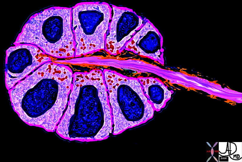
Cells organized in a Rosette


39939b03.800 exocrine gland epithelium ductule histology cytoplasmic granules nucleus Davidoff art


    [nodeType] => 1
    [parentNode] => (object value omitted)
    [childNodes] => (object value omitted)
    [firstChild] => (object value omitted)
    [lastChild] => (object value omitted)
    [previousSibling] => (object value omitted)
    [nextSibling] => (object value omitted)
    [attributes] => (object value omitted)
    [ownerDocument] => (object value omitted)
    [namespaceURI] =>
    [prefix] =>
    [localName] => table
    [baseURI] =>
    [textContent] =>


Cells organized in a Rosette


39939b03.800 exocrine gland epithelium ductule histology cytoplasmic granules nucleus Davidoff art


)
DOMElement Object
(
    [schemaTypeInfo] =>
    [tagName] => td
    [firstElementChild] => (object value omitted)
    [lastElementChild] => (object value omitted)
    [childElementCount] => 1
    [previousElementSibling] =>
    [nextElementSibling] =>
    [nodeName] => td
    [nodeValue] => 39939b03.800 exocrine gland epithelium ductule histology cytoplasmic granules nucleus Davidoff art
    [nodeType] => 1
    [parentNode] => (object value omitted)
    [childNodes] => (object value omitted)
    [firstChild] => (object value omitted)
    [lastChild] => (object value omitted)
    [previousSibling] => (object value omitted)
    [nextSibling] => (object value omitted)
    [attributes] => (object value omitted)
    [ownerDocument] => (object value omitted)
    [namespaceURI] =>
    [prefix] =>
    [localName] => td
    [baseURI] =>
    [textContent] => 39939b03.800 exocrine gland epithelium ductule histology cytoplasmic granules nucleus Davidoff art
)
DOMElement Object
(
    [schemaTypeInfo] =>
    [tagName] => td
    [firstElementChild] => (object value omitted)
    [lastElementChild] => (object value omitted)
    [childElementCount] => 2
    [previousElementSibling] =>
    [nextElementSibling] =>
    [nodeName] => td
    [nodeValue] =>
Cells organized in a Rosette
    [nodeType] => 1
    [parentNode] => (object value omitted)
    [childNodes] => (object value omitted)
    [firstChild] => (object value omitted)
    [lastChild] => (object value omitted)
    [previousSibling] => (object value omitted)
    [nextSibling] => (object value omitted)
    [attributes] => (object value omitted)
    [ownerDocument] => (object value omitted)
    [namespaceURI] =>
    [prefix] =>
    [localName] => td
    [baseURI] =>
    [textContent] =>
Cells organized in a Rosette
)
DOMElement Object
(
    [schemaTypeInfo] =>
    [tagName] => table
    [firstElementChild] => (object value omitted)
    [lastElementChild] => (object value omitted)
    [childElementCount] => 1
    [previousElementSibling] => (object value omitted)
    [nextElementSibling] => (object value omitted)
    [nodeName] => table
    [nodeValue] =>


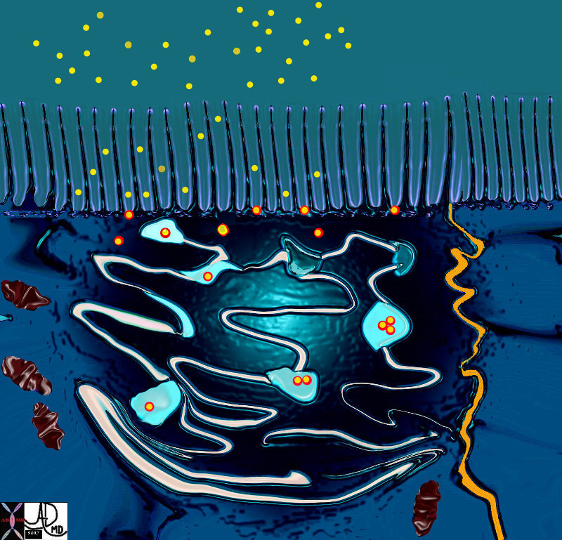
Surface of the Cell


71798b13b03.800 cell endoplasmic reticulum mitochondria fat droplet absorbtion by microvilli processing cisterna interdigitaion of cells pinocytic vesicle intercellular space function absorbtion Davidoff art Davidoff MD


    [nodeType] => 1
    [parentNode] => (object value omitted)
    [childNodes] => (object value omitted)
    [firstChild] => (object value omitted)
    [lastChild] => (object value omitted)
    [previousSibling] => (object value omitted)
    [nextSibling] => (object value omitted)
    [attributes] => (object value omitted)
    [ownerDocument] => (object value omitted)
    [namespaceURI] =>
    [prefix] =>
    [localName] => table
    [baseURI] =>
    [textContent] =>


Surface of the Cell


71798b13b03.800 cell endoplasmic reticulum mitochondria fat droplet absorbtion by microvilli processing cisterna interdigitaion of cells pinocytic vesicle intercellular space function absorbtion Davidoff art Davidoff MD


)
DOMElement Object
(
    [schemaTypeInfo] =>
    [tagName] => td
    [firstElementChild] => (object value omitted)
    [lastElementChild] => (object value omitted)
    [childElementCount] => 1
    [previousElementSibling] =>
    [nextElementSibling] =>
    [nodeName] => td
    [nodeValue] => 71798b13b03.800 cell endoplasmic reticulum mitochondria fat droplet absorbtion by microvilli processing cisterna interdigitaion of cells pinocytic vesicle intercellular space function absorbtion Davidoff art Davidoff MD
    [nodeType] => 1
    [parentNode] => (object value omitted)
    [childNodes] => (object value omitted)
    [firstChild] => (object value omitted)
    [lastChild] => (object value omitted)
    [previousSibling] => (object value omitted)
    [nextSibling] => (object value omitted)
    [attributes] => (object value omitted)
    [ownerDocument] => (object value omitted)
    [namespaceURI] =>
    [prefix] =>
    [localName] => td
    [baseURI] =>
    [textContent] => 71798b13b03.800 cell endoplasmic reticulum mitochondria fat droplet absorbtion by microvilli processing cisterna interdigitaion of cells pinocytic vesicle intercellular space function absorbtion Davidoff art Davidoff MD
)
DOMElement Object
(
    [schemaTypeInfo] =>
    [tagName] => td
    [firstElementChild] => (object value omitted)
    [lastElementChild] => (object value omitted)
    [childElementCount] => 2
    [previousElementSibling] =>
    [nextElementSibling] =>
    [nodeName] => td
    [nodeValue] =>
Surface of the Cell

    [nodeType] => 1
    [parentNode] => (object value omitted)
    [childNodes] => (object value omitted)
    [firstChild] => (object value omitted)
    [lastChild] => (object value omitted)
    [previousSibling] => (object value omitted)
    [nextSibling] => (object value omitted)
    [attributes] => (object value omitted)
    [ownerDocument] => (object value omitted)
    [namespaceURI] =>
    [prefix] =>
    [localName] => td
    [baseURI] =>
    [textContent] =>
Surface of the Cell

)
DOMElement Object
(
    [schemaTypeInfo] =>
    [tagName] => table
    [firstElementChild] => (object value omitted)
    [lastElementChild] => (object value omitted)
    [childElementCount] => 1
    [previousElementSibling] => (object value omitted)
    [nextElementSibling] => (object value omitted)
    [nodeName] => table
    [nodeValue] =>


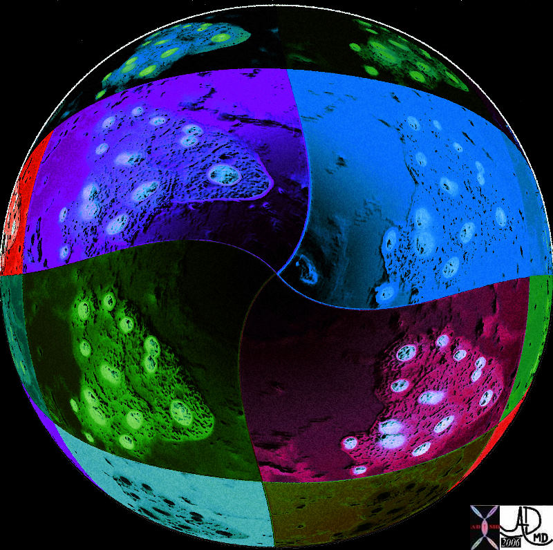
Liver cells on the moon


This artistic rendition of a group of histological group of liver cells that have been transformed into a ball with a crater like appearance of a colorful moon.
Davidoff art 13440c06i07


    [nodeType] => 1
    [parentNode] => (object value omitted)
    [childNodes] => (object value omitted)
    [firstChild] => (object value omitted)
    [lastChild] => (object value omitted)
    [previousSibling] => (object value omitted)
    [nextSibling] => (object value omitted)
    [attributes] => (object value omitted)
    [ownerDocument] => (object value omitted)
    [namespaceURI] =>
    [prefix] =>
    [localName] => table
    [baseURI] =>
    [textContent] =>


Liver cells on the moon


This artistic rendition of a group of histological group of liver cells that have been transformed into a ball with a crater like appearance of a colorful moon.
Davidoff art 13440c06i07


)
DOMElement Object
(
    [schemaTypeInfo] =>
    [tagName] => td
    [firstElementChild] => (object value omitted)
    [lastElementChild] => (object value omitted)
    [childElementCount] => 2
    [previousElementSibling] =>
    [nextElementSibling] =>
    [nodeName] => td
    [nodeValue] => This artistic rendition of a group of histological group of liver cells that have been transformed into a ball with a crater like appearance of a colorful moon.
Davidoff art 13440c06i07
    [nodeType] => 1
    [parentNode] => (object value omitted)
    [childNodes] => (object value omitted)
    [firstChild] => (object value omitted)
    [lastChild] => (object value omitted)
    [previousSibling] => (object value omitted)
    [nextSibling] => (object value omitted)
    [attributes] => (object value omitted)
    [ownerDocument] => (object value omitted)
    [namespaceURI] =>
    [prefix] =>
    [localName] => td
    [baseURI] =>
    [textContent] => This artistic rendition of a group of histological group of liver cells that have been transformed into a ball with a crater like appearance of a colorful moon.
Davidoff art 13440c06i07
)
DOMElement Object
(
    [schemaTypeInfo] =>
    [tagName] => td
    [firstElementChild] => (object value omitted)
    [lastElementChild] => (object value omitted)
    [childElementCount] => 2
    [previousElementSibling] =>
    [nextElementSibling] =>
    [nodeName] => td
    [nodeValue] =>
Liver cells on the moon
    [nodeType] => 1
    [parentNode] => (object value omitted)
    [childNodes] => (object value omitted)
    [firstChild] => (object value omitted)
    [lastChild] => (object value omitted)
    [previousSibling] => (object value omitted)
    [nextSibling] => (object value omitted)
    [attributes] => (object value omitted)
    [ownerDocument] => (object value omitted)
    [namespaceURI] =>
    [prefix] =>
    [localName] => td
    [baseURI] =>
    [textContent] =>
Liver cells on the moon
)
DOMElement Object
(
    [schemaTypeInfo] =>
    [tagName] => table
    [firstElementChild] => (object value omitted)
    [lastElementChild] => (object value omitted)
    [childElementCount] => 1
    [previousElementSibling] => (object value omitted)
    [nextElementSibling] => (object value omitted)
    [nodeName] => table
    [nodeValue] =>


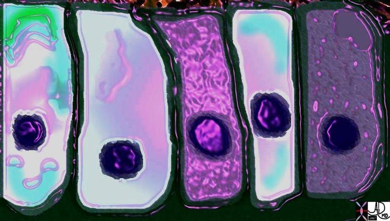
Macrophage


71680b04.800 reticuloendothelial system macrophage cell wbc protection immune scavenger leukocyte white cell Davidoff art Davidoff MD normal cytology


    [nodeType] => 1
    [parentNode] => (object value omitted)
    [childNodes] => (object value omitted)
    [firstChild] => (object value omitted)
    [lastChild] => (object value omitted)
    [previousSibling] => (object value omitted)
    [nextSibling] => (object value omitted)
    [attributes] => (object value omitted)
    [ownerDocument] => (object value omitted)
    [namespaceURI] =>
    [prefix] =>
    [localName] => table
    [baseURI] =>
    [textContent] =>


Macrophage


71680b04.800 reticuloendothelial system macrophage cell wbc protection immune scavenger leukocyte white cell Davidoff art Davidoff MD normal cytology


)
DOMElement Object
(
    [schemaTypeInfo] =>
    [tagName] => td
    [firstElementChild] => (object value omitted)
    [lastElementChild] => (object value omitted)
    [childElementCount] => 1
    [previousElementSibling] =>
    [nextElementSibling] =>
    [nodeName] => td
    [nodeValue] => 71680b04.800 reticuloendothelial system macrophage cell wbc protection immune scavenger leukocyte white cell Davidoff art Davidoff MD normal cytology
    [nodeType] => 1
    [parentNode] => (object value omitted)
    [childNodes] => (object value omitted)
    [firstChild] => (object value omitted)
    [lastChild] => (object value omitted)
    [previousSibling] => (object value omitted)
    [nextSibling] => (object value omitted)
    [attributes] => (object value omitted)
    [ownerDocument] => (object value omitted)
    [namespaceURI] =>
    [prefix] =>
    [localName] => td
    [baseURI] =>
    [textContent] => 71680b04.800 reticuloendothelial system macrophage cell wbc protection immune scavenger leukocyte white cell Davidoff art Davidoff MD normal cytology
)
DOMElement Object
(
    [schemaTypeInfo] =>
    [tagName] => td
    [firstElementChild] => (object value omitted)
    [lastElementChild] => (object value omitted)
    [childElementCount] => 2
    [previousElementSibling] =>
    [nextElementSibling] =>
    [nodeName] => td
    [nodeValue] =>
Macrophage

    [nodeType] => 1
    [parentNode] => (object value omitted)
    [childNodes] => (object value omitted)
    [firstChild] => (object value omitted)
    [lastChild] => (object value omitted)
    [previousSibling] => (object value omitted)
    [nextSibling] => (object value omitted)
    [attributes] => (object value omitted)
    [ownerDocument] => (object value omitted)
    [namespaceURI] =>
    [prefix] =>
    [localName] => td
    [baseURI] =>
    [textContent] =>
Macrophage

)
DOMElement Object
(
    [schemaTypeInfo] =>
    [tagName] => table
    [firstElementChild] => (object value omitted)
    [lastElementChild] => (object value omitted)
    [childElementCount] => 1
    [previousElementSibling] => (object value omitted)
    [nextElementSibling] =>
    [nodeName] => table
    [nodeValue] =>


Art of the Cell
The Common Vein
Copyright 2009


Macrophage


71680b04.800 reticuloendothelial system macrophage cell wbc protection immune scavenger leukocyte white cell Davidoff art Davidoff MD normal cytology


Liver cells on the moon


This artistic rendition of a group of histological group of liver cells that have been transformed into a ball with a crater like appearance of a colorful moon.
Davidoff art 13440c06i07


Surface of the Cell


71798b13b03.800 cell endoplasmic reticulum mitochondria fat droplet absorbtion by microvilli processing cisterna interdigitaion of cells pinocytic vesicle intercellular space function absorbtion Davidoff art Davidoff MD


Cells organized in a Rosette


39939b03.800 exocrine gland epithelium ductule histology cytoplasmic granules nucleus Davidoff art


Cells in Action


71798b13d01.800 interactions action reaction environment waves particles molecules cells forces order disorder energy structure Davidoff art Davidoff MD


Nerve Cells in Action


72046b04b12.8s presynaptic ending synaptic ceft post synaptic ending voltage gated calcium channel chemical gated ion channel neuroceptors synaptic vesicles containing neurotransmitters membrane voltage mitochondria nerve sensory berve synapse dorsal horn first order neuron second order neuron impulse propogation transmission voltage gated calcium channel releases calcium ions stimulates synaptic vesicles to release nurotransmitters into the cleft which attach and stimulate specifc receptors called chemical gated ion channel which results sodium entry into the cell and initiation of action potential normal physiology Davidoff art copyright 2008


Columnar Cells in Color


70043cD08.800 cells columnar epithelium food processing packing packaging digestion absorbtion excretion Davidoff art Davidoff MD


Problem with the Timing of OneCell


85198gc25.08 cells epithelium columnar epithelium nucleus cytoplasm cell time adenocarcinoma aberrant growth malignant cancer neoplasm death cycle normal life span age growth mucus dysplasia histopathology hyperchromatic nucleus increased nuclear to cytoplasmic ratio Davidoff art Courtesy Ashley Davidoff MD copyright 2009 all rights reserved


A Cancer Cell – Aberrant Sense of Time


The image represents the life of a single set of columnar cells showing a progressioon of generations as the cell lives dies and is regenerated. The orange secretions of the cell are seen in the background of the pink cytoplasm and the purple nucleus. The nucleus of the newesest generation and cell is seen as a clock that has become distorted and time has become disordered. This process is abnormal and is a forerunner of a malignant process.
histology time malignancy cancer columnar cell histopathology Davidoff art copyright 2009 all rights reserved  85198j03s.81s


A Cancer Cell – Aberrant Sense of Time


The image represents the life of a single set of columnar cells showing a progressioon of generations as the cell lives dies and is regenerated. The orange secretions of the cell are seen in the background of the pink cytoplasm and the purple nucleus. The nucleus of the newesest generation and cell is seen as a clock that has become distorted and time has become disordered. This process is abnormal and is a forerunner of a malignant process.  The background is a collage of cytopatholagical specimens obtained from the liver representing a variety of metastases from different primary sites but all matked by large nuclear to cytoplasmic ratio.
03183c05.8s cell hyperchromatic increased nuclear cytoplasmic ratio disordered time cytopathology blue malignant cancer Davidoff art Copyright 2009


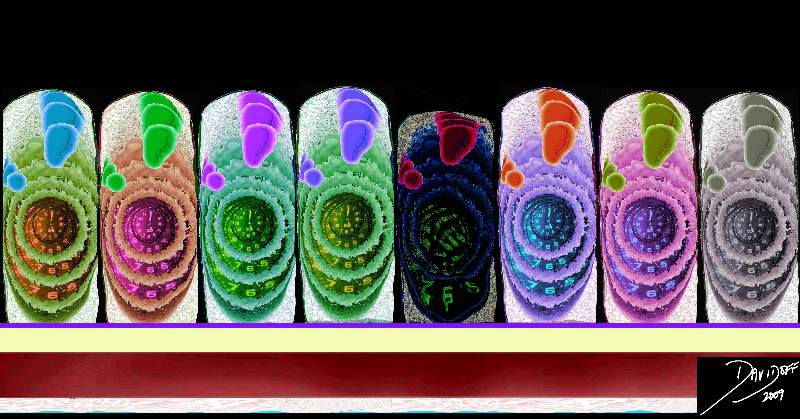
Cells in a factory line


32347b01 colon large bowel columnar cell colonic mucosa epithelium simple columnar epithelium rectangle shape Manhattan apartments Davidoff art Courtesy Ashley Davidoff MD


    [nodeType] => 1
    [parentNode] => (object value omitted)
    [childNodes] => (object value omitted)
    [firstChild] => (object value omitted)
    [lastChild] => (object value omitted)
    [previousSibling] => (object value omitted)
    [nextSibling] => (object value omitted)
    [attributes] => (object value omitted)
    [ownerDocument] => (object value omitted)
    [namespaceURI] =>
    [prefix] =>
    [localName] => table
    [baseURI] =>
    [textContent] =>


Art of the Cell
The Common Vein
Copyright 2009


Macrophage


71680b04.800 reticuloendothelial system macrophage cell wbc protection immune scavenger leukocyte white cell Davidoff art Davidoff MD normal cytology


Liver cells on the moon


This artistic rendition of a group of histological group of liver cells that have been transformed into a ball with a crater like appearance of a colorful moon.
Davidoff art 13440c06i07


Surface of the Cell


71798b13b03.800 cell endoplasmic reticulum mitochondria fat droplet absorbtion by microvilli processing cisterna interdigitaion of cells pinocytic vesicle intercellular space function absorbtion Davidoff art Davidoff MD


Cells organized in a Rosette


39939b03.800 exocrine gland epithelium ductule histology cytoplasmic granules nucleus Davidoff art


Cells in Action


71798b13d01.800 interactions action reaction environment waves particles molecules cells forces order disorder energy structure Davidoff art Davidoff MD


Nerve Cells in Action


72046b04b12.8s presynaptic ending synaptic ceft post synaptic ending voltage gated calcium channel chemical gated ion channel neuroceptors synaptic vesicles containing neurotransmitters membrane voltage mitochondria nerve sensory berve synapse dorsal horn first order neuron second order neuron impulse propogation transmission voltage gated calcium channel releases calcium ions stimulates synaptic vesicles to release nurotransmitters into the cleft which attach and stimulate specifc receptors called chemical gated ion channel which results sodium entry into the cell and initiation of action potential normal physiology Davidoff art copyright 2008


Columnar Cells in Color


70043cD08.800 cells columnar epithelium food processing packing packaging digestion absorbtion excretion Davidoff art Davidoff MD


Problem with the Timing of OneCell


85198gc25.08 cells epithelium columnar epithelium nucleus cytoplasm cell time adenocarcinoma aberrant growth malignant cancer neoplasm death cycle normal life span age growth mucus dysplasia histopathology hyperchromatic nucleus increased nuclear to cytoplasmic ratio Davidoff art Courtesy Ashley Davidoff MD copyright 2009 all rights reserved


A Cancer Cell – Aberrant Sense of Time


The image represents the life of a single set of columnar cells showing a progressioon of generations as the cell lives dies and is regenerated. The orange secretions of the cell are seen in the background of the pink cytoplasm and the purple nucleus. The nucleus of the newesest generation and cell is seen as a clock that has become distorted and time has become disordered. This process is abnormal and is a forerunner of a malignant process.
histology time malignancy cancer columnar cell histopathology Davidoff art copyright 2009 all rights reserved  85198j03s.81s


A Cancer Cell – Aberrant Sense of Time


The image represents the life of a single set of columnar cells showing a progressioon of generations as the cell lives dies and is regenerated. The orange secretions of the cell are seen in the background of the pink cytoplasm and the purple nucleus. The nucleus of the newesest generation and cell is seen as a clock that has become distorted and time has become disordered. This process is abnormal and is a forerunner of a malignant process.  The background is a collage of cytopatholagical specimens obtained from the liver representing a variety of metastases from different primary sites but all matked by large nuclear to cytoplasmic ratio.
03183c05.8s cell hyperchromatic increased nuclear cytoplasmic ratio disordered time cytopathology blue malignant cancer Davidoff art Copyright 2009


Cells in a factory line


32347b01 colon large bowel columnar cell colonic mucosa epithelium simple columnar epithelium rectangle shape Manhattan apartments Davidoff art Courtesy Ashley Davidoff MD


)
DOMElement Object
(
    [schemaTypeInfo] =>
    [tagName] => td
    [firstElementChild] => (object value omitted)
    [lastElementChild] => (object value omitted)
    [childElementCount] => 1
    [previousElementSibling] =>
    [nextElementSibling] =>
    [nodeName] => td
    [nodeValue] => 32347b01 colon large bowel columnar cell colonic mucosa epithelium simple columnar epithelium rectangle shape Manhattan apartments Davidoff art Courtesy Ashley Davidoff MD
    [nodeType] => 1
    [parentNode] => (object value omitted)
    [childNodes] => (object value omitted)
    [firstChild] => (object value omitted)
    [lastChild] => (object value omitted)
    [previousSibling] => (object value omitted)
    [nextSibling] => (object value omitted)
    [attributes] => (object value omitted)
    [ownerDocument] => (object value omitted)
    [namespaceURI] =>
    [prefix] =>
    [localName] => td
    [baseURI] =>
    [textContent] => 32347b01 colon large bowel columnar cell colonic mucosa epithelium simple columnar epithelium rectangle shape Manhattan apartments Davidoff art Courtesy Ashley Davidoff MD
)
DOMElement Object
(
    [schemaTypeInfo] =>
    [tagName] => td
    [firstElementChild] => (object value omitted)
    [lastElementChild] => (object value omitted)
    [childElementCount] => 2
    [previousElementSibling] =>
    [nextElementSibling] =>
    [nodeName] => td
    [nodeValue] =>
Cells in a factory line
    [nodeType] => 1
    [parentNode] => (object value omitted)
    [childNodes] => (object value omitted)
    [firstChild] => (object value omitted)
    [lastChild] => (object value omitted)
    [previousSibling] => (object value omitted)
    [nextSibling] => (object value omitted)
    [attributes] => (object value omitted)
    [ownerDocument] => (object value omitted)
    [namespaceURI] =>
    [prefix] =>
    [localName] => td
    [baseURI] =>
    [textContent] =>
Cells in a factory line
)
DOMElement Object
(
    [schemaTypeInfo] =>
    [tagName] => td
    [firstElementChild] => (object value omitted)
    [lastElementChild] => (object value omitted)
    [childElementCount] => 2
    [previousElementSibling] =>
    [nextElementSibling] =>
    [nodeName] => td
    [nodeValue] => The image represents the life of a single set of columnar cells showing a progressioon of generations as the cell lives dies and is regenerated. The orange secretions of the cell are seen in the background of the pink cytoplasm and the purple nucleus. The nucleus of the newesest generation and cell is seen as a clock that has become distorted and time has become disordered. This process is abnormal and is a forerunner of a malignant process.  The background is a collage of cytopatholagical specimens obtained from the liver representing a variety of metastases from different primary sites but all matked by large nuclear to cytoplasmic ratio.
03183c05.8s cell hyperchromatic increased nuclear cytoplasmic ratio disordered time cytopathology blue malignant cancer Davidoff art Copyright 2009
    [nodeType] => 1
    [parentNode] => (object value omitted)
    [childNodes] => (object value omitted)
    [firstChild] => (object value omitted)
    [lastChild] => (object value omitted)
    [previousSibling] => (object value omitted)
    [nextSibling] => (object value omitted)
    [attributes] => (object value omitted)
    [ownerDocument] => (object value omitted)
    [namespaceURI] =>
    [prefix] =>
    [localName] => td
    [baseURI] =>
    [textContent] => The image represents the life of a single set of columnar cells showing a progressioon of generations as the cell lives dies and is regenerated. The orange secretions of the cell are seen in the background of the pink cytoplasm and the purple nucleus. The nucleus of the newesest generation and cell is seen as a clock that has become distorted and time has become disordered. This process is abnormal and is a forerunner of a malignant process.  The background is a collage of cytopatholagical specimens obtained from the liver representing a variety of metastases from different primary sites but all matked by large nuclear to cytoplasmic ratio.
03183c05.8s cell hyperchromatic increased nuclear cytoplasmic ratio disordered time cytopathology blue malignant cancer Davidoff art Copyright 2009
)
DOMElement Object
(
    [schemaTypeInfo] =>
    [tagName] => td
    [firstElementChild] => (object value omitted)
    [lastElementChild] => (object value omitted)
    [childElementCount] => 2
    [previousElementSibling] =>
    [nextElementSibling] =>
    [nodeName] => td
    [nodeValue] =>
A Cancer Cell – Aberrant Sense of Time

    [nodeType] => 1
    [parentNode] => (object value omitted)
    [childNodes] => (object value omitted)
    [firstChild] => (object value omitted)
    [lastChild] => (object value omitted)
    [previousSibling] => (object value omitted)
    [nextSibling] => (object value omitted)
    [attributes] => (object value omitted)
    [ownerDocument] => (object value omitted)
    [namespaceURI] =>
    [prefix] =>
    [localName] => td
    [baseURI] =>
    [textContent] =>
A Cancer Cell – Aberrant Sense of Time

)
DOMElement Object
(
    [schemaTypeInfo] =>
    [tagName] => td
    [firstElementChild] => (object value omitted)
    [lastElementChild] => (object value omitted)
    [childElementCount] => 2
    [previousElementSibling] =>
    [nextElementSibling] =>
    [nodeName] => td
    [nodeValue] => The image represents the life of a single set of columnar cells showing a progressioon of generations as the cell lives dies and is regenerated. The orange secretions of the cell are seen in the background of the pink cytoplasm and the purple nucleus. The nucleus of the newesest generation and cell is seen as a clock that has become distorted and time has become disordered. This process is abnormal and is a forerunner of a malignant process.
histology time malignancy cancer columnar cell histopathology Davidoff art copyright 2009 all rights reserved  85198j03s.81s
    [nodeType] => 1
    [parentNode] => (object value omitted)
    [childNodes] => (object value omitted)
    [firstChild] => (object value omitted)
    [lastChild] => (object value omitted)
    [previousSibling] => (object value omitted)
    [nextSibling] => (object value omitted)
    [attributes] => (object value omitted)
    [ownerDocument] => (object value omitted)
    [namespaceURI] =>
    [prefix] =>
    [localName] => td
    [baseURI] =>
    [textContent] => The image represents the life of a single set of columnar cells showing a progressioon of generations as the cell lives dies and is regenerated. The orange secretions of the cell are seen in the background of the pink cytoplasm and the purple nucleus. The nucleus of the newesest generation and cell is seen as a clock that has become distorted and time has become disordered. This process is abnormal and is a forerunner of a malignant process.
histology time malignancy cancer columnar cell histopathology Davidoff art copyright 2009 all rights reserved  85198j03s.81s
)
DOMElement Object
(
    [schemaTypeInfo] =>
    [tagName] => td
    [firstElementChild] => (object value omitted)
    [lastElementChild] => (object value omitted)
    [childElementCount] => 2
    [previousElementSibling] =>
    [nextElementSibling] =>
    [nodeName] => td
    [nodeValue] =>
A Cancer Cell – Aberrant Sense of Time

    [nodeType] => 1
    [parentNode] => (object value omitted)
    [childNodes] => (object value omitted)
    [firstChild] => (object value omitted)
    [lastChild] => (object value omitted)
    [previousSibling] => (object value omitted)
    [nextSibling] => (object value omitted)
    [attributes] => (object value omitted)
    [ownerDocument] => (object value omitted)
    [namespaceURI] =>
    [prefix] =>
    [localName] => td
    [baseURI] =>
    [textContent] =>
A Cancer Cell – Aberrant Sense of Time

)
DOMElement Object
(
    [schemaTypeInfo] =>
    [tagName] => td
    [firstElementChild] => (object value omitted)
    [lastElementChild] => (object value omitted)
    [childElementCount] => 1
    [previousElementSibling] =>
    [nextElementSibling] =>
    [nodeName] => td
    [nodeValue] => 85198gc25.08 cells epithelium columnar epithelium nucleus cytoplasm cell time adenocarcinoma aberrant growth malignant cancer neoplasm death cycle normal life span age growth mucus dysplasia histopathology hyperchromatic nucleus increased nuclear to cytoplasmic ratio Davidoff art Courtesy Ashley Davidoff MD copyright 2009 all rights reserved
    [nodeType] => 1
    [parentNode] => (object value omitted)
    [childNodes] => (object value omitted)
    [firstChild] => (object value omitted)
    [lastChild] => (object value omitted)
    [previousSibling] => (object value omitted)
    [nextSibling] => (object value omitted)
    [attributes] => (object value omitted)
    [ownerDocument] => (object value omitted)
    [namespaceURI] =>
    [prefix] =>
    [localName] => td
    [baseURI] =>
    [textContent] => 85198gc25.08 cells epithelium columnar epithelium nucleus cytoplasm cell time adenocarcinoma aberrant growth malignant cancer neoplasm death cycle normal life span age growth mucus dysplasia histopathology hyperchromatic nucleus increased nuclear to cytoplasmic ratio Davidoff art Courtesy Ashley Davidoff MD copyright 2009 all rights reserved
)
DOMElement Object
(
    [schemaTypeInfo] =>
    [tagName] => td
    [firstElementChild] => (object value omitted)
    [lastElementChild] => (object value omitted)
    [childElementCount] => 2
    [previousElementSibling] =>
    [nextElementSibling] =>
    [nodeName] => td
    [nodeValue] =>
Problem with the Timing of OneCell

    [nodeType] => 1
    [parentNode] => (object value omitted)
    [childNodes] => (object value omitted)
    [firstChild] => (object value omitted)
    [lastChild] => (object value omitted)
    [previousSibling] => (object value omitted)
    [nextSibling] => (object value omitted)
    [attributes] => (object value omitted)
    [ownerDocument] => (object value omitted)
    [namespaceURI] =>
    [prefix] =>
    [localName] => td
    [baseURI] =>
    [textContent] =>
Problem with the Timing of OneCell

)
DOMElement Object
(
    [schemaTypeInfo] =>
    [tagName] => td
    [firstElementChild] => (object value omitted)
    [lastElementChild] => (object value omitted)
    [childElementCount] => 1
    [previousElementSibling] =>
    [nextElementSibling] =>
    [nodeName] => td
    [nodeValue] => 70043cD08.800 cells columnar epithelium food processing packing packaging digestion absorbtion excretion Davidoff art Davidoff MD
    [nodeType] => 1
    [parentNode] => (object value omitted)
    [childNodes] => (object value omitted)
    [firstChild] => (object value omitted)
    [lastChild] => (object value omitted)
    [previousSibling] => (object value omitted)
    [nextSibling] => (object value omitted)
    [attributes] => (object value omitted)
    [ownerDocument] => (object value omitted)
    [namespaceURI] =>
    [prefix] =>
    [localName] => td
    [baseURI] =>
    [textContent] => 70043cD08.800 cells columnar epithelium food processing packing packaging digestion absorbtion excretion Davidoff art Davidoff MD
)
DOMElement Object
(
    [schemaTypeInfo] =>
    [tagName] => td
    [firstElementChild] => (object value omitted)
    [lastElementChild] => (object value omitted)
    [childElementCount] => 2
    [previousElementSibling] =>
    [nextElementSibling] =>
    [nodeName] => td
    [nodeValue] =>
Columnar Cells in Color

    [nodeType] => 1
    [parentNode] => (object value omitted)
    [childNodes] => (object value omitted)
    [firstChild] => (object value omitted)
    [lastChild] => (object value omitted)
    [previousSibling] => (object value omitted)
    [nextSibling] => (object value omitted)
    [attributes] => (object value omitted)
    [ownerDocument] => (object value omitted)
    [namespaceURI] =>
    [prefix] =>
    [localName] => td
    [baseURI] =>
    [textContent] =>
Columnar Cells in Color

)
DOMElement Object
(
    [schemaTypeInfo] =>
    [tagName] => td
    [firstElementChild] => (object value omitted)
    [lastElementChild] => (object value omitted)
    [childElementCount] => 1
    [previousElementSibling] =>
    [nextElementSibling] =>
    [nodeName] => td
    [nodeValue] => 72046b04b12.8s presynaptic ending synaptic ceft post synaptic ending voltage gated calcium channel chemical gated ion channel neuroceptors synaptic vesicles containing neurotransmitters membrane voltage mitochondria nerve sensory berve synapse dorsal horn first order neuron second order neuron impulse propogation transmission voltage gated calcium channel releases calcium ions stimulates synaptic vesicles to release nurotransmitters into the cleft which attach and stimulate specifc receptors called chemical gated ion channel which results sodium entry into the cell and initiation of action potential normal physiology Davidoff art copyright 2008
    [nodeType] => 1
    [parentNode] => (object value omitted)
    [childNodes] => (object value omitted)
    [firstChild] => (object value omitted)
    [lastChild] => (object value omitted)
    [previousSibling] => (object value omitted)
    [nextSibling] => (object value omitted)
    [attributes] => (object value omitted)
    [ownerDocument] => (object value omitted)
    [namespaceURI] =>
    [prefix] =>
    [localName] => td
    [baseURI] =>
    [textContent] => 72046b04b12.8s presynaptic ending synaptic ceft post synaptic ending voltage gated calcium channel chemical gated ion channel neuroceptors synaptic vesicles containing neurotransmitters membrane voltage mitochondria nerve sensory berve synapse dorsal horn first order neuron second order neuron impulse propogation transmission voltage gated calcium channel releases calcium ions stimulates synaptic vesicles to release nurotransmitters into the cleft which attach and stimulate specifc receptors called chemical gated ion channel which results sodium entry into the cell and initiation of action potential normal physiology Davidoff art copyright 2008
)
DOMElement Object
(
    [schemaTypeInfo] =>
    [tagName] => td
    [firstElementChild] => (object value omitted)
    [lastElementChild] => (object value omitted)
    [childElementCount] => 2
    [previousElementSibling] =>
    [nextElementSibling] =>
    [nodeName] => td
    [nodeValue] =>
Nerve Cells in Action

    [nodeType] => 1
    [parentNode] => (object value omitted)
    [childNodes] => (object value omitted)
    [firstChild] => (object value omitted)
    [lastChild] => (object value omitted)
    [previousSibling] => (object value omitted)
    [nextSibling] => (object value omitted)
    [attributes] => (object value omitted)
    [ownerDocument] => (object value omitted)
    [namespaceURI] =>
    [prefix] =>
    [localName] => td
    [baseURI] =>
    [textContent] =>
Nerve Cells in Action

)
DOMElement Object
(
    [schemaTypeInfo] =>
    [tagName] => td
    [firstElementChild] => (object value omitted)
    [lastElementChild] => (object value omitted)
    [childElementCount] => 1
    [previousElementSibling] =>
    [nextElementSibling] =>
    [nodeName] => td
    [nodeValue] => 71798b13d01.800 interactions action reaction environment waves particles molecules cells forces order disorder energy structure Davidoff art Davidoff MD
    [nodeType] => 1
    [parentNode] => (object value omitted)
    [childNodes] => (object value omitted)
    [firstChild] => (object value omitted)
    [lastChild] => (object value omitted)
    [previousSibling] => (object value omitted)
    [nextSibling] => (object value omitted)
    [attributes] => (object value omitted)
    [ownerDocument] => (object value omitted)
    [namespaceURI] =>
    [prefix] =>
    [localName] => td
    [baseURI] =>
    [textContent] => 71798b13d01.800 interactions action reaction environment waves particles molecules cells forces order disorder energy structure Davidoff art Davidoff MD
)
DOMElement Object
(
    [schemaTypeInfo] =>
    [tagName] => td
    [firstElementChild] => (object value omitted)
    [lastElementChild] => (object value omitted)
    [childElementCount] => 2
    [previousElementSibling] =>
    [nextElementSibling] =>
    [nodeName] => td
    [nodeValue] =>
Cells in Action

    [nodeType] => 1
    [parentNode] => (object value omitted)
    [childNodes] => (object value omitted)
    [firstChild] => (object value omitted)
    [lastChild] => (object value omitted)
    [previousSibling] => (object value omitted)
    [nextSibling] => (object value omitted)
    [attributes] => (object value omitted)
    [ownerDocument] => (object value omitted)
    [namespaceURI] =>
    [prefix] =>
    [localName] => td
    [baseURI] =>
    [textContent] =>
Cells in Action

)
DOMElement Object
(
    [schemaTypeInfo] =>
    [tagName] => td
    [firstElementChild] => (object value omitted)
    [lastElementChild] => (object value omitted)
    [childElementCount] => 1
    [previousElementSibling] =>
    [nextElementSibling] =>
    [nodeName] => td
    [nodeValue] => 39939b03.800 exocrine gland epithelium ductule histology cytoplasmic granules nucleus Davidoff art
    [nodeType] => 1
    [parentNode] => (object value omitted)
    [childNodes] => (object value omitted)
    [firstChild] => (object value omitted)
    [lastChild] => (object value omitted)
    [previousSibling] => (object value omitted)
    [nextSibling] => (object value omitted)
    [attributes] => (object value omitted)
    [ownerDocument] => (object value omitted)
    [namespaceURI] =>
    [prefix] =>
    [localName] => td
    [baseURI] =>
    [textContent] => 39939b03.800 exocrine gland epithelium ductule histology cytoplasmic granules nucleus Davidoff art
)
DOMElement Object
(
    [schemaTypeInfo] =>
    [tagName] => td
    [firstElementChild] => (object value omitted)
    [lastElementChild] => (object value omitted)
    [childElementCount] => 2
    [previousElementSibling] =>
    [nextElementSibling] =>
    [nodeName] => td
    [nodeValue] =>
Cells organized in a Rosette
    [nodeType] => 1
    [parentNode] => (object value omitted)
    [childNodes] => (object value omitted)
    [firstChild] => (object value omitted)
    [lastChild] => (object value omitted)
    [previousSibling] => (object value omitted)
    [nextSibling] => (object value omitted)
    [attributes] => (object value omitted)
    [ownerDocument] => (object value omitted)
    [namespaceURI] =>
    [prefix] =>
    [localName] => td
    [baseURI] =>
    [textContent] =>
Cells organized in a Rosette
)
DOMElement Object
(
    [schemaTypeInfo] =>
    [tagName] => td
    [firstElementChild] => (object value omitted)
    [lastElementChild] => (object value omitted)
    [childElementCount] => 1
    [previousElementSibling] =>
    [nextElementSibling] =>
    [nodeName] => td
    [nodeValue] => 71798b13b03.800 cell endoplasmic reticulum mitochondria fat droplet absorbtion by microvilli processing cisterna interdigitaion of cells pinocytic vesicle intercellular space function absorbtion Davidoff art Davidoff MD
    [nodeType] => 1
    [parentNode] => (object value omitted)
    [childNodes] => (object value omitted)
    [firstChild] => (object value omitted)
    [lastChild] => (object value omitted)
    [previousSibling] => (object value omitted)
    [nextSibling] => (object value omitted)
    [attributes] => (object value omitted)
    [ownerDocument] => (object value omitted)
    [namespaceURI] =>
    [prefix] =>
    [localName] => td
    [baseURI] =>
    [textContent] => 71798b13b03.800 cell endoplasmic reticulum mitochondria fat droplet absorbtion by microvilli processing cisterna interdigitaion of cells pinocytic vesicle intercellular space function absorbtion Davidoff art Davidoff MD
)
DOMElement Object
(
    [schemaTypeInfo] =>
    [tagName] => td
    [firstElementChild] => (object value omitted)
    [lastElementChild] => (object value omitted)
    [childElementCount] => 3
    [previousElementSibling] =>
    [nextElementSibling] =>
    [nodeName] => td
    [nodeValue] =>
Surface of the Cell

    [nodeType] => 1
    [parentNode] => (object value omitted)
    [childNodes] => (object value omitted)
    [firstChild] => (object value omitted)
    [lastChild] => (object value omitted)
    [previousSibling] => (object value omitted)
    [nextSibling] => (object value omitted)
    [attributes] => (object value omitted)
    [ownerDocument] => (object value omitted)
    [namespaceURI] =>
    [prefix] =>
    [localName] => td
    [baseURI] =>
    [textContent] =>
Surface of the Cell

)
DOMElement Object
(
    [schemaTypeInfo] =>
    [tagName] => td
    [firstElementChild] => (object value omitted)
    [lastElementChild] => (object value omitted)
    [childElementCount] => 2
    [previousElementSibling] =>
    [nextElementSibling] =>
    [nodeName] => td
    [nodeValue] => This artistic rendition of a group of histological group of liver cells that have been transformed into a ball with a crater like appearance of a colorful moon.
Davidoff art 13440c06i07
    [nodeType] => 1
    [parentNode] => (object value omitted)
    [childNodes] => (object value omitted)
    [firstChild] => (object value omitted)
    [lastChild] => (object value omitted)
    [previousSibling] => (object value omitted)
    [nextSibling] => (object value omitted)
    [attributes] => (object value omitted)
    [ownerDocument] => (object value omitted)
    [namespaceURI] =>
    [prefix] =>
    [localName] => td
    [baseURI] =>
    [textContent] => This artistic rendition of a group of histological group of liver cells that have been transformed into a ball with a crater like appearance of a colorful moon.
Davidoff art 13440c06i07
)
DOMElement Object
(
    [schemaTypeInfo] =>
    [tagName] => td
    [firstElementChild] => (object value omitted)
    [lastElementChild] => (object value omitted)
    [childElementCount] => 2
    [previousElementSibling] =>
    [nextElementSibling] =>
    [nodeName] => td
    [nodeValue] =>
Liver cells on the moon
    [nodeType] => 1
    [parentNode] => (object value omitted)
    [childNodes] => (object value omitted)
    [firstChild] => (object value omitted)
    [lastChild] => (object value omitted)
    [previousSibling] => (object value omitted)
    [nextSibling] => (object value omitted)
    [attributes] => (object value omitted)
    [ownerDocument] => (object value omitted)
    [namespaceURI] =>
    [prefix] =>
    [localName] => td
    [baseURI] =>
    [textContent] =>
Liver cells on the moon
)
DOMElement Object
(
    [schemaTypeInfo] =>
    [tagName] => td
    [firstElementChild] => (object value omitted)
    [lastElementChild] => (object value omitted)
    [childElementCount] => 1
    [previousElementSibling] =>
    [nextElementSibling] =>
    [nodeName] => td
    [nodeValue] => 71680b04.800 reticuloendothelial system macrophage cell wbc protection immune scavenger leukocyte white cell Davidoff art Davidoff MD normal cytology
    [nodeType] => 1
    [parentNode] => (object value omitted)
    [childNodes] => (object value omitted)
    [firstChild] => (object value omitted)
    [lastChild] => (object value omitted)
    [previousSibling] => (object value omitted)
    [nextSibling] => (object value omitted)
    [attributes] => (object value omitted)
    [ownerDocument] => (object value omitted)
    [namespaceURI] =>
    [prefix] =>
    [localName] => td
    [baseURI] =>
    [textContent] => 71680b04.800 reticuloendothelial system macrophage cell wbc protection immune scavenger leukocyte white cell Davidoff art Davidoff MD normal cytology
)
DOMElement Object
(
    [schemaTypeInfo] =>
    [tagName] => td
    [firstElementChild] => (object value omitted)
    [lastElementChild] => (object value omitted)
    [childElementCount] => 2
    [previousElementSibling] =>
    [nextElementSibling] =>
    [nodeName] => td
    [nodeValue] =>
Macrophage

    [nodeType] => 1
    [parentNode] => (object value omitted)
    [childNodes] => (object value omitted)
    [firstChild] => (object value omitted)
    [lastChild] => (object value omitted)
    [previousSibling] => (object value omitted)
    [nextSibling] => (object value omitted)
    [attributes] => (object value omitted)
    [ownerDocument] => (object value omitted)
    [namespaceURI] =>
    [prefix] =>
    [localName] => td
    [baseURI] =>
    [textContent] =>
Macrophage

)
DOMElement Object
(
    [schemaTypeInfo] =>
    [tagName] => td
    [firstElementChild] => (object value omitted)
    [lastElementChild] => (object value omitted)
    [childElementCount] => 15
    [previousElementSibling] =>
    [nextElementSibling] =>
    [nodeName] => td
    [nodeValue] =>
Art of the Cell
The Common Vein
Copyright 2009


Macrophage


71680b04.800 reticuloendothelial system macrophage cell wbc protection immune scavenger leukocyte white cell Davidoff art Davidoff MD normal cytology


Liver cells on the moon


This artistic rendition of a group of histological group of liver cells that have been transformed into a ball with a crater like appearance of a colorful moon.
Davidoff art 13440c06i07


Surface of the Cell


71798b13b03.800 cell endoplasmic reticulum mitochondria fat droplet absorbtion by microvilli processing cisterna interdigitaion of cells pinocytic vesicle intercellular space function absorbtion Davidoff art Davidoff MD


Cells organized in a Rosette


39939b03.800 exocrine gland epithelium ductule histology cytoplasmic granules nucleus Davidoff art


Cells in Action


71798b13d01.800 interactions action reaction environment waves particles molecules cells forces order disorder energy structure Davidoff art Davidoff MD


Nerve Cells in Action


72046b04b12.8s presynaptic ending synaptic ceft post synaptic ending voltage gated calcium channel chemical gated ion channel neuroceptors synaptic vesicles containing neurotransmitters membrane voltage mitochondria nerve sensory berve synapse dorsal horn first order neuron second order neuron impulse propogation transmission voltage gated calcium channel releases calcium ions stimulates synaptic vesicles to release nurotransmitters into the cleft which attach and stimulate specifc receptors called chemical gated ion channel which results sodium entry into the cell and initiation of action potential normal physiology Davidoff art copyright 2008


Columnar Cells in Color


70043cD08.800 cells columnar epithelium food processing packing packaging digestion absorbtion excretion Davidoff art Davidoff MD


Problem with the Timing of OneCell


85198gc25.08 cells epithelium columnar epithelium nucleus cytoplasm cell time adenocarcinoma aberrant growth malignant cancer neoplasm death cycle normal life span age growth mucus dysplasia histopathology hyperchromatic nucleus increased nuclear to cytoplasmic ratio Davidoff art Courtesy Ashley Davidoff MD copyright 2009 all rights reserved


A Cancer Cell – Aberrant Sense of Time


The image represents the life of a single set of columnar cells showing a progressioon of generations as the cell lives dies and is regenerated. The orange secretions of the cell are seen in the background of the pink cytoplasm and the purple nucleus. The nucleus of the newesest generation and cell is seen as a clock that has become distorted and time has become disordered. This process is abnormal and is a forerunner of a malignant process.
histology time malignancy cancer columnar cell histopathology Davidoff art copyright 2009 all rights reserved  85198j03s.81s


A Cancer Cell – Aberrant Sense of Time


The image represents the life of a single set of columnar cells showing a progressioon of generations as the cell lives dies and is regenerated. The orange secretions of the cell are seen in the background of the pink cytoplasm and the purple nucleus. The nucleus of the newesest generation and cell is seen as a clock that has become distorted and time has become disordered. This process is abnormal and is a forerunner of a malignant process.  The background is a collage of cytopatholagical specimens obtained from the liver representing a variety of metastases from different primary sites but all matked by large nuclear to cytoplasmic ratio.
03183c05.8s cell hyperchromatic increased nuclear cytoplasmic ratio disordered time cytopathology blue malignant cancer Davidoff art Copyright 2009


Cells in a factory line


32347b01 colon large bowel columnar cell colonic mucosa epithelium simple columnar epithelium rectangle shape Manhattan apartments Davidoff art Courtesy Ashley Davidoff MD


    [nodeType] => 1
    [parentNode] => (object value omitted)
    [childNodes] => (object value omitted)
    [firstChild] => (object value omitted)
    [lastChild] => (object value omitted)
    [previousSibling] => (object value omitted)
    [nextSibling] => (object value omitted)
    [attributes] => (object value omitted)
    [ownerDocument] => (object value omitted)
    [namespaceURI] =>
    [prefix] =>
    [localName] => td
    [baseURI] =>
    [textContent] =>
Art of the Cell
The Common Vein
Copyright 2009


Macrophage


71680b04.800 reticuloendothelial system macrophage cell wbc protection immune scavenger leukocyte white cell Davidoff art Davidoff MD normal cytology


Liver cells on the moon


This artistic rendition of a group of histological group of liver cells that have been transformed into a ball with a crater like appearance of a colorful moon.
Davidoff art 13440c06i07


Surface of the Cell


71798b13b03.800 cell endoplasmic reticulum mitochondria fat droplet absorbtion by microvilli processing cisterna interdigitaion of cells pinocytic vesicle intercellular space function absorbtion Davidoff art Davidoff MD


Cells organized in a Rosette


39939b03.800 exocrine gland epithelium ductule histology cytoplasmic granules nucleus Davidoff art


Cells in Action


71798b13d01.800 interactions action reaction environment waves particles molecules cells forces order disorder energy structure Davidoff art Davidoff MD


Nerve Cells in Action


72046b04b12.8s presynaptic ending synaptic ceft post synaptic ending voltage gated calcium channel chemical gated ion channel neuroceptors synaptic vesicles containing neurotransmitters membrane voltage mitochondria nerve sensory berve synapse dorsal horn first order neuron second order neuron impulse propogation transmission voltage gated calcium channel releases calcium ions stimulates synaptic vesicles to release nurotransmitters into the cleft which attach and stimulate specifc receptors called chemical gated ion channel which results sodium entry into the cell and initiation of action potential normal physiology Davidoff art copyright 2008


Columnar Cells in Color


70043cD08.800 cells columnar epithelium food processing packing packaging digestion absorbtion excretion Davidoff art Davidoff MD


Problem with the Timing of OneCell


85198gc25.08 cells epithelium columnar epithelium nucleus cytoplasm cell time adenocarcinoma aberrant growth malignant cancer neoplasm death cycle normal life span age growth mucus dysplasia histopathology hyperchromatic nucleus increased nuclear to cytoplasmic ratio Davidoff art Courtesy Ashley Davidoff MD copyright 2009 all rights reserved


A Cancer Cell – Aberrant Sense of Time


The image represents the life of a single set of columnar cells showing a progressioon of generations as the cell lives dies and is regenerated. The orange secretions of the cell are seen in the background of the pink cytoplasm and the purple nucleus. The nucleus of the newesest generation and cell is seen as a clock that has become distorted and time has become disordered. This process is abnormal and is a forerunner of a malignant process.
histology time malignancy cancer columnar cell histopathology Davidoff art copyright 2009 all rights reserved  85198j03s.81s


A Cancer Cell – Aberrant Sense of Time


The image represents the life of a single set of columnar cells showing a progressioon of generations as the cell lives dies and is regenerated. The orange secretions of the cell are seen in the background of the pink cytoplasm and the purple nucleus. The nucleus of the newesest generation and cell is seen as a clock that has become distorted and time has become disordered. This process is abnormal and is a forerunner of a malignant process.  The background is a collage of cytopatholagical specimens obtained from the liver representing a variety of metastases from different primary sites but all matked by large nuclear to cytoplasmic ratio.
03183c05.8s cell hyperchromatic increased nuclear cytoplasmic ratio disordered time cytopathology blue malignant cancer Davidoff art Copyright 2009


Cells in a factory line


32347b01 colon large bowel columnar cell colonic mucosa epithelium simple columnar epithelium rectangle shape Manhattan apartments Davidoff art Courtesy Ashley Davidoff MD


)

