Copyright 2009
Definition
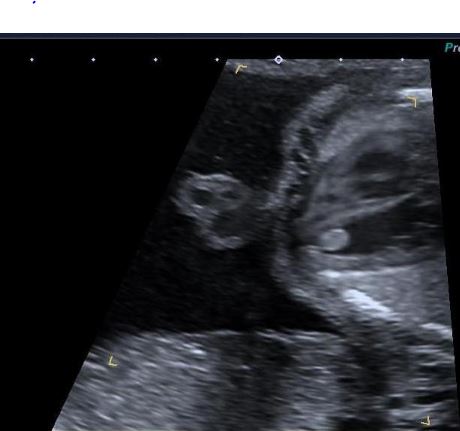
Cardiac rhabdomyoma is a hamartoma of the heart , most commonly associated with tuberous sclerosis (Bourneville’s disease). It is estimated that between 50-60% of patients with tuberous sclerosis develop cardiac rhabdomyomas.
It is usually present in the first years of life and sometimes identified in utero.
size May be multiple
shape round lobular
position LV or septum on walls or intramural
character echogenic/
- T1: isointense to adjacent myocardium
- T2: hyperintense to adjacent myocardium
The result in the heart are manifest with a mass or masses in the heart. They are most commonly situated in the ventricles, generally multiple, well-circumscribed tumours. They can be intramural or pedunculated and may encroach into the cavity. 30% of patients have associated with cardiac arrhythmias (eg Wolff-Parkinson-White syndrome)
Mechanical interference with cardiac and valve function can occur. Most tumors regress spontaneously.
The diagnosis is usually made by cardiac echo, but imaging with MRI and MDCT is also helpful.
Treatment is usually based on the clinical symptoms. If the patients are asymptomatic and the tumors are small they require follow up with echocardiography, and many spontaneously regress within 4 years. If there is mechanical interference with cardiac function, then surgery is indicated.
Rhabdomyomas are the most common primary cardiac tumor in children, Extensive growth and shrinkage are seen, and most of these tumors regress spontaneously (33). Thus, serial echocardiographic follow-up is key and often sufficient. Resection may be required if the cardiac mass causes obstructive symptoms or arrhythmias (33, 34).

