
Benign neoplasia ….. is …
characterized by …..
caused by ..etiology or predisposing factors
resulting in a pathological feature (structural change or functional change) or clinical feauture
Sometimes complicated by ….
Diagnosis is suspected clinically by … and confirmed by ….
Imaging includes the use of
Treatment options depend on …. but includes …..
Etymology if available
Principles
CNS
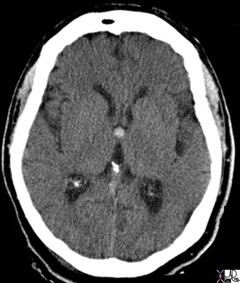
Colloid Cyst of Third Ventricle – Indication for Surgery of Benign Tumor |
| 70045.800 brain third ventricle foramen of Munro third ventricle headaches, vertigo, memory deficits, diplopia, and behavioral disturbances. smooth, round hyperdense endodermal origin, typically located at the foramen of Munro in the anterior aspect of the third ventricle. T benign filled with gelatinous material and cholesterol crystals cause hyperdensity potential complication can cause hydrocephalus by obstructing third ventricle usual Rx is surgery CTscan Davidoff MD |
CVS
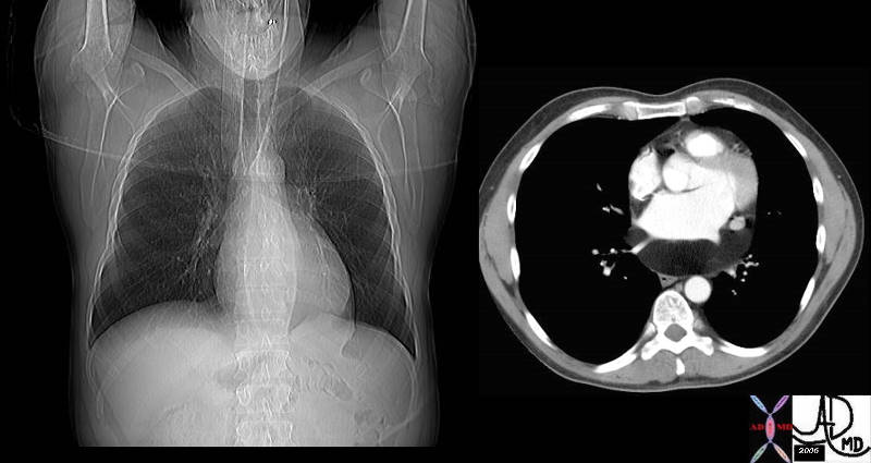
Lipoma of the Pericardium |
| 34800c01 cardiac heart pericardium fx lucent subcarinal angle altered dx lipoma of pericardium fat CTscan plain film scout Davidoff MD |
GUT
 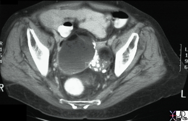
Mass with Fat, Calcium, Fluid Fluid Level – Dermoid of the Ovary |
| 24078 ovary fx mass fx fat fx calcification fx fluid-fluid level dx dermoid CTscan C- 24078 24077 Davidoff MD |
MSK
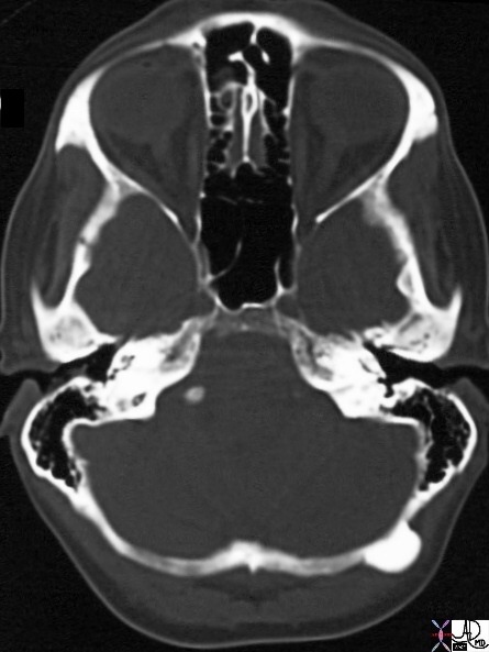
Osteoma |
| 29107 44F one occiput occipital bone fx mass ossified dx osteoma neoplasm benign CTscan Davidoff MD |
Uterus
Benign Adenomatous Endometrial Polyp |
| 72563c01 uterus endometrial cavity endometrium mucosa vascular mass benign neoplasm nodule mucosal lesion acute angles doppler flow through transmission balloon catheter dx benign adenomatous endometrial polyp sonohysterogram USscan Davidoff MD |
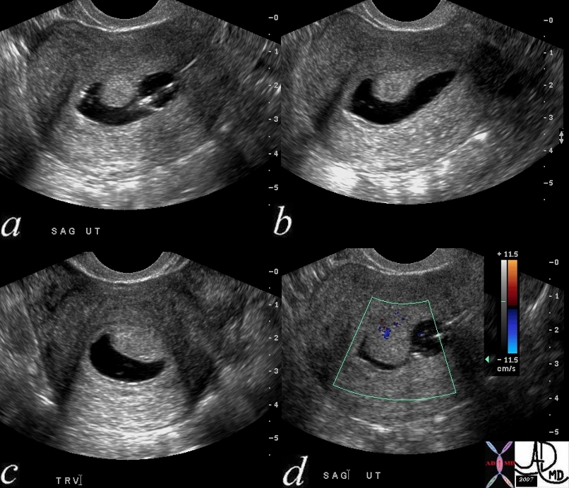
Submucosal Fibroid |
| The USscan (sonohysterogram) scan is from a 41year old female, who presents with dysfunctional uterine bleeding. The study reveals a 4mms submucosal nodule shown at pathology to be a submucosal fibroid.
uterus fibroid leiomyoma submucosal USscan sonohysterogram Courtesy Ashley Davidoff MD copyright 2009 all rights reserved 84299c041.8 |

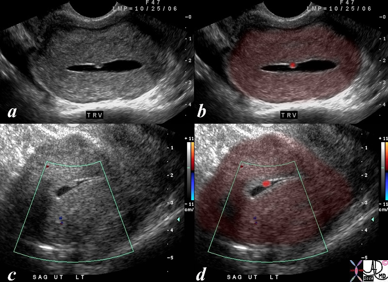
Hyperplastic Polyp |
| 84299c05.8s The USscan (hysterosonogram is from a 47year old female, with history metrorhagia dysfunctional uterine bleeding. The studies reveal a mass in the uterus within the endometrial cavity The polypwas shown to be a benign hyperplastic polyp. uterus endometrium mass polyp hyperplastic USscan hysterosonogram Courtesy Ashley Davidoff MD copyright 2009 all rights reserved |
DOMElement Object
(
    [schemaTypeInfo] =>
    [tagName] => table
    [firstElementChild] => (object value omitted)
    [lastElementChild] => (object value omitted)
    [childElementCount] => 1
    [previousElementSibling] => (object value omitted)
    [nextElementSibling] =>
    [nodeName] => table
    [nodeValue] =>


Hyperplastic Polyp


84299c05.8s The USscan (hysterosonogram is from a 47year old female, with history metrorhagia dysfunctional uterine bleeding. The studies reveal a mass in the uterus within the endometrial cavity The polypwas shown to be a benign hyperplastic polyp. uterus endometrium mass polyp hyperplastic USscan hysterosonogram Courtesy Ashley Davidoff MD copyright 2009 all rights reserved


    [nodeType] => 1
    [parentNode] => (object value omitted)
    [childNodes] => (object value omitted)
    [firstChild] => (object value omitted)
    [lastChild] => (object value omitted)
    [previousSibling] => (object value omitted)
    [nextSibling] => (object value omitted)
    [attributes] => (object value omitted)
    [ownerDocument] => (object value omitted)
    [namespaceURI] =>
    [prefix] =>
    [localName] => table
    [baseURI] =>
    [textContent] =>


Hyperplastic Polyp


84299c05.8s The USscan (hysterosonogram is from a 47year old female, with history metrorhagia dysfunctional uterine bleeding. The studies reveal a mass in the uterus within the endometrial cavity The polypwas shown to be a benign hyperplastic polyp. uterus endometrium mass polyp hyperplastic USscan hysterosonogram Courtesy Ashley Davidoff MD copyright 2009 all rights reserved


)
DOMElement Object
(
    [schemaTypeInfo] =>
    [tagName] => td
    [firstElementChild] => (object value omitted)
    [lastElementChild] => (object value omitted)
    [childElementCount] => 1
    [previousElementSibling] =>
    [nextElementSibling] =>
    [nodeName] => td
    [nodeValue] => 84299c05.8s The USscan (hysterosonogram is from a 47year old female, with history metrorhagia dysfunctional uterine bleeding. The studies reveal a mass in the uterus within the endometrial cavity The polypwas shown to be a benign hyperplastic polyp. uterus endometrium mass polyp hyperplastic USscan hysterosonogram Courtesy Ashley Davidoff MD copyright 2009 all rights reserved
    [nodeType] => 1
    [parentNode] => (object value omitted)
    [childNodes] => (object value omitted)
    [firstChild] => (object value omitted)
    [lastChild] => (object value omitted)
    [previousSibling] => (object value omitted)
    [nextSibling] => (object value omitted)
    [attributes] => (object value omitted)
    [ownerDocument] => (object value omitted)
    [namespaceURI] =>
    [prefix] =>
    [localName] => td
    [baseURI] =>
    [textContent] => 84299c05.8s The USscan (hysterosonogram is from a 47year old female, with history metrorhagia dysfunctional uterine bleeding. The studies reveal a mass in the uterus within the endometrial cavity The polypwas shown to be a benign hyperplastic polyp. uterus endometrium mass polyp hyperplastic USscan hysterosonogram Courtesy Ashley Davidoff MD copyright 2009 all rights reserved
)
DOMElement Object
(
    [schemaTypeInfo] =>
    [tagName] => td
    [firstElementChild] => (object value omitted)
    [lastElementChild] => (object value omitted)
    [childElementCount] => 2
    [previousElementSibling] =>
    [nextElementSibling] =>
    [nodeName] => td
    [nodeValue] =>
Hyperplastic Polyp

    [nodeType] => 1
    [parentNode] => (object value omitted)
    [childNodes] => (object value omitted)
    [firstChild] => (object value omitted)
    [lastChild] => (object value omitted)
    [previousSibling] => (object value omitted)
    [nextSibling] => (object value omitted)
    [attributes] => (object value omitted)
    [ownerDocument] => (object value omitted)
    [namespaceURI] =>
    [prefix] =>
    [localName] => td
    [baseURI] =>
    [textContent] =>
Hyperplastic Polyp

)
DOMElement Object
(
    [schemaTypeInfo] =>
    [tagName] => table
    [firstElementChild] => (object value omitted)
    [lastElementChild] => (object value omitted)
    [childElementCount] => 1
    [previousElementSibling] => (object value omitted)
    [nextElementSibling] => (object value omitted)
    [nodeName] => table
    [nodeValue] =>


Submucosal Fibroid


The USscan (sonohysterogram) scan is from a 41year old female, who presents with dysfunctional uterine bleeding. The study reveals a 4mms submucosal nodule shown at pathology to be a submucosal fibroid.
uterus fibroid leiomyoma submucosal USscan sonohysterogram Courtesy Ashley Davidoff MD copyright 2009 all rights reserved 84299c041.8


    [nodeType] => 1
    [parentNode] => (object value omitted)
    [childNodes] => (object value omitted)
    [firstChild] => (object value omitted)
    [lastChild] => (object value omitted)
    [previousSibling] => (object value omitted)
    [nextSibling] => (object value omitted)
    [attributes] => (object value omitted)
    [ownerDocument] => (object value omitted)
    [namespaceURI] =>
    [prefix] =>
    [localName] => table
    [baseURI] =>
    [textContent] =>


Submucosal Fibroid


The USscan (sonohysterogram) scan is from a 41year old female, who presents with dysfunctional uterine bleeding. The study reveals a 4mms submucosal nodule shown at pathology to be a submucosal fibroid.
uterus fibroid leiomyoma submucosal USscan sonohysterogram Courtesy Ashley Davidoff MD copyright 2009 all rights reserved 84299c041.8


)
DOMElement Object
(
    [schemaTypeInfo] =>
    [tagName] => td
    [firstElementChild] => (object value omitted)
    [lastElementChild] => (object value omitted)
    [childElementCount] => 2
    [previousElementSibling] =>
    [nextElementSibling] =>
    [nodeName] => td
    [nodeValue] => The USscan (sonohysterogram) scan is from a 41year old female, who presents with dysfunctional uterine bleeding. The study reveals a 4mms submucosal nodule shown at pathology to be a submucosal fibroid.
uterus fibroid leiomyoma submucosal USscan sonohysterogram Courtesy Ashley Davidoff MD copyright 2009 all rights reserved 84299c041.8
    [nodeType] => 1
    [parentNode] => (object value omitted)
    [childNodes] => (object value omitted)
    [firstChild] => (object value omitted)
    [lastChild] => (object value omitted)
    [previousSibling] => (object value omitted)
    [nextSibling] => (object value omitted)
    [attributes] => (object value omitted)
    [ownerDocument] => (object value omitted)
    [namespaceURI] =>
    [prefix] =>
    [localName] => td
    [baseURI] =>
    [textContent] => The USscan (sonohysterogram) scan is from a 41year old female, who presents with dysfunctional uterine bleeding. The study reveals a 4mms submucosal nodule shown at pathology to be a submucosal fibroid.
uterus fibroid leiomyoma submucosal USscan sonohysterogram Courtesy Ashley Davidoff MD copyright 2009 all rights reserved 84299c041.8
)
DOMElement Object
(
    [schemaTypeInfo] =>
    [tagName] => td
    [firstElementChild] => (object value omitted)
    [lastElementChild] => (object value omitted)
    [childElementCount] => 2
    [previousElementSibling] =>
    [nextElementSibling] =>
    [nodeName] => td
    [nodeValue] =>
Submucosal Fibroid

    [nodeType] => 1
    [parentNode] => (object value omitted)
    [childNodes] => (object value omitted)
    [firstChild] => (object value omitted)
    [lastChild] => (object value omitted)
    [previousSibling] => (object value omitted)
    [nextSibling] => (object value omitted)
    [attributes] => (object value omitted)
    [ownerDocument] => (object value omitted)
    [namespaceURI] =>
    [prefix] =>
    [localName] => td
    [baseURI] =>
    [textContent] =>
Submucosal Fibroid

)
DOMElement Object
(
    [schemaTypeInfo] =>
    [tagName] => table
    [firstElementChild] => (object value omitted)
    [lastElementChild] => (object value omitted)
    [childElementCount] => 1
    [previousElementSibling] => (object value omitted)
    [nextElementSibling] => (object value omitted)
    [nodeName] => table
    [nodeValue] =>


Benign Adenomatous Endometrial Polyp


72563c01 uterus endometrial cavity endometrium mucosa vascular mass benign neoplasm nodule mucosal lesion acute angles doppler flow through transmission balloon catheter dx benign adenomatous endometrial polyp sonohysterogram USscan Davidoff MD


    [nodeType] => 1
    [parentNode] => (object value omitted)
    [childNodes] => (object value omitted)
    [firstChild] => (object value omitted)
    [lastChild] => (object value omitted)
    [previousSibling] => (object value omitted)
    [nextSibling] => (object value omitted)
    [attributes] => (object value omitted)
    [ownerDocument] => (object value omitted)
    [namespaceURI] =>
    [prefix] =>
    [localName] => table
    [baseURI] =>
    [textContent] =>


Benign Adenomatous Endometrial Polyp


72563c01 uterus endometrial cavity endometrium mucosa vascular mass benign neoplasm nodule mucosal lesion acute angles doppler flow through transmission balloon catheter dx benign adenomatous endometrial polyp sonohysterogram USscan Davidoff MD


)
DOMElement Object
(
    [schemaTypeInfo] =>
    [tagName] => td
    [firstElementChild] => (object value omitted)
    [lastElementChild] => (object value omitted)
    [childElementCount] => 1
    [previousElementSibling] =>
    [nextElementSibling] =>
    [nodeName] => td
    [nodeValue] => 72563c01 uterus endometrial cavity endometrium mucosa vascular mass benign neoplasm nodule mucosal lesion acute angles doppler flow through transmission balloon catheter dx benign adenomatous endometrial polyp sonohysterogram USscan Davidoff MD
    [nodeType] => 1
    [parentNode] => (object value omitted)
    [childNodes] => (object value omitted)
    [firstChild] => (object value omitted)
    [lastChild] => (object value omitted)
    [previousSibling] => (object value omitted)
    [nextSibling] => (object value omitted)
    [attributes] => (object value omitted)
    [ownerDocument] => (object value omitted)
    [namespaceURI] =>
    [prefix] =>
    [localName] => td
    [baseURI] =>
    [textContent] => 72563c01 uterus endometrial cavity endometrium mucosa vascular mass benign neoplasm nodule mucosal lesion acute angles doppler flow through transmission balloon catheter dx benign adenomatous endometrial polyp sonohysterogram USscan Davidoff MD
)
DOMElement Object
(
    [schemaTypeInfo] =>
    [tagName] => td
    [firstElementChild] => (object value omitted)
    [lastElementChild] => (object value omitted)
    [childElementCount] => 2
    [previousElementSibling] =>
    [nextElementSibling] =>
    [nodeName] => td
    [nodeValue] =>
Benign Adenomatous Endometrial Polyp

    [nodeType] => 1
    [parentNode] => (object value omitted)
    [childNodes] => (object value omitted)
    [firstChild] => (object value omitted)
    [lastChild] => (object value omitted)
    [previousSibling] => (object value omitted)
    [nextSibling] => (object value omitted)
    [attributes] => (object value omitted)
    [ownerDocument] => (object value omitted)
    [namespaceURI] =>
    [prefix] =>
    [localName] => td
    [baseURI] =>
    [textContent] =>
Benign Adenomatous Endometrial Polyp

)
DOMElement Object
(
    [schemaTypeInfo] =>
    [tagName] => table
    [firstElementChild] => (object value omitted)
    [lastElementChild] => (object value omitted)
    [childElementCount] => 1
    [previousElementSibling] => (object value omitted)
    [nextElementSibling] => (object value omitted)
    [nodeName] => table
    [nodeValue] =>


Osteoma


29107 44F one occiput occipital bone fx mass ossified dx osteoma neoplasm benign CTscan Davidoff MD


    [nodeType] => 1
    [parentNode] => (object value omitted)
    [childNodes] => (object value omitted)
    [firstChild] => (object value omitted)
    [lastChild] => (object value omitted)
    [previousSibling] => (object value omitted)
    [nextSibling] => (object value omitted)
    [attributes] => (object value omitted)
    [ownerDocument] => (object value omitted)
    [namespaceURI] =>
    [prefix] =>
    [localName] => table
    [baseURI] =>
    [textContent] =>


Osteoma


29107 44F one occiput occipital bone fx mass ossified dx osteoma neoplasm benign CTscan Davidoff MD


)
DOMElement Object
(
    [schemaTypeInfo] =>
    [tagName] => td
    [firstElementChild] => (object value omitted)
    [lastElementChild] => (object value omitted)
    [childElementCount] => 1
    [previousElementSibling] =>
    [nextElementSibling] =>
    [nodeName] => td
    [nodeValue] => 29107 44F one occiput occipital bone fx mass ossified dx osteoma neoplasm benign CTscan Davidoff MD
    [nodeType] => 1
    [parentNode] => (object value omitted)
    [childNodes] => (object value omitted)
    [firstChild] => (object value omitted)
    [lastChild] => (object value omitted)
    [previousSibling] => (object value omitted)
    [nextSibling] => (object value omitted)
    [attributes] => (object value omitted)
    [ownerDocument] => (object value omitted)
    [namespaceURI] =>
    [prefix] =>
    [localName] => td
    [baseURI] =>
    [textContent] => 29107 44F one occiput occipital bone fx mass ossified dx osteoma neoplasm benign CTscan Davidoff MD
)
DOMElement Object
(
    [schemaTypeInfo] =>
    [tagName] => td
    [firstElementChild] => (object value omitted)
    [lastElementChild] => (object value omitted)
    [childElementCount] => 2
    [previousElementSibling] =>
    [nextElementSibling] =>
    [nodeName] => td
    [nodeValue] =>
Osteoma
    [nodeType] => 1
    [parentNode] => (object value omitted)
    [childNodes] => (object value omitted)
    [firstChild] => (object value omitted)
    [lastChild] => (object value omitted)
    [previousSibling] => (object value omitted)
    [nextSibling] => (object value omitted)
    [attributes] => (object value omitted)
    [ownerDocument] => (object value omitted)
    [namespaceURI] =>
    [prefix] =>
    [localName] => td
    [baseURI] =>
    [textContent] =>
Osteoma
)
DOMElement Object
(
    [schemaTypeInfo] =>
    [tagName] => table
    [firstElementChild] => (object value omitted)
    [lastElementChild] => (object value omitted)
    [childElementCount] => 1
    [previousElementSibling] => (object value omitted)
    [nextElementSibling] => (object value omitted)
    [nodeName] => table
    [nodeValue] =>


Mass with Fat, Calcium, Fluid Fluid Level – Dermoid of the Ovary


24078 ovary fx mass fx fat fx calcification fx fluid-fluid level dx dermoid CTscan C- 24078 24077 Davidoff MD


    [nodeType] => 1
    [parentNode] => (object value omitted)
    [childNodes] => (object value omitted)
    [firstChild] => (object value omitted)
    [lastChild] => (object value omitted)
    [previousSibling] => (object value omitted)
    [nextSibling] => (object value omitted)
    [attributes] => (object value omitted)
    [ownerDocument] => (object value omitted)
    [namespaceURI] =>
    [prefix] =>
    [localName] => table
    [baseURI] =>
    [textContent] =>


Mass with Fat, Calcium, Fluid Fluid Level – Dermoid of the Ovary


24078 ovary fx mass fx fat fx calcification fx fluid-fluid level dx dermoid CTscan C- 24078 24077 Davidoff MD


)
DOMElement Object
(
    [schemaTypeInfo] =>
    [tagName] => td
    [firstElementChild] => (object value omitted)
    [lastElementChild] => (object value omitted)
    [childElementCount] => 1
    [previousElementSibling] =>
    [nextElementSibling] =>
    [nodeName] => td
    [nodeValue] => 24078 ovary fx mass fx fat fx calcification fx fluid-fluid level dx dermoid CTscan C- 24078 24077 Davidoff MD
    [nodeType] => 1
    [parentNode] => (object value omitted)
    [childNodes] => (object value omitted)
    [firstChild] => (object value omitted)
    [lastChild] => (object value omitted)
    [previousSibling] => (object value omitted)
    [nextSibling] => (object value omitted)
    [attributes] => (object value omitted)
    [ownerDocument] => (object value omitted)
    [namespaceURI] =>
    [prefix] =>
    [localName] => td
    [baseURI] =>
    [textContent] => 24078 ovary fx mass fx fat fx calcification fx fluid-fluid level dx dermoid CTscan C- 24078 24077 Davidoff MD
)
DOMElement Object
(
    [schemaTypeInfo] =>
    [tagName] => td
    [firstElementChild] => (object value omitted)
    [lastElementChild] => (object value omitted)
    [childElementCount] => 2
    [previousElementSibling] =>
    [nextElementSibling] =>
    [nodeName] => td
    [nodeValue] =>
Mass with Fat, Calcium, Fluid Fluid Level – Dermoid of the Ovary

    [nodeType] => 1
    [parentNode] => (object value omitted)
    [childNodes] => (object value omitted)
    [firstChild] => (object value omitted)
    [lastChild] => (object value omitted)
    [previousSibling] => (object value omitted)
    [nextSibling] => (object value omitted)
    [attributes] => (object value omitted)
    [ownerDocument] => (object value omitted)
    [namespaceURI] =>
    [prefix] =>
    [localName] => td
    [baseURI] =>
    [textContent] =>
Mass with Fat, Calcium, Fluid Fluid Level – Dermoid of the Ovary

)
DOMElement Object
(
    [schemaTypeInfo] =>
    [tagName] => table
    [firstElementChild] => (object value omitted)
    [lastElementChild] => (object value omitted)
    [childElementCount] => 1
    [previousElementSibling] => (object value omitted)
    [nextElementSibling] => (object value omitted)
    [nodeName] => table
    [nodeValue] =>


Lipoma of the Pericardium


34800c01 cardiac heart pericardium fx lucent subcarinal angle altered dx lipoma of pericardium fat CTscan plain film scout Davidoff MD


    [nodeType] => 1
    [parentNode] => (object value omitted)
    [childNodes] => (object value omitted)
    [firstChild] => (object value omitted)
    [lastChild] => (object value omitted)
    [previousSibling] => (object value omitted)
    [nextSibling] => (object value omitted)
    [attributes] => (object value omitted)
    [ownerDocument] => (object value omitted)
    [namespaceURI] =>
    [prefix] =>
    [localName] => table
    [baseURI] =>
    [textContent] =>


Lipoma of the Pericardium


34800c01 cardiac heart pericardium fx lucent subcarinal angle altered dx lipoma of pericardium fat CTscan plain film scout Davidoff MD


)
DOMElement Object
(
    [schemaTypeInfo] =>
    [tagName] => td
    [firstElementChild] => (object value omitted)
    [lastElementChild] => (object value omitted)
    [childElementCount] => 1
    [previousElementSibling] =>
    [nextElementSibling] =>
    [nodeName] => td
    [nodeValue] => 34800c01 cardiac heart pericardium fx lucent subcarinal angle altered dx lipoma of pericardium fat CTscan plain film scout Davidoff MD
    [nodeType] => 1
    [parentNode] => (object value omitted)
    [childNodes] => (object value omitted)
    [firstChild] => (object value omitted)
    [lastChild] => (object value omitted)
    [previousSibling] => (object value omitted)
    [nextSibling] => (object value omitted)
    [attributes] => (object value omitted)
    [ownerDocument] => (object value omitted)
    [namespaceURI] =>
    [prefix] =>
    [localName] => td
    [baseURI] =>
    [textContent] => 34800c01 cardiac heart pericardium fx lucent subcarinal angle altered dx lipoma of pericardium fat CTscan plain film scout Davidoff MD
)
DOMElement Object
(
    [schemaTypeInfo] =>
    [tagName] => td
    [firstElementChild] => (object value omitted)
    [lastElementChild] => (object value omitted)
    [childElementCount] => 2
    [previousElementSibling] =>
    [nextElementSibling] =>
    [nodeName] => td
    [nodeValue] =>
Lipoma of the Pericardium
    [nodeType] => 1
    [parentNode] => (object value omitted)
    [childNodes] => (object value omitted)
    [firstChild] => (object value omitted)
    [lastChild] => (object value omitted)
    [previousSibling] => (object value omitted)
    [nextSibling] => (object value omitted)
    [attributes] => (object value omitted)
    [ownerDocument] => (object value omitted)
    [namespaceURI] =>
    [prefix] =>
    [localName] => td
    [baseURI] =>
    [textContent] =>
Lipoma of the Pericardium
)
DOMElement Object
(
    [schemaTypeInfo] =>
    [tagName] => table
    [firstElementChild] => (object value omitted)
    [lastElementChild] => (object value omitted)
    [childElementCount] => 1
    [previousElementSibling] => (object value omitted)
    [nextElementSibling] => (object value omitted)
    [nodeName] => table
    [nodeValue] =>


Colloid Cyst of Third Ventricle  – Indication for Surgery of Benign Tumor


70045.800 brain third ventricle foramen of Munro third ventricle headaches, vertigo, memory deficits, diplopia, and behavioral disturbances. smooth, round hyperdense endodermal origin, typically located at the foramen of Munro in the anterior aspect of the third ventricle. T benign filled with gelatinous material and cholesterol crystals cause hyperdensity potential complication can cause hydrocephalus by obstructing third ventricle usual Rx is surgery CTscan Davidoff MD


    [nodeType] => 1
    [parentNode] => (object value omitted)
    [childNodes] => (object value omitted)
    [firstChild] => (object value omitted)
    [lastChild] => (object value omitted)
    [previousSibling] => (object value omitted)
    [nextSibling] => (object value omitted)
    [attributes] => (object value omitted)
    [ownerDocument] => (object value omitted)
    [namespaceURI] =>
    [prefix] =>
    [localName] => table
    [baseURI] =>
    [textContent] =>


Colloid Cyst of Third Ventricle  – Indication for Surgery of Benign Tumor


70045.800 brain third ventricle foramen of Munro third ventricle headaches, vertigo, memory deficits, diplopia, and behavioral disturbances. smooth, round hyperdense endodermal origin, typically located at the foramen of Munro in the anterior aspect of the third ventricle. T benign filled with gelatinous material and cholesterol crystals cause hyperdensity potential complication can cause hydrocephalus by obstructing third ventricle usual Rx is surgery CTscan Davidoff MD


)
DOMElement Object
(
    [schemaTypeInfo] =>
    [tagName] => td
    [firstElementChild] => (object value omitted)
    [lastElementChild] => (object value omitted)
    [childElementCount] => 1
    [previousElementSibling] =>
    [nextElementSibling] =>
    [nodeName] => td
    [nodeValue] => 70045.800 brain third ventricle foramen of Munro third ventricle headaches, vertigo, memory deficits, diplopia, and behavioral disturbances. smooth, round hyperdense endodermal origin, typically located at the foramen of Munro in the anterior aspect of the third ventricle. T benign filled with gelatinous material and cholesterol crystals cause hyperdensity potential complication can cause hydrocephalus by obstructing third ventricle usual Rx is surgery CTscan Davidoff MD
    [nodeType] => 1
    [parentNode] => (object value omitted)
    [childNodes] => (object value omitted)
    [firstChild] => (object value omitted)
    [lastChild] => (object value omitted)
    [previousSibling] => (object value omitted)
    [nextSibling] => (object value omitted)
    [attributes] => (object value omitted)
    [ownerDocument] => (object value omitted)
    [namespaceURI] =>
    [prefix] =>
    [localName] => td
    [baseURI] =>
    [textContent] => 70045.800 brain third ventricle foramen of Munro third ventricle headaches, vertigo, memory deficits, diplopia, and behavioral disturbances. smooth, round hyperdense endodermal origin, typically located at the foramen of Munro in the anterior aspect of the third ventricle. T benign filled with gelatinous material and cholesterol crystals cause hyperdensity potential complication can cause hydrocephalus by obstructing third ventricle usual Rx is surgery CTscan Davidoff MD
)
DOMElement Object
(
    [schemaTypeInfo] =>
    [tagName] => td
    [firstElementChild] => (object value omitted)
    [lastElementChild] => (object value omitted)
    [childElementCount] => 2
    [previousElementSibling] =>
    [nextElementSibling] =>
    [nodeName] => td
    [nodeValue] =>
Colloid Cyst of Third Ventricle  – Indication for Surgery of Benign Tumor

    [nodeType] => 1
    [parentNode] => (object value omitted)
    [childNodes] => (object value omitted)
    [firstChild] => (object value omitted)
    [lastChild] => (object value omitted)
    [previousSibling] => (object value omitted)
    [nextSibling] => (object value omitted)
    [attributes] => (object value omitted)
    [ownerDocument] => (object value omitted)
    [namespaceURI] =>
    [prefix] =>
    [localName] => td
    [baseURI] =>
    [textContent] =>
Colloid Cyst of Third Ventricle  – Indication for Surgery of Benign Tumor

)