Ashley Davidoff MD
Copyright The Common
Definition
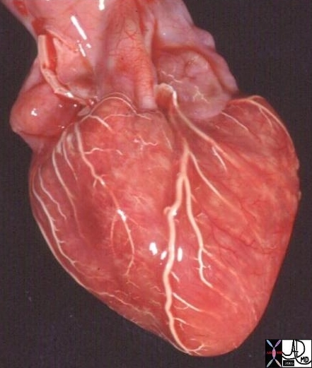
15009c cardiac heart coronary artery normal anatomy gross pathology LAD diagonal artery acute marginal artery conal artery arc of Vieussens Courtesy Ashley Davidoff MD
The heart is a muscular pump that is part of the cardiovascular system. The component parts include paired atria, ventricles, atrioventricular valves and paired outflow valves. It is surrounded by the two layered pericardium. It is connected to the pulmonary circulation via the pulmonary artery and pulmonary veins, and to the systemic circulation via the aorta and vena cava. It is characterized by its pumping function.
Structural features include its fist size, location in the chest, asymmetric symmetry of the chambers and great vessels and the and the syncytial nature of the myocardium.
Functional characteristics include its contractile nature, and ability to adapt to a variety of physiological states with beat to beat variations.
Common diseases include ischemic heart disease, infections, and valvular disorders.
Clinical history examination and EKG are key first steps in the diagnostic workup. Cardiac enzymes have been an extremely useful in the evaluation of acute myocardial infarction. Echocardiography is the mainstay of imaging techniques. Nuclear medicine has also been an important component in cardiac assessment. Newer techniques including multidetector CT and MRI are evolving.
Therapy includes pharmaceuticals that are evolving, but mainstays include digoxin and diuretics. Minimally invasive therapies, and surgery are advancing and improving management of mechanical problems of the heart. Heart transplant was pioneered in 1967 , and remains a surgical option for appropriate candidates.
The Heart – Symmetric Asymmetry
The heart is a muscular pump that is part of the cardiovascular system. The component parts include paired atria, ventricles, atrioventricular valves and paired outflow valves. It is surrounded by the two layered pericardium. It is connected to the pulmonary circulation via the pulmonary artery, to the systemic circulation via the aorta. Systemic venous circulation is connected to the heart via the superior and inferior vena cavae, while the pulmonary circulation returns via the pulmonary veins. It is characterized by its position in the chest cavity, and coordinated pumping ability.
Structural features include its fist size, asymmetric symmetry of the chambers and great vessels and the and the syncytial nature of the myocardium. Functional characteristics include its contractile nature, and ability to adapt to a variety of physiological states. Common diseases include ischemic heart disease, infections, and valvular disorders. Clinical history examination and EKG are key first steps in the diagnostic workup. Cardiac enzymes have been an extremely useful in the evaluation of acute myocardial infarction. Echocardiography is the mainstay of imaging techniques. Nuclear medicine has also been an important component in cardiac assessment. Newer techniques including multidetector CT and MRI are evolving. Treatment depends on the cause but includes a variety of medical and surgical options. Minimally invasive techniques have had a major influence in the treatment of coronary artery disease.
Systole and Diastole


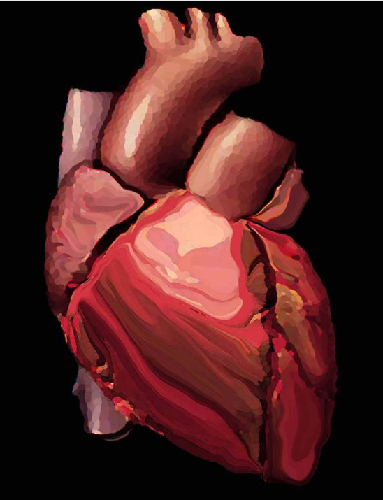
Pulling Together in the Heart “Pulling Together in the Heart” is an artistic rendering of the heart derived from a histological section of the cellular makeup of the heart. The syncytial morphology is an open door design allowing free communication between the cells. This design is particularly necessary in the heart and allows for efficient, coordinated, and collaborative communication and contraction between the cells. It is a great example of units to unity.Ashley Davidoff MD