The shape of the heart is quite complex and can be described in many ways based on which professional is evaluating the heart and from which angle it is viewed. The shape is of extreme importance in imaging since a subtle change may be the key to the diagnosis. Right ventricular dysplasia, for example can cause life threatening arrhythmias and sometimes, the only clue to the diagnosis is a subtle bulge of the right ventricular outflow tract. Thus although simple in concept, shape of structure is a key element to diagnosis.
The shape of the heart has been variably described as conical, triangular and ovoid. It is rather difficult to provide a single descriptor that accurately describes the shape of the heart mostly because of the variety of facets or angles from which it can be evaluated
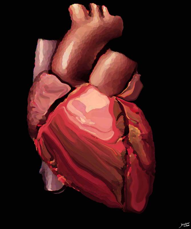
Ambivalence in Shape of the Heart
The artistic rendition of the heart attempts to reveal the ambivalence in the shape of the heart as either a triangular structure or an oval on its side, and it seems to satisfy both shapes in this view. The right ventricle dominates the anterior view and the left ventricle peeks around the left border of the heart holding its power as its trump card behind the right ventricle.
davidoff art copyright 2009 Courtesy Ashley Davidoff MD 87180b11.8s
The Anatomy
The anatomist, and pathologist will have a sense of the shape with the heart devoid of life and blood while the surgeon and the imager will have a different perspective with a heart filled with life and blood that changes in shape every millisecond. On the other hand the detail of muscle bundles, chordae, as well as either micro components are part of the secrets of the heart available only to the anatomist and pathologist, but not yet visible to the imager at this time.
The angle from which the heart is viewed also affects the perspective. The frontal view is different from the lateral which will be different from all the other projections that are used to view this organ including the oblique, coronal, long axis, and short axis. In each of these many views the shape will invoke different subjective descriptions, and most importantly will help define whether the heart is normal or not.
Each of the chambers is quite unique in individual structural characteristics and shape is no exception. The first general description will relate to the anatomical view- how it appears to anatomist or pathologist.
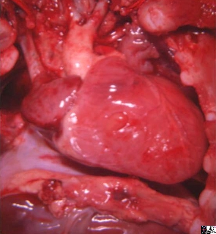
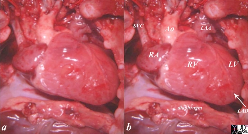
Post Mortem In Situ View of the Heart
The post mortem view of the heart in the chest cavity shows a horizontal orientation of the heart, since the lungs are deflated the heart is more horizontally oriented and has an almost rectangular shape with the longest axis from right to left. The anterior view is dominated by the right ventricle which tends to be triangular. The inferior border of the heart is straight while the rightward, superior and leftward borders are minimally rounded.
Courtesy Ashley Davidoff MD copyright 2009 all rights reserved 06925c02.8s 06925.8s.jpg
The shapes on the inner surface of the heart are unique to each chamber. The right atrium has a combination of a smooth areas, with both large and smaller muscular bundles, the left atrium is mostly smooth, while the left ventricle is smooth but for its set of papillary muscles, and the right ventricle tends to be heavily trabeculated. The overall external surface is smooth.
The overall external surface is smooth.
The shapes on the inner surface of the heart are unique to each chamber. The right atrium has a combination of smooth areas, with both large and smaller muscular bundles; the left atrium is mostly smooth, while the left ventricle is smooth but for its set of papillary muscles, and the right ventricle tends to be heavily trabeculated.
Clinical and Imaging
The shape of the heart cannot be evaluated by clinical examination. However in imaging it is a key and major factor in diagnosis.
In vivo, during life, the heart on a chest X-ray is usually imaged during inspiration and the heart tends to be more elongated. This is in stark difference to the heart as seen in the in situ image portrayed above.
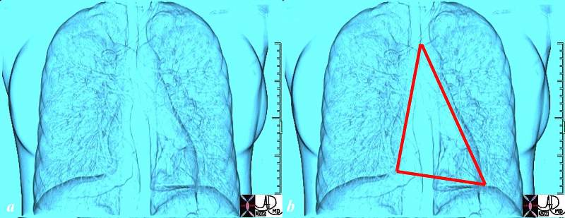
The Shape of the Normal Heart from Frontal View
Perception as a Triangle
The normal heart lies as a gracile structure in the middle of the chest and takes up about 25% of the chest volume. Its shape in this view is interpreted by some as being more or less triangular with the base on the diaphragm and apex in the superior mediastinum. At its base on the diaphragm, the left ventricular apex is tipped a little more inferiorly than the right side. it does differ among individuals. Also if a patient does not take a deep breath, then the heart becomes more horizontal in orientation.
Courtesy Ashley Davidoff MD Davidoff art 45774c01.8s
Others perceive the overall shape in this view as an oblong, perhaps an egg on its side, or a football waiting to be punted.
Perception as a Oval
The shape of the heart in this view could also be interpreted as an oval on its side and for some viewers this makes sense, with the inferior aspect pointing down and to the left and the other superiorly and right.
Courtesy Ashley Davidoff MD Davidoff art copyright 2009 all rights reserved 45774c01.8s
The perception as to whether the heart is shaped like a triangle or an oval depend on multiple variables including the viewers perception, the phase of the respiration, the body habitus of the patient, the overall structure of the individual heart.
In truth though the heart has both triangular features and ovoid features. The right ventricle tends to be triangular and the left ventricle more oval, and this theme will be revisited a number of time throughout this module.
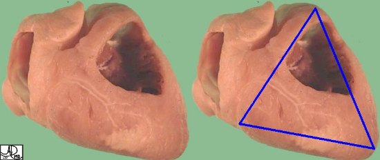
Shape of the RV and LV This is an external and frontal view of the heart. The vessel that you see coursing straight down the center of the heart is the left anterior descending artery, (LAD) that runs anterior to the ventricular septum (vertical limb of the cross) separating the right and left ventricles. Conceptualize the triangular shape of the right ventricle, and the ovoid almost football shape of the left ventricle. Courtesy of Ashley Davidoff M.D. 32078
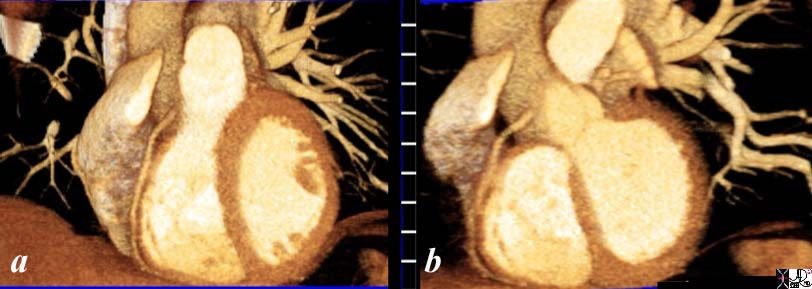
A coronal view of the heart as viewed by CT or MRI imaging portrays the deeper coronal shapes of the heart its muscle and and cavities. In these views the triangle of the RV and the oval of the LV are maintained.
RV shape The right ventricle is less sporty in its shape. It is triangular. (Images courtesy of Ashley Davidoff M.D.32080
Coronal Images through the RV and LV
The CT scan of the heart has been reconstructed in the coronal view with image (a) being slightly anterior to image b. In image a, the triangular nature of the RV is noted and in image b the ovoid nature of the LV is noted.
Courtesy Ashley Davidoff copyright 2009 all rights reserved 31235c.81s
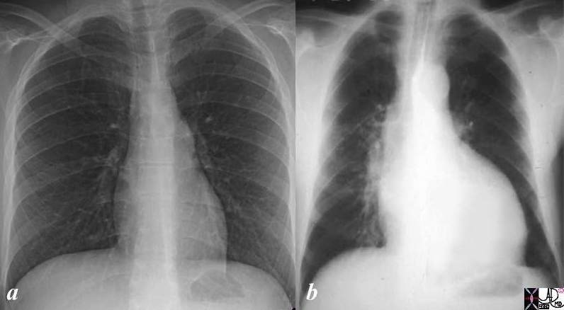
The CXR and EKG used to be standard and first line methods of evaluation of the heart. The shape of the heart on CXR was a means of evaluating its size. Although it is still important, and practical, it is subjective. The enlarged right ventricle is reflected in triangular cardiomediastinal silhouette while the enlarged left ventricle is reflected as an ovoid shape on the frontal view.
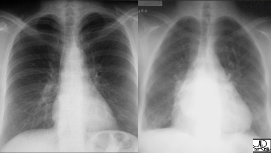
Normal heart and Enlarged Right Heart
Two different patients showing the normal patient on the left and the CXR of a patient with an enlarged triangular shaped heart on the right. The triangular heart with upturned apex is characteristic of right ventricular enlargement, and the elevation of the carina and a double density seen on the right side of the heart suggests left atrial enlargement. These findings are consistent with rheumatic mitral stenosis.
Courtesy of Ashley Davidoff M.D. 32113 Copyright 2009
Aortic regurgitation and Left Ventricular Enlargement The Oval Heart Dominates The normal chest X-ray (a) is placed besides the CXR of a patient with an enlarged left ventricle. The oval shape of the left ventricle dominates the appearance together with a cardiothoracic ratio that is greater than 50% Courtesy Ashley Davidoff MD copyright 2009 08136c01.81s
Shapes of the Heart in Congenital Heart Disease
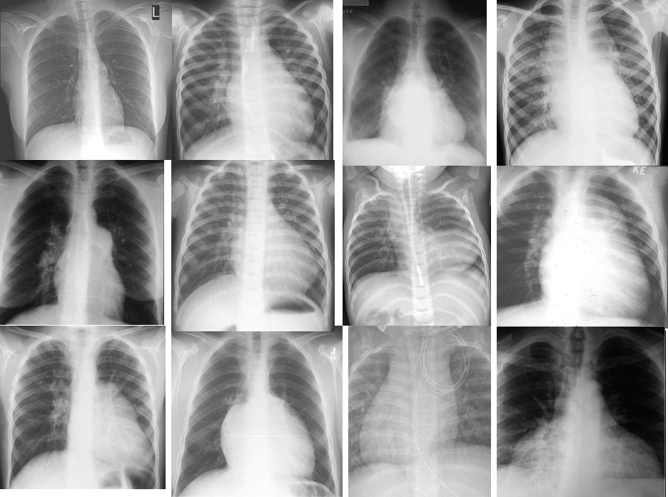
Shapes of the Heart in Congenital Heart Disease on CXR
1 = normal heart 2 = LV enlargement 3 = RV enlargement 4 large mediastinum = snowman TAPVR 5 = small mediastinum = D’TGA TA Pulmonary atresia 6 = pulmonary bay P Atresia Tetralogy truncus TGA Coronal LTGA is 07197 heart shape cardiac CXR plain films Some of the images have been downloaded from the web with sources unknown and are used for education purposes alone 86774 s
The Shapes of the Heart in Health and Disease
The collage of frontal views of the heart is a combination of images from a personal collection and borrowed from the internet for educational purposes only.
From top left and across the rows they are
The normal heart , the “football” of LV enlargement the “triangle” or “proud breast” of RV enlargement, “snowman” of total anomalous pulmonary venous return, “egg on its side” of D transposition of the great vessels, the big mogul of skiing down the left heart border in pulmonary stenosis or pulmonary hypertension, “boot shaped” heart seen in both pulmonary atresia and Tetralogy of Fallot, the long smooth mogul that has a differential diagnosis of L transposition, absence of the pericardium, and juxtaposition of the atrial appendages, the box shaped large heart of Ebstein’s anomaly, the D loop where the genetic instruction of keep right ” seems likely, and the water bottle” heart of a large pericardial effusion.
07197 Some of the images have been downloaded from the web with sources unknown and are used for educational purposes alone 86774b02
Axial Imaging
In this projection the triangular nature of the RV is mostly retained, but because the non orthogonal nature of routine cross sectional imaging the apex of the LV is not usually imaged orthogonally so the oval appearance of the LV is not consistent. The shape of the atria are best defined and assessed in the axial projection.
The Atria
In the axial projection the atria have flattened free borders- the lateral border of the right atrium and the posterior border of the left atrium. When it enlarges the borders become rotund indicating an increase in pressure or volume.
Shapes of the Chambers in Axial View
This axial image of the heart is through right atrium and left atria and are normal and about the same size. The left ventricle with the papillary muscles and the right ventricle with its papillary muscle are well seen and in this view both have a triangular shape. Note that the normal atria have flattened outside walls.
Courtesy Ashley Davidoff MD copyright 2009 all rights reserved 27531d01.8s
Rotund Left Atrium and Right Atrium
Both the right atrium and left atrium in this axial CT study are enlarged. Not only is their absolute size increased, as witnessed by the less than 1:1 ratio of the aorta to the LA, but the outer borders of both atria ar rotund instead of being flat.
Courtesy Ashley Davidoff MD Copyright 2009 48079
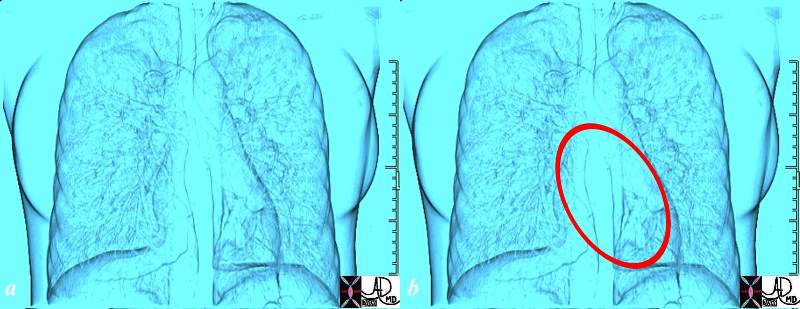
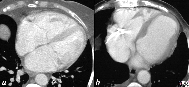
An aneurysm of the left ventricular apex is a common occurrence and is recognized by both a change in shape and size of the apex. These changes are accentuated in systole but in the non gated study as shown below, the abnormal size and shape are easy to recognize
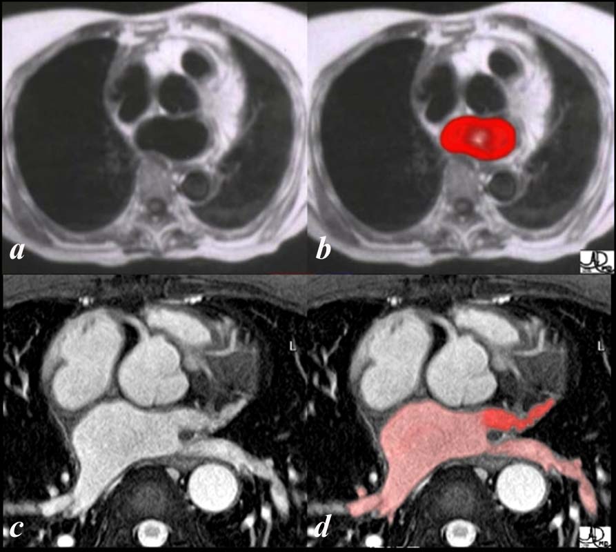
Normal heart (a) and Left Ventricular Aneurysm (b)
Axial images through the chest show a normal patient (a) and a patient with an aneurysm of the left ventricle (b). In the normal image of the ventricles the shape of the apex of the LV is slightly pointed . The bulbous shape of the apex of the left ventricle in the second image becomes obviously abnormal and is diagnostic of a left ventricular aneurysm.
Courtesy Ashley Davidoff copyright 2009 all rights reserved 26336cc01.8s
Other Examples
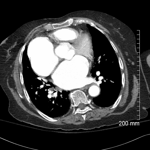
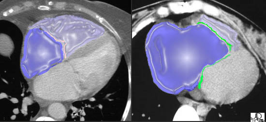
Changes in the Shape of the Right Atrium Reflecting Enlargement
Right Atrium
Normal and Enlarged
These two images are enhanced CT images through the tricuspid valve. The first image is normal, while the second image demonstrates a TV that lies too far forward and low associated with a huge RA and a diminutive RV. This appearance is classical of a congenital condition called Ebstein’s anomaly. In this disease, the posterior leaflet is stuck down to the posterior wall of the RV and the anterior leaflet is larger than normal often giving a flapping sound to the listening ear. The anterior leaflet behaves like a flapping sail in the wind of the blood flow.
In the overlays, the RA is in royal blue and the RV is in light purple overlay. The normal TV in the first image is in pink while the malformed valve in the second image is in green. Note how large the RA is and how small the RV is in Ebstein’s anomaly.
Courtesy of Ashley Davidoff M.D. 32102 32101 copyright 2009
The Right Ventricle
The right ventricle consists of two parts that in conglomerate do make up a triangle. The two parts are the inflow and the out flow and . The shapes are sometimes better appreciated in the abnormal heart – the triangle tends to bulge outward
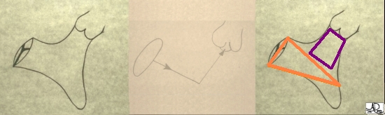
Inflow and Outflow Components of the Right Ventricle
The first diagram is a simple drawing of the right ventricle as seen in a frontal projection. The tricuspid valve is to the left and inferior and the pulmonary valve to the right and superior. The arrows of the second diagram show the inflow portion of the right ventricle and the outflow portion. The third and last diagram shows the two chambers that make up the right ventricle. The right ventricular inflow chamber also called the RV sinus is triangular and in orange while the outflow chamber is more tubular or cylindrical and has been called many names – but somehow it does not seem to care. Right ventricular outflow tract (RVOT), and infundibulum seem to be the most popular.
Courtesy of Ashley Davidoff M.D. 32087 copyright 2009
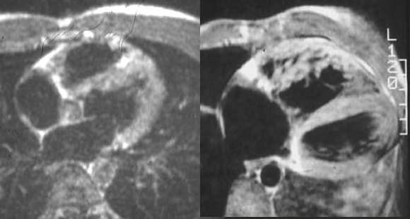
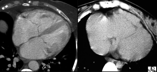
Normal and Abnormal RV Axial images through the heart using black blood imaging shows a normal MRI on the left, alongside an hypertrophied right ventricle (right). Note the thickening and heavy trabeculation of the anterior wall of the right ventricle in the second image . Note also the ovoid shape of the normal LV and the triangular shape of the RV. Courtesy of Ashley Davidoff M.D. 2019 32076
The Left Atrium
The left atrium is almost a rectangular (rectanguloid may be a good term) but develops rounded borders when it enlarges.
Change in Shape with Enlargement
The axial CT scan shows a change in shape of both the RA and LA which have become rotund indicating too much pressure or too much volume .
48079 Courtesy Ashley Davidoff MD copyright 2009
The Right Atrium
Complex Shapes in the Right Atrium
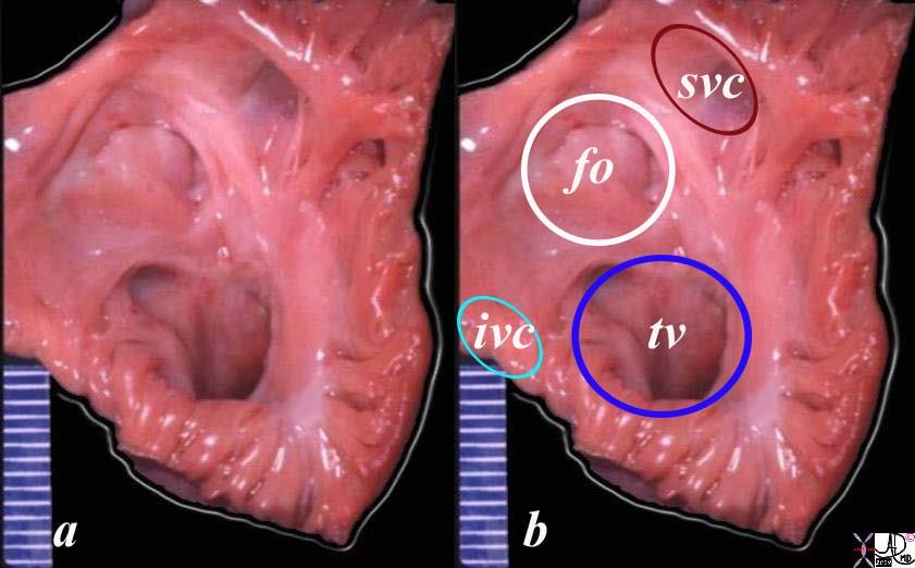
The shapes of the inside of the right atrium are intriguing, and contain stories of ancient history and more modern evolution. The normal right atrium in this post mortem specimen has been opened , revealing the complexity of shapes and types of muscle bundles in the right atrium. they range from broad bands of the tenia sagittalis and septum secundum, to the finer bands of the pectinate muscles, to the smoothness of the fossa ovalis. The rings in image b are for orientation with the white ring representing the fossa ovalis, maroon the svc, the teal, the inferior vena cava and the blue the tricuspid valve
Courtesy Ashley Davidoff MD copyright 2009 all rights reserved AFD 01653c02.8s
The Right Ventricle
Complex Shapes of the Right Ventricle
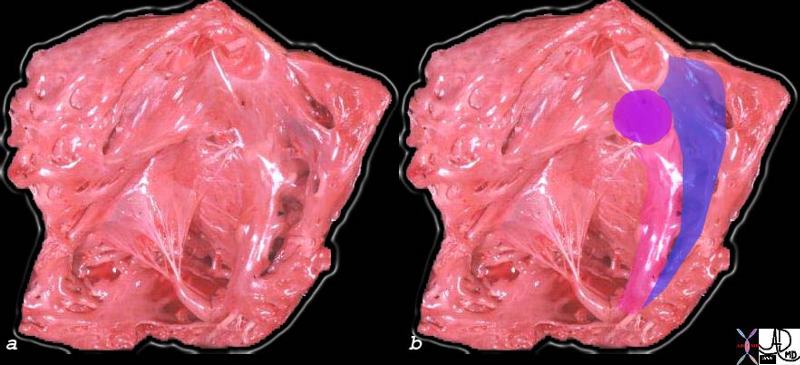
The complex shapes of the right ventricle are just as intriguing as those of the right atrium. The right ventricle has been opened with septal side to the right with free wall side to the left. The septal band is a prominent muscle (pink ) that houses the right bundle branch, and extends inferiorly onto the anterior papillary muscle via the moderator band. The blue band is the trabeculated portion of the septum. The round purple muscle is the conal septum.
06409c02 Courtesy Ashley Davidoff copyright 2009 all rights reserved
Left Atrium
The Shapes of the Left Atrium
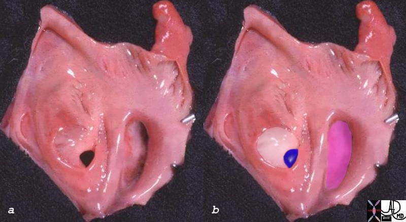
The left atrium does have curves to its inside surface but not to the extent of the right atrium, and it is mostly smooth. The septum primum (white) and foramen ovale (blue) ar in the center. The mitral valve is overlaid in pink. Fine pectinate muscles are seen at the mout of the finger like left atrial appendage.
06425c01 Courtesy Ashley Davidoff MD copyright 2009 all rights reserved
Left Ventricle
Shapes of the LV Interior
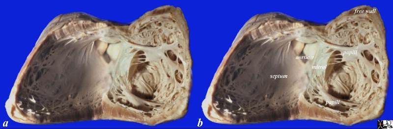
The left ventricle is far less interesting as well when compared to the shapes of the right ventricle. The two major groups of papillary muscles dominate the otherwise flat surfaced left ventricle. The left ventricle has been opened along the interventricular septum along the course of the LAD and PDA so that septal side is to the left and the free wall is to the right. The muscle bundles are confined to two major sets of papillary muscles, and some fine bundles that are not visible to imaging technologies.
Courtesy Ashley Davidoff MD copyright 2009 All rights reserved 15389c05.8s
The Right Atrium
In the axial projection the right atrium tends to be round with a flattened right lateral border. When it enlarges the lateral border rounds out indicating an increase in pressure or volume.
Shapes of the Chambers in Axial View
Normal Right Atrium and Left Atrium
This axial image of the heart is through the mitral (right ) and tricuspid valve (left) right atrium and left atrium (right) which are normal and about the same size. The left ventricle with the papillary muscles and the right ventricle with its papillary muscle are well seen. Note the right sided structures tend to be anterior and left sided structures tend to be posterior. Note also that the right atrium and the left atrium are about the same size and shape in this view with flat walls.
Courtesy Ashley Davidoff MD copyright 2009 all rights reserved 27531d01.8s
Changes in the Shape of the Right Atrium Reflecting Enlargement
Right Atrium
Normal and Enlarged
These two images are enhanced CT images through the tricuspid valve. The first image is normal, while the second image demonstrates a TV that lies too far forward and low associated with a huge RA and a diminutive RV. This appearance is classical of a congenital condition called Ebstein’s anomaly. In this disease, the posterior leaflet is stuck down to the posterior wall of the RV and the anterior leaflet is larger than normal often giving a flapping sound to the listening ear. The anterior leaflet behaves like a flapping sail in the wind of the blood flow.
In the overlays, the RA is in royal blue and the RV is in light purple overlay. The normal TV in the first image is in pink while the malformed valve in the second image is in green. Note how large the RA is and how small the RV is in Ebstein’s anomaly.
Courtesy of Ashley Davidoff M.D. 32102 32101 copyright 2009
The Right Ventricle
The right ventricle consists of two parts that in conglomerate do make up a triangle. The two parts are the inflow and the out flow and . The shapes are sometimes better appreciated in the abnormal heart – the triangle tends to bulge outward
Inflow and Outflow Components of the Right Ventricle
The first diagram is a simple drawing of the right ventricle as seen in a frontal projection. The tricuspid valve is to the left and inferior and the pulmonary valve to the right and superior. The arrows of the second diagram show the inflow portion of the right ventricle and the outflow portion. The third and last diagram shows the two chambers that make up the right ventricle. The right ventricular inflow chamber also called the RV sinus is triangular and in orange while the outflow chamber is more tubular or cylindrical and has been called many names – but somehow it does not seem to care. Right ventricular outflow tract (RVOT), and infundibulum seem to be the most popular.
Courtesy of Ashley Davidoff M.D. 32087 copyright 2009
Triangles and Ovals in Disease These axial image of a T1 weighted MRI shows a hypertrophied right and left ventricle exemplifying the normal innate shapes of both ventricles even in the axial view and in disease. The triangular RV is in blue and the ovoid “rugby-football” of the left ventricle is in red. Courtesy of Ashley Davidoff M.D. Copyright 2009 32076
The Left Atrium
The left atrium is almost a rectangular (rectanguloid may be a good term) but develops rounded borders when it enlarges.
Change in Shape with Enlargement
The axial CT scan shows a change in shape of both the RA and LA which have become rotund indicating too much pressure or too much volume .
48079 Courtesy Ashley Davidoff MD copyright 2009
The Left Ventricle
In the axial projection the left ventricle varies between a triangle and an oval depending on how the apex is oriented and and whether it is sectioned orthogonal to the apex or at an oblique angle. In the former situation it will appear as a oval, and the latter as a triangle.
The shape of the heart is of supreme importance and even more so are its individual components. The complexity of the shapes of the parts will be expanded in the “parts” section of the module.
DOMElement Object
(
    [schemaTypeInfo] =>
    [tagName] => table
    [firstElementChild] => (object value omitted)
    [lastElementChild] => (object value omitted)
    [childElementCount] => 1
    [previousElementSibling] => (object value omitted)
    [nextElementSibling] => (object value omitted)
    [nodeName] => table
    [nodeValue] =>


Change in Shape with Enlargement


The axial CT scan shows a change in shape of both the RA and LA which have become rotund indicating too much pressure or too much volume .
48079 Courtesy Ashley Davidoff MD copyright 2009


    [nodeType] => 1
    [parentNode] => (object value omitted)
    [childNodes] => (object value omitted)
    [firstChild] => (object value omitted)
    [lastChild] => (object value omitted)
    [previousSibling] => (object value omitted)
    [nextSibling] => (object value omitted)
    [attributes] => (object value omitted)
    [ownerDocument] => (object value omitted)
    [namespaceURI] =>
    [prefix] =>
    [localName] => table
    [baseURI] =>
    [textContent] =>


Change in Shape with Enlargement


The axial CT scan shows a change in shape of both the RA and LA which have become rotund indicating too much pressure or too much volume .
48079 Courtesy Ashley Davidoff MD copyright 2009


)
DOMElement Object
(
    [schemaTypeInfo] =>
    [tagName] => td
    [firstElementChild] => (object value omitted)
    [lastElementChild] => (object value omitted)
    [childElementCount] => 1
    [previousElementSibling] =>
    [nextElementSibling] =>
    [nodeName] => td
    [nodeValue] => The axial CT scan shows a change in shape of both the RA and LA which have become rotund indicating too much pressure or too much volume .
48079 Courtesy Ashley Davidoff MD copyright 2009

    [nodeType] => 1
    [parentNode] => (object value omitted)
    [childNodes] => (object value omitted)
    [firstChild] => (object value omitted)
    [lastChild] => (object value omitted)
    [previousSibling] => (object value omitted)
    [nextSibling] => (object value omitted)
    [attributes] => (object value omitted)
    [ownerDocument] => (object value omitted)
    [namespaceURI] =>
    [prefix] =>
    [localName] => td
    [baseURI] =>
    [textContent] => The axial CT scan shows a change in shape of both the RA and LA which have become rotund indicating too much pressure or too much volume .
48079 Courtesy Ashley Davidoff MD copyright 2009

)
DOMElement Object
(
    [schemaTypeInfo] =>
    [tagName] => td
    [firstElementChild] => (object value omitted)
    [lastElementChild] => (object value omitted)
    [childElementCount] => 2
    [previousElementSibling] =>
    [nextElementSibling] =>
    [nodeName] => td
    [nodeValue] =>
Change in Shape with Enlargement

    [nodeType] => 1
    [parentNode] => (object value omitted)
    [childNodes] => (object value omitted)
    [firstChild] => (object value omitted)
    [lastChild] => (object value omitted)
    [previousSibling] => (object value omitted)
    [nextSibling] => (object value omitted)
    [attributes] => (object value omitted)
    [ownerDocument] => (object value omitted)
    [namespaceURI] =>
    [prefix] =>
    [localName] => td
    [baseURI] =>
    [textContent] =>
Change in Shape with Enlargement

)
DOMElement Object
(
    [schemaTypeInfo] =>
    [tagName] => table
    [firstElementChild] => (object value omitted)
    [lastElementChild] => (object value omitted)
    [childElementCount] => 1
    [previousElementSibling] => (object value omitted)
    [nextElementSibling] => (object value omitted)
    [nodeName] => table
    [nodeValue] =>


Inflow and Outflow Components of the Right Ventricle


The first diagram is a simple drawing of the right ventricle as seen in a frontal projection. The tricuspid valve is to the left and inferior and the pulmonary valve to the right and superior. The arrows of the second diagram show the inflow portion of the right ventricle and the outflow portion. The third and last diagram shows the two chambers that make up the right ventricle. The right ventricular inflow chamber also called the RV sinus is triangular and in orange while the outflow chamber is more tubular or cylindrical and has been called many names – but somehow it does not seem to care. Right ventricular outflow tract (RVOT), and infundibulum seem to be the most popular.
Courtesy of Ashley Davidoff M.D. 32087 copyright 2009


    [nodeType] => 1
    [parentNode] => (object value omitted)
    [childNodes] => (object value omitted)
    [firstChild] => (object value omitted)
    [lastChild] => (object value omitted)
    [previousSibling] => (object value omitted)
    [nextSibling] => (object value omitted)
    [attributes] => (object value omitted)
    [ownerDocument] => (object value omitted)
    [namespaceURI] =>
    [prefix] =>
    [localName] => table
    [baseURI] =>
    [textContent] =>


Inflow and Outflow Components of the Right Ventricle


The first diagram is a simple drawing of the right ventricle as seen in a frontal projection. The tricuspid valve is to the left and inferior and the pulmonary valve to the right and superior. The arrows of the second diagram show the inflow portion of the right ventricle and the outflow portion. The third and last diagram shows the two chambers that make up the right ventricle. The right ventricular inflow chamber also called the RV sinus is triangular and in orange while the outflow chamber is more tubular or cylindrical and has been called many names – but somehow it does not seem to care. Right ventricular outflow tract (RVOT), and infundibulum seem to be the most popular.
Courtesy of Ashley Davidoff M.D. 32087 copyright 2009


)
DOMElement Object
(
    [schemaTypeInfo] =>
    [tagName] => td
    [firstElementChild] => (object value omitted)
    [lastElementChild] => (object value omitted)
    [childElementCount] => 2
    [previousElementSibling] =>
    [nextElementSibling] =>
    [nodeName] => td
    [nodeValue] =>
The first diagram is a simple drawing of the right ventricle as seen in a frontal projection. The tricuspid valve is to the left and inferior and the pulmonary valve to the right and superior. The arrows of the second diagram show the inflow portion of the right ventricle and the outflow portion. The third and last diagram shows the two chambers that make up the right ventricle. The right ventricular inflow chamber also called the RV sinus is triangular and in orange while the outflow chamber is more tubular or cylindrical and has been called many names – but somehow it does not seem to care. Right ventricular outflow tract (RVOT), and infundibulum seem to be the most popular.
Courtesy of Ashley Davidoff M.D. 32087 copyright 2009

    [nodeType] => 1
    [parentNode] => (object value omitted)
    [childNodes] => (object value omitted)
    [firstChild] => (object value omitted)
    [lastChild] => (object value omitted)
    [previousSibling] => (object value omitted)
    [nextSibling] => (object value omitted)
    [attributes] => (object value omitted)
    [ownerDocument] => (object value omitted)
    [namespaceURI] =>
    [prefix] =>
    [localName] => td
    [baseURI] =>
    [textContent] =>
The first diagram is a simple drawing of the right ventricle as seen in a frontal projection. The tricuspid valve is to the left and inferior and the pulmonary valve to the right and superior. The arrows of the second diagram show the inflow portion of the right ventricle and the outflow portion. The third and last diagram shows the two chambers that make up the right ventricle. The right ventricular inflow chamber also called the RV sinus is triangular and in orange while the outflow chamber is more tubular or cylindrical and has been called many names – but somehow it does not seem to care. Right ventricular outflow tract (RVOT), and infundibulum seem to be the most popular.
Courtesy of Ashley Davidoff M.D. 32087 copyright 2009

)
DOMElement Object
(
    [schemaTypeInfo] =>
    [tagName] => td
    [firstElementChild] => (object value omitted)
    [lastElementChild] => (object value omitted)
    [childElementCount] => 2
    [previousElementSibling] =>
    [nextElementSibling] =>
    [nodeName] => td
    [nodeValue] =>
Inflow and Outflow Components of the Right Ventricle
    [nodeType] => 1
    [parentNode] => (object value omitted)
    [childNodes] => (object value omitted)
    [firstChild] => (object value omitted)
    [lastChild] => (object value omitted)
    [previousSibling] => (object value omitted)
    [nextSibling] => (object value omitted)
    [attributes] => (object value omitted)
    [ownerDocument] => (object value omitted)
    [namespaceURI] =>
    [prefix] =>
    [localName] => td
    [baseURI] =>
    [textContent] =>
Inflow and Outflow Components of the Right Ventricle
)
DOMElement Object
(
    [schemaTypeInfo] =>
    [tagName] => table
    [firstElementChild] => (object value omitted)
    [lastElementChild] => (object value omitted)
    [childElementCount] => 1
    [previousElementSibling] => (object value omitted)
    [nextElementSibling] => (object value omitted)
    [nodeName] => table
    [nodeValue] =>


Changes in the Shape of the Right Atrium Reflecting Enlargement
Right Atrium
Normal and  Enlarged


These two images are enhanced CT images through the tricuspid valve. The first image is normal, while the second image demonstrates a TV that lies too far forward and low associated with a huge RA and a diminutive RV. This appearance is classical of a congenital condition called Ebstein’s anomaly. In this disease, the posterior leaflet is stuck down to the posterior wall of the RV and the anterior leaflet is larger than normal often giving a flapping sound to the listening ear. The anterior leaflet behaves like a flapping sail in the wind of the blood flow.
In the overlays, the RA is in royal blue and the RV is in light purple overlay. The normal TV in the first image is in pink while the malformed valve in the second image is in green. Note how large the RA is and how small the RV is in Ebstein’s anomaly.
Courtesy of Ashley Davidoff M.D. 32102  32101 copyright 2009


    [nodeType] => 1
    [parentNode] => (object value omitted)
    [childNodes] => (object value omitted)
    [firstChild] => (object value omitted)
    [lastChild] => (object value omitted)
    [previousSibling] => (object value omitted)
    [nextSibling] => (object value omitted)
    [attributes] => (object value omitted)
    [ownerDocument] => (object value omitted)
    [namespaceURI] =>
    [prefix] =>
    [localName] => table
    [baseURI] =>
    [textContent] =>


Changes in the Shape of the Right Atrium Reflecting Enlargement
Right Atrium
Normal and  Enlarged


These two images are enhanced CT images through the tricuspid valve. The first image is normal, while the second image demonstrates a TV that lies too far forward and low associated with a huge RA and a diminutive RV. This appearance is classical of a congenital condition called Ebstein’s anomaly. In this disease, the posterior leaflet is stuck down to the posterior wall of the RV and the anterior leaflet is larger than normal often giving a flapping sound to the listening ear. The anterior leaflet behaves like a flapping sail in the wind of the blood flow.
In the overlays, the RA is in royal blue and the RV is in light purple overlay. The normal TV in the first image is in pink while the malformed valve in the second image is in green. Note how large the RA is and how small the RV is in Ebstein’s anomaly.
Courtesy of Ashley Davidoff M.D. 32102  32101 copyright 2009


)
DOMElement Object
(
    [schemaTypeInfo] =>
    [tagName] => td
    [firstElementChild] => (object value omitted)
    [lastElementChild] => (object value omitted)
    [childElementCount] => 3
    [previousElementSibling] =>
    [nextElementSibling] =>
    [nodeName] => td
    [nodeValue] =>
These two images are enhanced CT images through the tricuspid valve. The first image is normal, while the second image demonstrates a TV that lies too far forward and low associated with a huge RA and a diminutive RV. This appearance is classical of a congenital condition called Ebstein’s anomaly. In this disease, the posterior leaflet is stuck down to the posterior wall of the RV and the anterior leaflet is larger than normal often giving a flapping sound to the listening ear. The anterior leaflet behaves like a flapping sail in the wind of the blood flow.
In the overlays, the RA is in royal blue and the RV is in light purple overlay. The normal TV in the first image is in pink while the malformed valve in the second image is in green. Note how large the RA is and how small the RV is in Ebstein’s anomaly.
Courtesy of Ashley Davidoff M.D. 32102  32101 copyright 2009

    [nodeType] => 1
    [parentNode] => (object value omitted)
    [childNodes] => (object value omitted)
    [firstChild] => (object value omitted)
    [lastChild] => (object value omitted)
    [previousSibling] => (object value omitted)
    [nextSibling] => (object value omitted)
    [attributes] => (object value omitted)
    [ownerDocument] => (object value omitted)
    [namespaceURI] =>
    [prefix] =>
    [localName] => td
    [baseURI] =>
    [textContent] =>
These two images are enhanced CT images through the tricuspid valve. The first image is normal, while the second image demonstrates a TV that lies too far forward and low associated with a huge RA and a diminutive RV. This appearance is classical of a congenital condition called Ebstein’s anomaly. In this disease, the posterior leaflet is stuck down to the posterior wall of the RV and the anterior leaflet is larger than normal often giving a flapping sound to the listening ear. The anterior leaflet behaves like a flapping sail in the wind of the blood flow.
In the overlays, the RA is in royal blue and the RV is in light purple overlay. The normal TV in the first image is in pink while the malformed valve in the second image is in green. Note how large the RA is and how small the RV is in Ebstein’s anomaly.
Courtesy of Ashley Davidoff M.D. 32102  32101 copyright 2009

)
DOMElement Object
(
    [schemaTypeInfo] =>
    [tagName] => td
    [firstElementChild] => (object value omitted)
    [lastElementChild] => (object value omitted)
    [childElementCount] => 5
    [previousElementSibling] =>
    [nextElementSibling] =>
    [nodeName] => td
    [nodeValue] =>

Changes in the Shape of the Right Atrium Reflecting Enlargement
Right Atrium
Normal and  Enlarged
    [nodeType] => 1
    [parentNode] => (object value omitted)
    [childNodes] => (object value omitted)
    [firstChild] => (object value omitted)
    [lastChild] => (object value omitted)
    [previousSibling] => (object value omitted)
    [nextSibling] => (object value omitted)
    [attributes] => (object value omitted)
    [ownerDocument] => (object value omitted)
    [namespaceURI] =>
    [prefix] =>
    [localName] => td
    [baseURI] =>
    [textContent] =>

Changes in the Shape of the Right Atrium Reflecting Enlargement
Right Atrium
Normal and  Enlarged
)
DOMElement Object
(
    [schemaTypeInfo] =>
    [tagName] => table
    [firstElementChild] => (object value omitted)
    [lastElementChild] => (object value omitted)
    [childElementCount] => 1
    [previousElementSibling] => (object value omitted)
    [nextElementSibling] => (object value omitted)
    [nodeName] => table
    [nodeValue] =>


Shapes of the Chambers in Axial View
Normal Right Atrium and Left Atrium


This axial image of the heart is through the mitral (right ) and tricuspid valve (left) right atrium and left atrium (right) which are normal and about the same size. The left ventricle with the papillary muscles and the right ventricle with its papillary muscle are well seen. Note the right sided structures tend to be anterior and left sided structures tend to be posterior. Note also that the right atrium and the left atrium are about the same size and shape in this view with flat walls.
Courtesy Ashley Davidoff MD copyright 2009 all rights reserved 27531d01.8s


    [nodeType] => 1
    [parentNode] => (object value omitted)
    [childNodes] => (object value omitted)
    [firstChild] => (object value omitted)
    [lastChild] => (object value omitted)
    [previousSibling] => (object value omitted)
    [nextSibling] => (object value omitted)
    [attributes] => (object value omitted)
    [ownerDocument] => (object value omitted)
    [namespaceURI] =>
    [prefix] =>
    [localName] => table
    [baseURI] =>
    [textContent] =>


Shapes of the Chambers in Axial View
Normal Right Atrium and Left Atrium


This axial image of the heart is through the mitral (right ) and tricuspid valve (left) right atrium and left atrium (right) which are normal and about the same size. The left ventricle with the papillary muscles and the right ventricle with its papillary muscle are well seen. Note the right sided structures tend to be anterior and left sided structures tend to be posterior. Note also that the right atrium and the left atrium are about the same size and shape in this view with flat walls.
Courtesy Ashley Davidoff MD copyright 2009 all rights reserved 27531d01.8s


)
DOMElement Object
(
    [schemaTypeInfo] =>
    [tagName] => td
    [firstElementChild] => (object value omitted)
    [lastElementChild] => (object value omitted)
    [childElementCount] => 2
    [previousElementSibling] =>
    [nextElementSibling] =>
    [nodeName] => td
    [nodeValue] =>
This axial image of the heart is through the mitral (right ) and tricuspid valve (left) right atrium and left atrium (right) which are normal and about the same size. The left ventricle with the papillary muscles and the right ventricle with its papillary muscle are well seen. Note the right sided structures tend to be anterior and left sided structures tend to be posterior. Note also that the right atrium and the left atrium are about the same size and shape in this view with flat walls.
Courtesy Ashley Davidoff MD copyright 2009 all rights reserved 27531d01.8s

    [nodeType] => 1
    [parentNode] => (object value omitted)
    [childNodes] => (object value omitted)
    [firstChild] => (object value omitted)
    [lastChild] => (object value omitted)
    [previousSibling] => (object value omitted)
    [nextSibling] => (object value omitted)
    [attributes] => (object value omitted)
    [ownerDocument] => (object value omitted)
    [namespaceURI] =>
    [prefix] =>
    [localName] => td
    [baseURI] =>
    [textContent] =>
This axial image of the heart is through the mitral (right ) and tricuspid valve (left) right atrium and left atrium (right) which are normal and about the same size. The left ventricle with the papillary muscles and the right ventricle with its papillary muscle are well seen. Note the right sided structures tend to be anterior and left sided structures tend to be posterior. Note also that the right atrium and the left atrium are about the same size and shape in this view with flat walls.
Courtesy Ashley Davidoff MD copyright 2009 all rights reserved 27531d01.8s

)
DOMElement Object
(
    [schemaTypeInfo] =>
    [tagName] => td
    [firstElementChild] => (object value omitted)
    [lastElementChild] => (object value omitted)
    [childElementCount] => 3
    [previousElementSibling] =>
    [nextElementSibling] =>
    [nodeName] => td
    [nodeValue] =>
Shapes of the Chambers in Axial View
Normal Right Atrium and Left Atrium
    [nodeType] => 1
    [parentNode] => (object value omitted)
    [childNodes] => (object value omitted)
    [firstChild] => (object value omitted)
    [lastChild] => (object value omitted)
    [previousSibling] => (object value omitted)
    [nextSibling] => (object value omitted)
    [attributes] => (object value omitted)
    [ownerDocument] => (object value omitted)
    [namespaceURI] =>
    [prefix] =>
    [localName] => td
    [baseURI] =>
    [textContent] =>
Shapes of the Chambers in Axial View
Normal Right Atrium and Left Atrium
)
DOMElement Object
(
    [schemaTypeInfo] =>
    [tagName] => table
    [firstElementChild] => (object value omitted)
    [lastElementChild] => (object value omitted)
    [childElementCount] => 1
    [previousElementSibling] => (object value omitted)
    [nextElementSibling] => (object value omitted)
    [nodeName] => table
    [nodeValue] =>


Shapes of the LV Interior


The left ventricle is far less interesting as well when compared to the shapes of the right ventricle.  The two major groups of papillary muscles dominate the otherwise flat surfaced left ventricle.   The left ventricle has been opened along the interventricular septum along the course of the LAD and PDA so that septal side is to the left and the free wall is to the right. The muscle bundles are confined to two major sets of papillary muscles, and some fine bundles that are not visible to imaging technologies.
Courtesy Ashley Davidoff MD copyright 2009 All rights reserved 15389c05.8s


    [nodeType] => 1
    [parentNode] => (object value omitted)
    [childNodes] => (object value omitted)
    [firstChild] => (object value omitted)
    [lastChild] => (object value omitted)
    [previousSibling] => (object value omitted)
    [nextSibling] => (object value omitted)
    [attributes] => (object value omitted)
    [ownerDocument] => (object value omitted)
    [namespaceURI] =>
    [prefix] =>
    [localName] => table
    [baseURI] =>
    [textContent] =>


Shapes of the LV Interior


The left ventricle is far less interesting as well when compared to the shapes of the right ventricle.  The two major groups of papillary muscles dominate the otherwise flat surfaced left ventricle.   The left ventricle has been opened along the interventricular septum along the course of the LAD and PDA so that septal side is to the left and the free wall is to the right. The muscle bundles are confined to two major sets of papillary muscles, and some fine bundles that are not visible to imaging technologies.
Courtesy Ashley Davidoff MD copyright 2009 All rights reserved 15389c05.8s


)
DOMElement Object
(
    [schemaTypeInfo] =>
    [tagName] => td
    [firstElementChild] => (object value omitted)
    [lastElementChild] => (object value omitted)
    [childElementCount] => 1
    [previousElementSibling] =>
    [nextElementSibling] =>
    [nodeName] => td
    [nodeValue] => The left ventricle is far less interesting as well when compared to the shapes of the right ventricle.  The two major groups of papillary muscles dominate the otherwise flat surfaced left ventricle.   The left ventricle has been opened along the interventricular septum along the course of the LAD and PDA so that septal side is to the left and the free wall is to the right. The muscle bundles are confined to two major sets of papillary muscles, and some fine bundles that are not visible to imaging technologies.
Courtesy Ashley Davidoff MD copyright 2009 All rights reserved 15389c05.8s

    [nodeType] => 1
    [parentNode] => (object value omitted)
    [childNodes] => (object value omitted)
    [firstChild] => (object value omitted)
    [lastChild] => (object value omitted)
    [previousSibling] => (object value omitted)
    [nextSibling] => (object value omitted)
    [attributes] => (object value omitted)
    [ownerDocument] => (object value omitted)
    [namespaceURI] =>
    [prefix] =>
    [localName] => td
    [baseURI] =>
    [textContent] => The left ventricle is far less interesting as well when compared to the shapes of the right ventricle.  The two major groups of papillary muscles dominate the otherwise flat surfaced left ventricle.   The left ventricle has been opened along the interventricular septum along the course of the LAD and PDA so that septal side is to the left and the free wall is to the right. The muscle bundles are confined to two major sets of papillary muscles, and some fine bundles that are not visible to imaging technologies.
Courtesy Ashley Davidoff MD copyright 2009 All rights reserved 15389c05.8s

)
DOMElement Object
(
    [schemaTypeInfo] =>
    [tagName] => td
    [firstElementChild] => (object value omitted)
    [lastElementChild] => (object value omitted)
    [childElementCount] => 2
    [previousElementSibling] =>
    [nextElementSibling] =>
    [nodeName] => td
    [nodeValue] =>
Shapes of the LV Interior

    [nodeType] => 1
    [parentNode] => (object value omitted)
    [childNodes] => (object value omitted)
    [firstChild] => (object value omitted)
    [lastChild] => (object value omitted)
    [previousSibling] => (object value omitted)
    [nextSibling] => (object value omitted)
    [attributes] => (object value omitted)
    [ownerDocument] => (object value omitted)
    [namespaceURI] =>
    [prefix] =>
    [localName] => td
    [baseURI] =>
    [textContent] =>
Shapes of the LV Interior

)
DOMElement Object
(
    [schemaTypeInfo] =>
    [tagName] => table
    [firstElementChild] => (object value omitted)
    [lastElementChild] => (object value omitted)
    [childElementCount] => 1
    [previousElementSibling] => (object value omitted)
    [nextElementSibling] => (object value omitted)
    [nodeName] => table
    [nodeValue] =>


The Shapes of the Left Atrium


The left atrium does have curves to its inside surface but not to the extent of the right atrium, and it is mostly smooth.  The septum primum (white) and foramen ovale (blue) ar in the center.  The mitral valve is overlaid in pink.  Fine pectinate muscles are seen at the mout of the finger like left atrial appendage.
06425c01 Courtesy Ashley Davidoff MD copyright 2009 all rights reserved


    [nodeType] => 1
    [parentNode] => (object value omitted)
    [childNodes] => (object value omitted)
    [firstChild] => (object value omitted)
    [lastChild] => (object value omitted)
    [previousSibling] => (object value omitted)
    [nextSibling] => (object value omitted)
    [attributes] => (object value omitted)
    [ownerDocument] => (object value omitted)
    [namespaceURI] =>
    [prefix] =>
    [localName] => table
    [baseURI] =>
    [textContent] =>


The Shapes of the Left Atrium


The left atrium does have curves to its inside surface but not to the extent of the right atrium, and it is mostly smooth.  The septum primum (white) and foramen ovale (blue) ar in the center.  The mitral valve is overlaid in pink.  Fine pectinate muscles are seen at the mout of the finger like left atrial appendage.
06425c01 Courtesy Ashley Davidoff MD copyright 2009 all rights reserved


)
DOMElement Object
(
    [schemaTypeInfo] =>
    [tagName] => td
    [firstElementChild] => (object value omitted)
    [lastElementChild] => (object value omitted)
    [childElementCount] => 1
    [previousElementSibling] =>
    [nextElementSibling] =>
    [nodeName] => td
    [nodeValue] => The left atrium does have curves to its inside surface but not to the extent of the right atrium, and it is mostly smooth.  The septum primum (white) and foramen ovale (blue) ar in the center.  The mitral valve is overlaid in pink.  Fine pectinate muscles are seen at the mout of the finger like left atrial appendage.
06425c01 Courtesy Ashley Davidoff MD copyright 2009 all rights reserved

    [nodeType] => 1
    [parentNode] => (object value omitted)
    [childNodes] => (object value omitted)
    [firstChild] => (object value omitted)
    [lastChild] => (object value omitted)
    [previousSibling] => (object value omitted)
    [nextSibling] => (object value omitted)
    [attributes] => (object value omitted)
    [ownerDocument] => (object value omitted)
    [namespaceURI] =>
    [prefix] =>
    [localName] => td
    [baseURI] =>
    [textContent] => The left atrium does have curves to its inside surface but not to the extent of the right atrium, and it is mostly smooth.  The septum primum (white) and foramen ovale (blue) ar in the center.  The mitral valve is overlaid in pink.  Fine pectinate muscles are seen at the mout of the finger like left atrial appendage.
06425c01 Courtesy Ashley Davidoff MD copyright 2009 all rights reserved

)
DOMElement Object
(
    [schemaTypeInfo] =>
    [tagName] => td
    [firstElementChild] => (object value omitted)
    [lastElementChild] => (object value omitted)
    [childElementCount] => 2
    [previousElementSibling] =>
    [nextElementSibling] =>
    [nodeName] => td
    [nodeValue] =>
The Shapes of the Left Atrium

    [nodeType] => 1
    [parentNode] => (object value omitted)
    [childNodes] => (object value omitted)
    [firstChild] => (object value omitted)
    [lastChild] => (object value omitted)
    [previousSibling] => (object value omitted)
    [nextSibling] => (object value omitted)
    [attributes] => (object value omitted)
    [ownerDocument] => (object value omitted)
    [namespaceURI] =>
    [prefix] =>
    [localName] => td
    [baseURI] =>
    [textContent] =>
The Shapes of the Left Atrium

)
DOMElement Object
(
    [schemaTypeInfo] =>
    [tagName] => table
    [firstElementChild] => (object value omitted)
    [lastElementChild] => (object value omitted)
    [childElementCount] => 1
    [previousElementSibling] => (object value omitted)
    [nextElementSibling] => (object value omitted)
    [nodeName] => table
    [nodeValue] =>


Complex Shapes of the Right Ventricle


The complex shapes of the right ventricle are just as intriguing as those of the right atrium.  The right ventricle has been opened with septal side to the right with free wall side to the left.  The septal band is a prominent muscle  (pink ) that houses the right bundle branch, and extends inferiorly onto the anterior papillary muscle via the moderator band.  The  blue band is  the trabeculated portion of the septum.  The round purple muscle is the conal septum.
06409c02 Courtesy Ashley Davidoff copyright 2009 all rights reserved


    [nodeType] => 1
    [parentNode] => (object value omitted)
    [childNodes] => (object value omitted)
    [firstChild] => (object value omitted)
    [lastChild] => (object value omitted)
    [previousSibling] => (object value omitted)
    [nextSibling] => (object value omitted)
    [attributes] => (object value omitted)
    [ownerDocument] => (object value omitted)
    [namespaceURI] =>
    [prefix] =>
    [localName] => table
    [baseURI] =>
    [textContent] =>


Complex Shapes of the Right Ventricle


The complex shapes of the right ventricle are just as intriguing as those of the right atrium.  The right ventricle has been opened with septal side to the right with free wall side to the left.  The septal band is a prominent muscle  (pink ) that houses the right bundle branch, and extends inferiorly onto the anterior papillary muscle via the moderator band.  The  blue band is  the trabeculated portion of the septum.  The round purple muscle is the conal septum.
06409c02 Courtesy Ashley Davidoff copyright 2009 all rights reserved


)
DOMElement Object
(
    [schemaTypeInfo] =>
    [tagName] => td
    [firstElementChild] => (object value omitted)
    [lastElementChild] => (object value omitted)
    [childElementCount] => 1
    [previousElementSibling] =>
    [nextElementSibling] =>
    [nodeName] => td
    [nodeValue] => The complex shapes of the right ventricle are just as intriguing as those of the right atrium.  The right ventricle has been opened with septal side to the right with free wall side to the left.  The septal band is a prominent muscle  (pink ) that houses the right bundle branch, and extends inferiorly onto the anterior papillary muscle via the moderator band.  The  blue band is  the trabeculated portion of the septum.  The round purple muscle is the conal septum.
06409c02 Courtesy Ashley Davidoff copyright 2009 all rights reserved

    [nodeType] => 1
    [parentNode] => (object value omitted)
    [childNodes] => (object value omitted)
    [firstChild] => (object value omitted)
    [lastChild] => (object value omitted)
    [previousSibling] => (object value omitted)
    [nextSibling] => (object value omitted)
    [attributes] => (object value omitted)
    [ownerDocument] => (object value omitted)
    [namespaceURI] =>
    [prefix] =>
    [localName] => td
    [baseURI] =>
    [textContent] => The complex shapes of the right ventricle are just as intriguing as those of the right atrium.  The right ventricle has been opened with septal side to the right with free wall side to the left.  The septal band is a prominent muscle  (pink ) that houses the right bundle branch, and extends inferiorly onto the anterior papillary muscle via the moderator band.  The  blue band is  the trabeculated portion of the septum.  The round purple muscle is the conal septum.
06409c02 Courtesy Ashley Davidoff copyright 2009 all rights reserved

)
DOMElement Object
(
    [schemaTypeInfo] =>
    [tagName] => td
    [firstElementChild] => (object value omitted)
    [lastElementChild] => (object value omitted)
    [childElementCount] => 2
    [previousElementSibling] =>
    [nextElementSibling] =>
    [nodeName] => td
    [nodeValue] =>
Complex Shapes of the Right Ventricle

    [nodeType] => 1
    [parentNode] => (object value omitted)
    [childNodes] => (object value omitted)
    [firstChild] => (object value omitted)
    [lastChild] => (object value omitted)
    [previousSibling] => (object value omitted)
    [nextSibling] => (object value omitted)
    [attributes] => (object value omitted)
    [ownerDocument] => (object value omitted)
    [namespaceURI] =>
    [prefix] =>
    [localName] => td
    [baseURI] =>
    [textContent] =>
Complex Shapes of the Right Ventricle

)
DOMElement Object
(
    [schemaTypeInfo] =>
    [tagName] => table
    [firstElementChild] => (object value omitted)
    [lastElementChild] => (object value omitted)
    [childElementCount] => 1
    [previousElementSibling] => (object value omitted)
    [nextElementSibling] => (object value omitted)
    [nodeName] => table
    [nodeValue] =>


Complex Shapes in the Right Atrium


The shapes of the inside of the right atrium are intriguing, and contain stories of ancient history and more modern evolution. The normal right atrium in this post mortem specimen has been opened , revealing the complexity of shapes and types of muscle bundles in the right atrium. they range from broad bands of the tenia sagittalis and septum secundum, to the finer bands of the pectinate muscles, to the smoothness of the fossa ovalis. The rings in image b are for orientation with the white ring representing the fossa ovalis, maroon the svc, the teal, the inferior vena cava and the blue the tricuspid valve
 Courtesy Ashley Davidoff MD copyright 2009 all rights reserved AFD 01653c02.8s


    [nodeType] => 1
    [parentNode] => (object value omitted)
    [childNodes] => (object value omitted)
    [firstChild] => (object value omitted)
    [lastChild] => (object value omitted)
    [previousSibling] => (object value omitted)
    [nextSibling] => (object value omitted)
    [attributes] => (object value omitted)
    [ownerDocument] => (object value omitted)
    [namespaceURI] =>
    [prefix] =>
    [localName] => table
    [baseURI] =>
    [textContent] =>


Complex Shapes in the Right Atrium


The shapes of the inside of the right atrium are intriguing, and contain stories of ancient history and more modern evolution. The normal right atrium in this post mortem specimen has been opened , revealing the complexity of shapes and types of muscle bundles in the right atrium. they range from broad bands of the tenia sagittalis and septum secundum, to the finer bands of the pectinate muscles, to the smoothness of the fossa ovalis. The rings in image b are for orientation with the white ring representing the fossa ovalis, maroon the svc, the teal, the inferior vena cava and the blue the tricuspid valve
 Courtesy Ashley Davidoff MD copyright 2009 all rights reserved AFD 01653c02.8s


)
DOMElement Object
(
    [schemaTypeInfo] =>
    [tagName] => td
    [firstElementChild] => (object value omitted)
    [lastElementChild] => (object value omitted)
    [childElementCount] => 1
    [previousElementSibling] =>
    [nextElementSibling] =>
    [nodeName] => td
    [nodeValue] => The shapes of the inside of the right atrium are intriguing, and contain stories of ancient history and more modern evolution. The normal right atrium in this post mortem specimen has been opened , revealing the complexity of shapes and types of muscle bundles in the right atrium. they range from broad bands of the tenia sagittalis and septum secundum, to the finer bands of the pectinate muscles, to the smoothness of the fossa ovalis. The rings in image b are for orientation with the white ring representing the fossa ovalis, maroon the svc, the teal, the inferior vena cava and the blue the tricuspid valve
 Courtesy Ashley Davidoff MD copyright 2009 all rights reserved AFD 01653c02.8s

    [nodeType] => 1
    [parentNode] => (object value omitted)
    [childNodes] => (object value omitted)
    [firstChild] => (object value omitted)
    [lastChild] => (object value omitted)
    [previousSibling] => (object value omitted)
    [nextSibling] => (object value omitted)
    [attributes] => (object value omitted)
    [ownerDocument] => (object value omitted)
    [namespaceURI] =>
    [prefix] =>
    [localName] => td
    [baseURI] =>
    [textContent] => The shapes of the inside of the right atrium are intriguing, and contain stories of ancient history and more modern evolution. The normal right atrium in this post mortem specimen has been opened , revealing the complexity of shapes and types of muscle bundles in the right atrium. they range from broad bands of the tenia sagittalis and septum secundum, to the finer bands of the pectinate muscles, to the smoothness of the fossa ovalis. The rings in image b are for orientation with the white ring representing the fossa ovalis, maroon the svc, the teal, the inferior vena cava and the blue the tricuspid valve
 Courtesy Ashley Davidoff MD copyright 2009 all rights reserved AFD 01653c02.8s

)
DOMElement Object
(
    [schemaTypeInfo] =>
    [tagName] => td
    [firstElementChild] => (object value omitted)
    [lastElementChild] => (object value omitted)
    [childElementCount] => 2
    [previousElementSibling] =>
    [nextElementSibling] =>
    [nodeName] => td
    [nodeValue] =>
Complex Shapes in the Right Atrium

    [nodeType] => 1
    [parentNode] => (object value omitted)
    [childNodes] => (object value omitted)
    [firstChild] => (object value omitted)
    [lastChild] => (object value omitted)
    [previousSibling] => (object value omitted)
    [nextSibling] => (object value omitted)
    [attributes] => (object value omitted)
    [ownerDocument] => (object value omitted)
    [namespaceURI] =>
    [prefix] =>
    [localName] => td
    [baseURI] =>
    [textContent] =>
Complex Shapes in the Right Atrium

)
DOMElement Object
(
    [schemaTypeInfo] =>
    [tagName] => table
    [firstElementChild] => (object value omitted)
    [lastElementChild] => (object value omitted)
    [childElementCount] => 1
    [previousElementSibling] => (object value omitted)
    [nextElementSibling] => (object value omitted)
    [nodeName] => table
    [nodeValue] =>


Change in Shape with Enlargement


The axial CT scan shows a change in shape of both the RA and LA which have become rotund indicating too much pressure or too much volume .
48079 Courtesy Ashley Davidoff MD copyright 2009


    [nodeType] => 1
    [parentNode] => (object value omitted)
    [childNodes] => (object value omitted)
    [firstChild] => (object value omitted)
    [lastChild] => (object value omitted)
    [previousSibling] => (object value omitted)
    [nextSibling] => (object value omitted)
    [attributes] => (object value omitted)
    [ownerDocument] => (object value omitted)
    [namespaceURI] =>
    [prefix] =>
    [localName] => table
    [baseURI] =>
    [textContent] =>


Change in Shape with Enlargement


The axial CT scan shows a change in shape of both the RA and LA which have become rotund indicating too much pressure or too much volume .
48079 Courtesy Ashley Davidoff MD copyright 2009


)
DOMElement Object
(
    [schemaTypeInfo] =>
    [tagName] => td
    [firstElementChild] => (object value omitted)
    [lastElementChild] => (object value omitted)
    [childElementCount] => 1
    [previousElementSibling] =>
    [nextElementSibling] =>
    [nodeName] => td
    [nodeValue] => The axial CT scan shows a change in shape of both the RA and LA which have become rotund indicating too much pressure or too much volume .
48079 Courtesy Ashley Davidoff MD copyright 2009
    [nodeType] => 1
    [parentNode] => (object value omitted)
    [childNodes] => (object value omitted)
    [firstChild] => (object value omitted)
    [lastChild] => (object value omitted)
    [previousSibling] => (object value omitted)
    [nextSibling] => (object value omitted)
    [attributes] => (object value omitted)
    [ownerDocument] => (object value omitted)
    [namespaceURI] =>
    [prefix] =>
    [localName] => td
    [baseURI] =>
    [textContent] => The axial CT scan shows a change in shape of both the RA and LA which have become rotund indicating too much pressure or too much volume .
48079 Courtesy Ashley Davidoff MD copyright 2009
)
DOMElement Object
(
    [schemaTypeInfo] =>
    [tagName] => td
    [firstElementChild] => (object value omitted)
    [lastElementChild] => (object value omitted)
    [childElementCount] => 2
    [previousElementSibling] =>
    [nextElementSibling] =>
    [nodeName] => td
    [nodeValue] =>
Change in Shape with Enlargement
    [nodeType] => 1
    [parentNode] => (object value omitted)
    [childNodes] => (object value omitted)
    [firstChild] => (object value omitted)
    [lastChild] => (object value omitted)
    [previousSibling] => (object value omitted)
    [nextSibling] => (object value omitted)
    [attributes] => (object value omitted)
    [ownerDocument] => (object value omitted)
    [namespaceURI] =>
    [prefix] =>
    [localName] => td
    [baseURI] =>
    [textContent] =>
Change in Shape with Enlargement
)
DOMElement Object
(
    [schemaTypeInfo] =>
    [tagName] => table
    [firstElementChild] => (object value omitted)
    [lastElementChild] => (object value omitted)
    [childElementCount] => 1
    [previousElementSibling] => (object value omitted)
    [nextElementSibling] => (object value omitted)
    [nodeName] => table
    [nodeValue] =>


Inflow and Outflow Components of the Right Ventricle


The first diagram is a simple drawing of the right ventricle as seen in a frontal projection. The tricuspid valve is to the left and inferior and the pulmonary valve to the right and superior. The arrows of the second diagram show the inflow portion of the right ventricle and the outflow portion. The third and last diagram shows the two chambers that make up the right ventricle. The right ventricular inflow chamber also called the RV sinus is triangular and in orange while the outflow chamber is more tubular or cylindrical and has been called many names – but somehow it does not seem to care. Right ventricular outflow tract (RVOT), and infundibulum seem to be the most popular.
Courtesy of Ashley Davidoff M.D. 32087 copyright 2009


    [nodeType] => 1
    [parentNode] => (object value omitted)
    [childNodes] => (object value omitted)
    [firstChild] => (object value omitted)
    [lastChild] => (object value omitted)
    [previousSibling] => (object value omitted)
    [nextSibling] => (object value omitted)
    [attributes] => (object value omitted)
    [ownerDocument] => (object value omitted)
    [namespaceURI] =>
    [prefix] =>
    [localName] => table
    [baseURI] =>
    [textContent] =>


Inflow and Outflow Components of the Right Ventricle


The first diagram is a simple drawing of the right ventricle as seen in a frontal projection. The tricuspid valve is to the left and inferior and the pulmonary valve to the right and superior. The arrows of the second diagram show the inflow portion of the right ventricle and the outflow portion. The third and last diagram shows the two chambers that make up the right ventricle. The right ventricular inflow chamber also called the RV sinus is triangular and in orange while the outflow chamber is more tubular or cylindrical and has been called many names – but somehow it does not seem to care. Right ventricular outflow tract (RVOT), and infundibulum seem to be the most popular.
Courtesy of Ashley Davidoff M.D. 32087 copyright 2009


)
DOMElement Object
(
    [schemaTypeInfo] =>
    [tagName] => td
    [firstElementChild] => (object value omitted)
    [lastElementChild] => (object value omitted)
    [childElementCount] => 1
    [previousElementSibling] =>
    [nextElementSibling] =>
    [nodeName] => td
    [nodeValue] => The first diagram is a simple drawing of the right ventricle as seen in a frontal projection. The tricuspid valve is to the left and inferior and the pulmonary valve to the right and superior. The arrows of the second diagram show the inflow portion of the right ventricle and the outflow portion. The third and last diagram shows the two chambers that make up the right ventricle. The right ventricular inflow chamber also called the RV sinus is triangular and in orange while the outflow chamber is more tubular or cylindrical and has been called many names – but somehow it does not seem to care. Right ventricular outflow tract (RVOT), and infundibulum seem to be the most popular.
Courtesy of Ashley Davidoff M.D. 32087 copyright 2009
    [nodeType] => 1
    [parentNode] => (object value omitted)
    [childNodes] => (object value omitted)
    [firstChild] => (object value omitted)
    [lastChild] => (object value omitted)
    [previousSibling] => (object value omitted)
    [nextSibling] => (object value omitted)
    [attributes] => (object value omitted)
    [ownerDocument] => (object value omitted)
    [namespaceURI] =>
    [prefix] =>
    [localName] => td
    [baseURI] =>
    [textContent] => The first diagram is a simple drawing of the right ventricle as seen in a frontal projection. The tricuspid valve is to the left and inferior and the pulmonary valve to the right and superior. The arrows of the second diagram show the inflow portion of the right ventricle and the outflow portion. The third and last diagram shows the two chambers that make up the right ventricle. The right ventricular inflow chamber also called the RV sinus is triangular and in orange while the outflow chamber is more tubular or cylindrical and has been called many names – but somehow it does not seem to care. Right ventricular outflow tract (RVOT), and infundibulum seem to be the most popular.
Courtesy of Ashley Davidoff M.D. 32087 copyright 2009
)
DOMElement Object
(
    [schemaTypeInfo] =>
    [tagName] => td
    [firstElementChild] => (object value omitted)
    [lastElementChild] => (object value omitted)
    [childElementCount] => 2
    [previousElementSibling] =>
    [nextElementSibling] =>
    [nodeName] => td
    [nodeValue] =>
Inflow and Outflow Components of the Right Ventricle
    [nodeType] => 1
    [parentNode] => (object value omitted)
    [childNodes] => (object value omitted)
    [firstChild] => (object value omitted)
    [lastChild] => (object value omitted)
    [previousSibling] => (object value omitted)
    [nextSibling] => (object value omitted)
    [attributes] => (object value omitted)
    [ownerDocument] => (object value omitted)
    [namespaceURI] =>
    [prefix] =>
    [localName] => td
    [baseURI] =>
    [textContent] =>
Inflow and Outflow Components of the Right Ventricle
)
DOMElement Object
(
    [schemaTypeInfo] =>
    [tagName] => table
    [firstElementChild] => (object value omitted)
    [lastElementChild] => (object value omitted)
    [childElementCount] => 1
    [previousElementSibling] => (object value omitted)
    [nextElementSibling] => (object value omitted)
    [nodeName] => table
    [nodeValue] =>


Changes in the Shape of the Right Atrium Reflecting Enlargement
Right Atrium
Normal and  Enlarged


These two images are enhanced CT images through the tricuspid valve. The first image is normal, while the second image demonstrates a TV that lies too far forward and low associated with a huge RA and a diminutive RV. This appearance is classical of a congenital condition called Ebstein’s anomaly. In this disease, the posterior leaflet is stuck down to the posterior wall of the RV and the anterior leaflet is larger than normal often giving a flapping sound to the listening ear. The anterior leaflet behaves like a flapping sail in the wind of the blood flow.
In the overlays, the RA is in royal blue and the RV is in light purple overlay. The normal TV in the first image is in pink while the malformed valve in the second image is in green. Note how large the RA is and how small the RV is in Ebstein’s anomaly.
Courtesy of Ashley Davidoff M.D. 32102  32101 copyright 2009


    [nodeType] => 1
    [parentNode] => (object value omitted)
    [childNodes] => (object value omitted)
    [firstChild] => (object value omitted)
    [lastChild] => (object value omitted)
    [previousSibling] => (object value omitted)
    [nextSibling] => (object value omitted)
    [attributes] => (object value omitted)
    [ownerDocument] => (object value omitted)
    [namespaceURI] =>
    [prefix] =>
    [localName] => table
    [baseURI] =>
    [textContent] =>


Changes in the Shape of the Right Atrium Reflecting Enlargement
Right Atrium
Normal and  Enlarged


These two images are enhanced CT images through the tricuspid valve. The first image is normal, while the second image demonstrates a TV that lies too far forward and low associated with a huge RA and a diminutive RV. This appearance is classical of a congenital condition called Ebstein’s anomaly. In this disease, the posterior leaflet is stuck down to the posterior wall of the RV and the anterior leaflet is larger than normal often giving a flapping sound to the listening ear. The anterior leaflet behaves like a flapping sail in the wind of the blood flow.
In the overlays, the RA is in royal blue and the RV is in light purple overlay. The normal TV in the first image is in pink while the malformed valve in the second image is in green. Note how large the RA is and how small the RV is in Ebstein’s anomaly.
Courtesy of Ashley Davidoff M.D. 32102  32101 copyright 2009


)
DOMElement Object
(
    [schemaTypeInfo] =>
    [tagName] => td
    [firstElementChild] => (object value omitted)
    [lastElementChild] => (object value omitted)
    [childElementCount] => 3
    [previousElementSibling] =>
    [nextElementSibling] =>
    [nodeName] => td
    [nodeValue] =>
These two images are enhanced CT images through the tricuspid valve. The first image is normal, while the second image demonstrates a TV that lies too far forward and low associated with a huge RA and a diminutive RV. This appearance is classical of a congenital condition called Ebstein’s anomaly. In this disease, the posterior leaflet is stuck down to the posterior wall of the RV and the anterior leaflet is larger than normal often giving a flapping sound to the listening ear. The anterior leaflet behaves like a flapping sail in the wind of the blood flow.
In the overlays, the RA is in royal blue and the RV is in light purple overlay. The normal TV in the first image is in pink while the malformed valve in the second image is in green. Note how large the RA is and how small the RV is in Ebstein’s anomaly.
Courtesy of Ashley Davidoff M.D. 32102  32101 copyright 2009

    [nodeType] => 1
    [parentNode] => (object value omitted)
    [childNodes] => (object value omitted)
    [firstChild] => (object value omitted)
    [lastChild] => (object value omitted)
    [previousSibling] => (object value omitted)
    [nextSibling] => (object value omitted)
    [attributes] => (object value omitted)
    [ownerDocument] => (object value omitted)
    [namespaceURI] =>
    [prefix] =>
    [localName] => td
    [baseURI] =>
    [textContent] =>
These two images are enhanced CT images through the tricuspid valve. The first image is normal, while the second image demonstrates a TV that lies too far forward and low associated with a huge RA and a diminutive RV. This appearance is classical of a congenital condition called Ebstein’s anomaly. In this disease, the posterior leaflet is stuck down to the posterior wall of the RV and the anterior leaflet is larger than normal often giving a flapping sound to the listening ear. The anterior leaflet behaves like a flapping sail in the wind of the blood flow.
In the overlays, the RA is in royal blue and the RV is in light purple overlay. The normal TV in the first image is in pink while the malformed valve in the second image is in green. Note how large the RA is and how small the RV is in Ebstein’s anomaly.
Courtesy of Ashley Davidoff M.D. 32102  32101 copyright 2009

)
DOMElement Object
(
    [schemaTypeInfo] =>
    [tagName] => td
    [firstElementChild] => (object value omitted)
    [lastElementChild] => (object value omitted)
    [childElementCount] => 5
    [previousElementSibling] =>
    [nextElementSibling] =>
    [nodeName] => td
    [nodeValue] =>


Changes in the Shape of the Right Atrium Reflecting Enlargement
Right Atrium
Normal and  Enlarged
    [nodeType] => 1
    [parentNode] => (object value omitted)
    [childNodes] => (object value omitted)
    [firstChild] => (object value omitted)
    [lastChild] => (object value omitted)
    [previousSibling] => (object value omitted)
    [nextSibling] => (object value omitted)
    [attributes] => (object value omitted)
    [ownerDocument] => (object value omitted)
    [namespaceURI] =>
    [prefix] =>
    [localName] => td
    [baseURI] =>
    [textContent] =>


Changes in the Shape of the Right Atrium Reflecting Enlargement
Right Atrium
Normal and  Enlarged
)
DOMElement Object
(
    [schemaTypeInfo] =>
    [tagName] => table
    [firstElementChild] => (object value omitted)
    [lastElementChild] => (object value omitted)
    [childElementCount] => 1
    [previousElementSibling] => (object value omitted)
    [nextElementSibling] => (object value omitted)
    [nodeName] => table
    [nodeValue] =>


Normal heart (a) and Left Ventricular Aneurysm (b)


Axial images through the chest show a normal patient (a) and a patient with an aneurysm of the left ventricle (b). In the normal image of the ventricles the shape of the apex of the LV is slightly pointed . The bulbous shape of the apex of the left ventricle in the second image becomes obviously abnormal and is diagnostic of a left ventricular aneurysm.
Courtesy Ashley Davidoff copyright 2009 all rights reserved 26336cc01.8s


    [nodeType] => 1
    [parentNode] => (object value omitted)
    [childNodes] => (object value omitted)
    [firstChild] => (object value omitted)
    [lastChild] => (object value omitted)
    [previousSibling] => (object value omitted)
    [nextSibling] => (object value omitted)
    [attributes] => (object value omitted)
    [ownerDocument] => (object value omitted)
    [namespaceURI] =>
    [prefix] =>
    [localName] => table
    [baseURI] =>
    [textContent] =>


Normal heart (a) and Left Ventricular Aneurysm (b)


Axial images through the chest show a normal patient (a) and a patient with an aneurysm of the left ventricle (b). In the normal image of the ventricles the shape of the apex of the LV is slightly pointed . The bulbous shape of the apex of the left ventricle in the second image becomes obviously abnormal and is diagnostic of a left ventricular aneurysm.
Courtesy Ashley Davidoff copyright 2009 all rights reserved 26336cc01.8s


)
DOMElement Object
(
    [schemaTypeInfo] =>
    [tagName] => td
    [firstElementChild] => (object value omitted)
    [lastElementChild] => (object value omitted)
    [childElementCount] => 2
    [previousElementSibling] =>
    [nextElementSibling] =>
    [nodeName] => td
    [nodeValue] =>
Axial images through the chest show a normal patient (a) and a patient with an aneurysm of the left ventricle (b). In the normal image of the ventricles the shape of the apex of the LV is slightly pointed . The bulbous shape of the apex of the left ventricle in the second image becomes obviously abnormal and is diagnostic of a left ventricular aneurysm.
Courtesy Ashley Davidoff copyright 2009 all rights reserved 26336cc01.8s

    [nodeType] => 1
    [parentNode] => (object value omitted)
    [childNodes] => (object value omitted)
    [firstChild] => (object value omitted)
    [lastChild] => (object value omitted)
    [previousSibling] => (object value omitted)
    [nextSibling] => (object value omitted)
    [attributes] => (object value omitted)
    [ownerDocument] => (object value omitted)
    [namespaceURI] =>
    [prefix] =>
    [localName] => td
    [baseURI] =>
    [textContent] =>
Axial images through the chest show a normal patient (a) and a patient with an aneurysm of the left ventricle (b). In the normal image of the ventricles the shape of the apex of the LV is slightly pointed . The bulbous shape of the apex of the left ventricle in the second image becomes obviously abnormal and is diagnostic of a left ventricular aneurysm.
Courtesy Ashley Davidoff copyright 2009 all rights reserved 26336cc01.8s

)
DOMElement Object
(
    [schemaTypeInfo] =>
    [tagName] => td
    [firstElementChild] => (object value omitted)
    [lastElementChild] => (object value omitted)
    [childElementCount] => 2
    [previousElementSibling] =>
    [nextElementSibling] =>
    [nodeName] => td
    [nodeValue] =>
Normal heart (a) and Left Ventricular Aneurysm (b)

    [nodeType] => 1
    [parentNode] => (object value omitted)
    [childNodes] => (object value omitted)
    [firstChild] => (object value omitted)
    [lastChild] => (object value omitted)
    [previousSibling] => (object value omitted)
    [nextSibling] => (object value omitted)
    [attributes] => (object value omitted)
    [ownerDocument] => (object value omitted)
    [namespaceURI] =>
    [prefix] =>
    [localName] => td
    [baseURI] =>
    [textContent] =>
Normal heart (a) and Left Ventricular Aneurysm (b)

)
DOMElement Object
(
    [schemaTypeInfo] =>
    [tagName] => table
    [firstElementChild] => (object value omitted)
    [lastElementChild] => (object value omitted)
    [childElementCount] => 1
    [previousElementSibling] => (object value omitted)
    [nextElementSibling] => (object value omitted)
    [nodeName] => table
    [nodeValue] =>


Rotund Left Atrium and Right Atrium


Both the right atrium and left atrium in this axial CT study are enlarged.  Not only is their absolute size increased, as witnessed by the less than 1:1 ratio of the aorta to the LA, but the outer borders of both atria ar rotund instead of being flat.
Courtesy Ashley Davidoff MD Copyright 2009 48079


    [nodeType] => 1
    [parentNode] => (object value omitted)
    [childNodes] => (object value omitted)
    [firstChild] => (object value omitted)
    [lastChild] => (object value omitted)
    [previousSibling] => (object value omitted)
    [nextSibling] => (object value omitted)
    [attributes] => (object value omitted)
    [ownerDocument] => (object value omitted)
    [namespaceURI] =>
    [prefix] =>
    [localName] => table
    [baseURI] =>
    [textContent] =>


Rotund Left Atrium and Right Atrium


Both the right atrium and left atrium in this axial CT study are enlarged.  Not only is their absolute size increased, as witnessed by the less than 1:1 ratio of the aorta to the LA, but the outer borders of both atria ar rotund instead of being flat.
Courtesy Ashley Davidoff MD Copyright 2009 48079


)
DOMElement Object
(
    [schemaTypeInfo] =>
    [tagName] => td
    [firstElementChild] => (object value omitted)
    [lastElementChild] => (object value omitted)
    [childElementCount] => 1
    [previousElementSibling] =>
    [nextElementSibling] =>
    [nodeName] => td
    [nodeValue] => Both the right atrium and left atrium in this axial CT study are enlarged.  Not only is their absolute size increased, as witnessed by the less than 1:1 ratio of the aorta to the LA, but the outer borders of both atria ar rotund instead of being flat.
Courtesy Ashley Davidoff MD Copyright 2009 48079
    [nodeType] => 1
    [parentNode] => (object value omitted)
    [childNodes] => (object value omitted)
    [firstChild] => (object value omitted)
    [lastChild] => (object value omitted)
    [previousSibling] => (object value omitted)
    [nextSibling] => (object value omitted)
    [attributes] => (object value omitted)
    [ownerDocument] => (object value omitted)
    [namespaceURI] =>
    [prefix] =>
    [localName] => td
    [baseURI] =>
    [textContent] => Both the right atrium and left atrium in this axial CT study are enlarged.  Not only is their absolute size increased, as witnessed by the less than 1:1 ratio of the aorta to the LA, but the outer borders of both atria ar rotund instead of being flat.
Courtesy Ashley Davidoff MD Copyright 2009 48079
)
DOMElement Object
(
    [schemaTypeInfo] =>
    [tagName] => td
    [firstElementChild] => (object value omitted)
    [lastElementChild] => (object value omitted)
    [childElementCount] => 2
    [previousElementSibling] =>
    [nextElementSibling] =>
    [nodeName] => td
    [nodeValue] =>
Rotund Left Atrium and Right Atrium

    [nodeType] => 1
    [parentNode] => (object value omitted)
    [childNodes] => (object value omitted)
    [firstChild] => (object value omitted)
    [lastChild] => (object value omitted)
    [previousSibling] => (object value omitted)
    [nextSibling] => (object value omitted)
    [attributes] => (object value omitted)
    [ownerDocument] => (object value omitted)
    [namespaceURI] =>
    [prefix] =>
    [localName] => td
    [baseURI] =>
    [textContent] =>
Rotund Left Atrium and Right Atrium

)
DOMElement Object
(
    [schemaTypeInfo] =>
    [tagName] => table
    [firstElementChild] => (object value omitted)
    [lastElementChild] => (object value omitted)
    [childElementCount] => 1
    [previousElementSibling] => (object value omitted)
    [nextElementSibling] => (object value omitted)
    [nodeName] => table
    [nodeValue] =>


Shapes of the Chambers in Axial View


This axial image of the heart is through right atrium and left atria and are normal and about the same size. The left ventricle with the papillary muscles and the right ventricle with its papillary muscle are well seen and in this view both have a triangular shape. Note that the normal atria have flattened outside walls.
Courtesy Ashley Davidoff MD copyright 2009 all rights reserved 27531d01.8s


    [nodeType] => 1
    [parentNode] => (object value omitted)
    [childNodes] => (object value omitted)
    [firstChild] => (object value omitted)
    [lastChild] => (object value omitted)
    [previousSibling] => (object value omitted)
    [nextSibling] => (object value omitted)
    [attributes] => (object value omitted)
    [ownerDocument] => (object value omitted)
    [namespaceURI] =>
    [prefix] =>
    [localName] => table
    [baseURI] =>
    [textContent] =>


Shapes of the Chambers in Axial View


This axial image of the heart is through right atrium and left atria and are normal and about the same size. The left ventricle with the papillary muscles and the right ventricle with its papillary muscle are well seen and in this view both have a triangular shape. Note that the normal atria have flattened outside walls.
Courtesy Ashley Davidoff MD copyright 2009 all rights reserved 27531d01.8s


)
DOMElement Object
(
    [schemaTypeInfo] =>
    [tagName] => td
    [firstElementChild] => (object value omitted)
    [lastElementChild] => (object value omitted)
    [childElementCount] => 2
    [previousElementSibling] =>
    [nextElementSibling] =>
    [nodeName] => td
    [nodeValue] =>
This axial image of the heart is through right atrium and left atria and are normal and about the same size. The left ventricle with the papillary muscles and the right ventricle with its papillary muscle are well seen and in this view both have a triangular shape. Note that the normal atria have flattened outside walls.
Courtesy Ashley Davidoff MD copyright 2009 all rights reserved 27531d01.8s

    [nodeType] => 1
    [parentNode] => (object value omitted)
    [childNodes] => (object value omitted)
    [firstChild] => (object value omitted)
    [lastChild] => (object value omitted)
    [previousSibling] => (object value omitted)
    [nextSibling] => (object value omitted)
    [attributes] => (object value omitted)
    [ownerDocument] => (object value omitted)
    [namespaceURI] =>
    [prefix] =>
    [localName] => td
    [baseURI] =>
    [textContent] =>
This axial image of the heart is through right atrium and left atria and are normal and about the same size. The left ventricle with the papillary muscles and the right ventricle with its papillary muscle are well seen and in this view both have a triangular shape. Note that the normal atria have flattened outside walls.
Courtesy Ashley Davidoff MD copyright 2009 all rights reserved 27531d01.8s

)
DOMElement Object
(
    [schemaTypeInfo] =>
    [tagName] => td
    [firstElementChild] => (object value omitted)
    [lastElementChild] => (object value omitted)
    [childElementCount] => 2
    [previousElementSibling] =>
    [nextElementSibling] =>
    [nodeName] => td
    [nodeValue] =>

Shapes of the Chambers in Axial View
    [nodeType] => 1
    [parentNode] => (object value omitted)
    [childNodes] => (object value omitted)
    [firstChild] => (object value omitted)
    [lastChild] => (object value omitted)
    [previousSibling] => (object value omitted)
    [nextSibling] => (object value omitted)
    [attributes] => (object value omitted)
    [ownerDocument] => (object value omitted)
    [namespaceURI] =>
    [prefix] =>
    [localName] => td
    [baseURI] =>
    [textContent] =>

Shapes of the Chambers in Axial View
)
DOMElement Object
(
    [schemaTypeInfo] =>
    [tagName] => table
    [firstElementChild] => (object value omitted)
    [lastElementChild] => (object value omitted)
    [childElementCount] => 1
    [previousElementSibling] => (object value omitted)
    [nextElementSibling] => (object value omitted)
    [nodeName] => table
    [nodeValue] =>


The Shapes of the Heart in Health and Disease


The collage of frontal views of the heart is a combination of images from a personal collection and borrowed from the internet for educational purposes only.
From top left and across the rows they are
The normal heart , the “football” of LV enlargement the “triangle” or “proud breast” of RV enlargement, “snowman” of total anomalous pulmonary venous return,  “egg on its side” of D transposition of the great vessels, the big mogul of skiing down the left heart border in pulmonary stenosis or pulmonary hypertension,  “boot shaped” heart seen in both pulmonary atresia and Tetralogy of Fallot, the long smooth mogul that has a differential diagnosis of L transposition, absence of the pericardium, and juxtaposition of the atrial appendages, the box shaped large heart of Ebstein’s anomaly, the D loop where the genetic instruction of keep right ” seems likely, and the water bottle” heart of a large pericardial effusion.
07197 Some of the images have been downloaded from the web with sources unknown and are used for educational  purposes alone 86774b02


    [nodeType] => 1
    [parentNode] => (object value omitted)
    [childNodes] => (object value omitted)
    [firstChild] => (object value omitted)
    [lastChild] => (object value omitted)
    [previousSibling] => (object value omitted)
    [nextSibling] => (object value omitted)
    [attributes] => (object value omitted)
    [ownerDocument] => (object value omitted)
    [namespaceURI] =>
    [prefix] =>
    [localName] => table
    [baseURI] =>
    [textContent] =>


The Shapes of the Heart in Health and Disease


The collage of frontal views of the heart is a combination of images from a personal collection and borrowed from the internet for educational purposes only.
From top left and across the rows they are
The normal heart , the “football” of LV enlargement the “triangle” or “proud breast” of RV enlargement, “snowman” of total anomalous pulmonary venous return,  “egg on its side” of D transposition of the great vessels, the big mogul of skiing down the left heart border in pulmonary stenosis or pulmonary hypertension,  “boot shaped” heart seen in both pulmonary atresia and Tetralogy of Fallot, the long smooth mogul that has a differential diagnosis of L transposition, absence of the pericardium, and juxtaposition of the atrial appendages, the box shaped large heart of Ebstein’s anomaly, the D loop where the genetic instruction of keep right ” seems likely, and the water bottle” heart of a large pericardial effusion.
07197 Some of the images have been downloaded from the web with sources unknown and are used for educational  purposes alone 86774b02


)
DOMElement Object
(
    [schemaTypeInfo] =>
    [tagName] => td
    [firstElementChild] => (object value omitted)
    [lastElementChild] => (object value omitted)
    [childElementCount] => 4
    [previousElementSibling] =>
    [nextElementSibling] =>
    [nodeName] => td
    [nodeValue] =>
The collage of frontal views of the heart is a combination of images from a personal collection and borrowed from the internet for educational purposes only.
From top left and across the rows they are
The normal heart , the “football” of LV enlargement the “triangle” or “proud breast” of RV enlargement, “snowman” of total anomalous pulmonary venous return,  “egg on its side” of D transposition of the great vessels, the big mogul of skiing down the left heart border in pulmonary stenosis or pulmonary hypertension,  “boot shaped” heart seen in both pulmonary atresia and Tetralogy of Fallot, the long smooth mogul that has a differential diagnosis of L transposition, absence of the pericardium, and juxtaposition of the atrial appendages, the box shaped large heart of Ebstein’s anomaly, the D loop where the genetic instruction of keep right ” seems likely, and the water bottle” heart of a large pericardial effusion.
07197 Some of the images have been downloaded from the web with sources unknown and are used for educational  purposes alone 86774b02

    [nodeType] => 1
    [parentNode] => (object value omitted)
    [childNodes] => (object value omitted)
    [firstChild] => (object value omitted)
    [lastChild] => (object value omitted)
    [previousSibling] => (object value omitted)
    [nextSibling] => (object value omitted)
    [attributes] => (object value omitted)
    [ownerDocument] => (object value omitted)
    [namespaceURI] =>
    [prefix] =>
    [localName] => td
    [baseURI] =>
    [textContent] =>
The collage of frontal views of the heart is a combination of images from a personal collection and borrowed from the internet for educational purposes only.
From top left and across the rows they are
The normal heart , the “football” of LV enlargement the “triangle” or “proud breast” of RV enlargement, “snowman” of total anomalous pulmonary venous return,  “egg on its side” of D transposition of the great vessels, the big mogul of skiing down the left heart border in pulmonary stenosis or pulmonary hypertension,  “boot shaped” heart seen in both pulmonary atresia and Tetralogy of Fallot, the long smooth mogul that has a differential diagnosis of L transposition, absence of the pericardium, and juxtaposition of the atrial appendages, the box shaped large heart of Ebstein’s anomaly, the D loop where the genetic instruction of keep right ” seems likely, and the water bottle” heart of a large pericardial effusion.
07197 Some of the images have been downloaded from the web with sources unknown and are used for educational  purposes alone 86774b02

)
DOMElement Object
(
    [schemaTypeInfo] =>
    [tagName] => td
    [firstElementChild] => (object value omitted)
    [lastElementChild] => (object value omitted)
    [childElementCount] => 2
    [previousElementSibling] =>
    [nextElementSibling] =>
    [nodeName] => td
    [nodeValue] =>

The Shapes of the Heart in Health and Disease

    [nodeType] => 1
    [parentNode] => (object value omitted)
    [childNodes] => (object value omitted)
    [firstChild] => (object value omitted)
    [lastChild] => (object value omitted)
    [previousSibling] => (object value omitted)
    [nextSibling] => (object value omitted)
    [attributes] => (object value omitted)
    [ownerDocument] => (object value omitted)
    [namespaceURI] =>
    [prefix] =>
    [localName] => td
    [baseURI] =>
    [textContent] =>

The Shapes of the Heart in Health and Disease

)
DOMElement Object
(
    [schemaTypeInfo] =>
    [tagName] => table
    [firstElementChild] => (object value omitted)
    [lastElementChild] => (object value omitted)
    [childElementCount] => 1
    [previousElementSibling] => (object value omitted)
    [nextElementSibling] => (object value omitted)
    [nodeName] => table
    [nodeValue] =>


Shapes of the Heart in Congenital Heart Disease on CXR


1 = normal heart 2 = LV enlargement 3 = RV enlargement 4 large mediastinum = snowman TAPVR 5 = small mediastinum = D’TGA TA Pulmonary atresia 6 = pulmonary bay P Atresia Tetralogy truncus TGA Coronal LTGA is 07197 heart shape cardiac CXR plain films Some of the images have been downloaded from the web with sources unknown and are used for education purposes alone 86774 s


    [nodeType] => 1
    [parentNode] => (object value omitted)
    [childNodes] => (object value omitted)
    [firstChild] => (object value omitted)
    [lastChild] => (object value omitted)
    [previousSibling] => (object value omitted)
    [nextSibling] => (object value omitted)
    [attributes] => (object value omitted)
    [ownerDocument] => (object value omitted)
    [namespaceURI] =>
    [prefix] =>
    [localName] => table
    [baseURI] =>
    [textContent] =>


Shapes of the Heart in Congenital Heart Disease on CXR


1 = normal heart 2 = LV enlargement 3 = RV enlargement 4 large mediastinum = snowman TAPVR 5 = small mediastinum = D’TGA TA Pulmonary atresia 6 = pulmonary bay P Atresia Tetralogy truncus TGA Coronal LTGA is 07197 heart shape cardiac CXR plain films Some of the images have been downloaded from the web with sources unknown and are used for education purposes alone 86774 s


)
DOMElement Object
(
    [schemaTypeInfo] =>
    [tagName] => td
    [firstElementChild] => (object value omitted)
    [lastElementChild] => (object value omitted)
    [childElementCount] => 1
    [previousElementSibling] =>
    [nextElementSibling] =>
    [nodeName] => td
    [nodeValue] => 1 = normal heart 2 = LV enlargement 3 = RV enlargement 4 large mediastinum = snowman TAPVR 5 = small mediastinum = D’TGA TA Pulmonary atresia 6 = pulmonary bay P Atresia Tetralogy truncus TGA Coronal LTGA is 07197 heart shape cardiac CXR plain films Some of the images have been downloaded from the web with sources unknown and are used for education purposes alone 86774 s
    [nodeType] => 1
    [parentNode] => (object value omitted)
    [childNodes] => (object value omitted)
    [firstChild] => (object value omitted)
    [lastChild] => (object value omitted)
    [previousSibling] => (object value omitted)
    [nextSibling] => (object value omitted)
    [attributes] => (object value omitted)
    [ownerDocument] => (object value omitted)
    [namespaceURI] =>
    [prefix] =>
    [localName] => td
    [baseURI] =>
    [textContent] => 1 = normal heart 2 = LV enlargement 3 = RV enlargement 4 large mediastinum = snowman TAPVR 5 = small mediastinum = D’TGA TA Pulmonary atresia 6 = pulmonary bay P Atresia Tetralogy truncus TGA Coronal LTGA is 07197 heart shape cardiac CXR plain films Some of the images have been downloaded from the web with sources unknown and are used for education purposes alone 86774 s
)
DOMElement Object
(
    [schemaTypeInfo] =>
    [tagName] => td
    [firstElementChild] => (object value omitted)
    [lastElementChild] => (object value omitted)
    [childElementCount] => 2
    [previousElementSibling] =>
    [nextElementSibling] =>
    [nodeName] => td
    [nodeValue] =>

Shapes of the Heart in Congenital Heart Disease on CXR

    [nodeType] => 1
    [parentNode] => (object value omitted)
    [childNodes] => (object value omitted)
    [firstChild] => (object value omitted)
    [lastChild] => (object value omitted)
    [previousSibling] => (object value omitted)
    [nextSibling] => (object value omitted)
    [attributes] => (object value omitted)
    [ownerDocument] => (object value omitted)
    [namespaceURI] =>
    [prefix] =>
    [localName] => td
    [baseURI] =>
    [textContent] =>

Shapes of the Heart in Congenital Heart Disease on CXR

)
DOMElement Object
(
    [schemaTypeInfo] =>
    [tagName] => table
    [firstElementChild] => (object value omitted)
    [lastElementChild] => (object value omitted)
    [childElementCount] => 1
    [previousElementSibling] => (object value omitted)
    [nextElementSibling] => (object value omitted)
    [nodeName] => table
    [nodeValue] =>


Normal heart and Enlarged Right Heart


Two different patients showing the normal patient on the left and the CXR of a patient with an enlarged triangular shaped heart on the right.  The triangular heart with upturned apex is characteristic of right ventricular enlargement, and the elevation of the carina and a double density seen on the right side of the heart suggests left atrial enlargement.  These findings are consistent with rheumatic mitral stenosis.
Courtesy of Ashley Davidoff M.D. 32113 Copyright 2009


    [nodeType] => 1
    [parentNode] => (object value omitted)
    [childNodes] => (object value omitted)
    [firstChild] => (object value omitted)
    [lastChild] => (object value omitted)
    [previousSibling] => (object value omitted)
    [nextSibling] => (object value omitted)
    [attributes] => (object value omitted)
    [ownerDocument] => (object value omitted)
    [namespaceURI] =>
    [prefix] =>
    [localName] => table
    [baseURI] =>
    [textContent] =>


Normal heart and Enlarged Right Heart


Two different patients showing the normal patient on the left and the CXR of a patient with an enlarged triangular shaped heart on the right.  The triangular heart with upturned apex is characteristic of right ventricular enlargement, and the elevation of the carina and a double density seen on the right side of the heart suggests left atrial enlargement.  These findings are consistent with rheumatic mitral stenosis.
Courtesy of Ashley Davidoff M.D. 32113 Copyright 2009


)
DOMElement Object
(
    [schemaTypeInfo] =>
    [tagName] => td
    [firstElementChild] => (object value omitted)
    [lastElementChild] => (object value omitted)
    [childElementCount] => 2
    [previousElementSibling] =>
    [nextElementSibling] =>
    [nodeName] => td
    [nodeValue] =>
Two different patients showing the normal patient on the left and the CXR of a patient with an enlarged triangular shaped heart on the right.  The triangular heart with upturned apex is characteristic of right ventricular enlargement, and the elevation of the carina and a double density seen on the right side of the heart suggests left atrial enlargement.  These findings are consistent with rheumatic mitral stenosis.
Courtesy of Ashley Davidoff M.D. 32113 Copyright 2009

    [nodeType] => 1
    [parentNode] => (object value omitted)
    [childNodes] => (object value omitted)
    [firstChild] => (object value omitted)
    [lastChild] => (object value omitted)
    [previousSibling] => (object value omitted)
    [nextSibling] => (object value omitted)
    [attributes] => (object value omitted)
    [ownerDocument] => (object value omitted)
    [namespaceURI] =>
    [prefix] =>
    [localName] => td
    [baseURI] =>
    [textContent] =>
Two different patients showing the normal patient on the left and the CXR of a patient with an enlarged triangular shaped heart on the right.  The triangular heart with upturned apex is characteristic of right ventricular enlargement, and the elevation of the carina and a double density seen on the right side of the heart suggests left atrial enlargement.  These findings are consistent with rheumatic mitral stenosis.
Courtesy of Ashley Davidoff M.D. 32113 Copyright 2009

)
DOMElement Object
(
    [schemaTypeInfo] =>
    [tagName] => td
    [firstElementChild] => (object value omitted)
    [lastElementChild] => (object value omitted)
    [childElementCount] => 2
    [previousElementSibling] =>
    [nextElementSibling] =>
    [nodeName] => td
    [nodeValue] =>
Normal heart and Enlarged Right Heart

    [nodeType] => 1
    [parentNode] => (object value omitted)
    [childNodes] => (object value omitted)
    [firstChild] => (object value omitted)
    [lastChild] => (object value omitted)
    [previousSibling] => (object value omitted)
    [nextSibling] => (object value omitted)
    [attributes] => (object value omitted)
    [ownerDocument] => (object value omitted)
    [namespaceURI] =>
    [prefix] =>
    [localName] => td
    [baseURI] =>
    [textContent] =>
Normal heart and Enlarged Right Heart

)
DOMElement Object
(
    [schemaTypeInfo] =>
    [tagName] => table
    [firstElementChild] => (object value omitted)
    [lastElementChild] => (object value omitted)
    [childElementCount] => 1
    [previousElementSibling] => (object value omitted)
    [nextElementSibling] => (object value omitted)
    [nodeName] => table
    [nodeValue] =>


Coronal Images through the RV and LV


The CT scan of the heart has been reconstructed in the coronal view with image (a) being slightly anterior to image b. In image a, the triangular nature of the RV is noted and in image b the ovoid nature of the LV is noted.
Courtesy Ashley Davidoff copyright 2009 all rights reserved 31235c.81s


    [nodeType] => 1
    [parentNode] => (object value omitted)
    [childNodes] => (object value omitted)
    [firstChild] => (object value omitted)
    [lastChild] => (object value omitted)
    [previousSibling] => (object value omitted)
    [nextSibling] => (object value omitted)
    [attributes] => (object value omitted)
    [ownerDocument] => (object value omitted)
    [namespaceURI] =>
    [prefix] =>
    [localName] => table
    [baseURI] =>
    [textContent] =>


Coronal Images through the RV and LV


The CT scan of the heart has been reconstructed in the coronal view with image (a) being slightly anterior to image b. In image a, the triangular nature of the RV is noted and in image b the ovoid nature of the LV is noted.
Courtesy Ashley Davidoff copyright 2009 all rights reserved 31235c.81s


)
DOMElement Object
(
    [schemaTypeInfo] =>
    [tagName] => td
    [firstElementChild] => (object value omitted)
    [lastElementChild] => (object value omitted)
    [childElementCount] => 2
    [previousElementSibling] =>
    [nextElementSibling] =>
    [nodeName] => td
    [nodeValue] =>
The CT scan of the heart has been reconstructed in the coronal view with image (a) being slightly anterior to image b. In image a, the triangular nature of the RV is noted and in image b the ovoid nature of the LV is noted.
Courtesy Ashley Davidoff copyright 2009 all rights reserved 31235c.81s

    [nodeType] => 1
    [parentNode] => (object value omitted)
    [childNodes] => (object value omitted)
    [firstChild] => (object value omitted)
    [lastChild] => (object value omitted)
    [previousSibling] => (object value omitted)
    [nextSibling] => (object value omitted)
    [attributes] => (object value omitted)
    [ownerDocument] => (object value omitted)
    [namespaceURI] =>
    [prefix] =>
    [localName] => td
    [baseURI] =>
    [textContent] =>
The CT scan of the heart has been reconstructed in the coronal view with image (a) being slightly anterior to image b. In image a, the triangular nature of the RV is noted and in image b the ovoid nature of the LV is noted.
Courtesy Ashley Davidoff copyright 2009 all rights reserved 31235c.81s

)
DOMElement Object
(
    [schemaTypeInfo] =>
    [tagName] => td
    [firstElementChild] => (object value omitted)
    [lastElementChild] => (object value omitted)
    [childElementCount] => 2
    [previousElementSibling] =>
    [nextElementSibling] =>
    [nodeName] => td
    [nodeValue] =>
Coronal Images through the RV and LV
    [nodeType] => 1
    [parentNode] => (object value omitted)
    [childNodes] => (object value omitted)
    [firstChild] => (object value omitted)
    [lastChild] => (object value omitted)
    [previousSibling] => (object value omitted)
    [nextSibling] => (object value omitted)
    [attributes] => (object value omitted)
    [ownerDocument] => (object value omitted)
    [namespaceURI] =>
    [prefix] =>
    [localName] => td
    [baseURI] =>
    [textContent] =>
Coronal Images through the RV and LV
)
DOMElement Object
(
    [schemaTypeInfo] =>
    [tagName] => table
    [firstElementChild] => (object value omitted)
    [lastElementChild] => (object value omitted)
    [childElementCount] => 1
    [previousElementSibling] => (object value omitted)
    [nextElementSibling] => (object value omitted)
    [nodeName] => table
    [nodeValue] =>


Perception as a Oval


The shape of the heart in this view could also be interpreted as an oval on its side and for some viewers this makes sense, with the inferior aspect pointing down and to the left and the other superiorly and right.
Courtesy Ashley Davidoff MD Davidoff art copyright 2009 all rights reserved 45774c01.8s


    [nodeType] => 1
    [parentNode] => (object value omitted)
    [childNodes] => (object value omitted)
    [firstChild] => (object value omitted)
    [lastChild] => (object value omitted)
    [previousSibling] => (object value omitted)
    [nextSibling] => (object value omitted)
    [attributes] => (object value omitted)
    [ownerDocument] => (object value omitted)
    [namespaceURI] =>
    [prefix] =>
    [localName] => table
    [baseURI] =>
    [textContent] =>


Perception as a Oval


The shape of the heart in this view could also be interpreted as an oval on its side and for some viewers this makes sense, with the inferior aspect pointing down and to the left and the other superiorly and right.
Courtesy Ashley Davidoff MD Davidoff art copyright 2009 all rights reserved 45774c01.8s


)
DOMElement Object
(
    [schemaTypeInfo] =>
    [tagName] => td
    [firstElementChild] => (object value omitted)
    [lastElementChild] => (object value omitted)
    [childElementCount] => 2
    [previousElementSibling] =>
    [nextElementSibling] =>
    [nodeName] => td
    [nodeValue] =>
The shape of the heart in this view could also be interpreted as an oval on its side and for some viewers this makes sense, with the inferior aspect pointing down and to the left and the other superiorly and right.
Courtesy Ashley Davidoff MD Davidoff art copyright 2009 all rights reserved 45774c01.8s

    [nodeType] => 1
    [parentNode] => (object value omitted)
    [childNodes] => (object value omitted)
    [firstChild] => (object value omitted)
    [lastChild] => (object value omitted)
    [previousSibling] => (object value omitted)
    [nextSibling] => (object value omitted)
    [attributes] => (object value omitted)
    [ownerDocument] => (object value omitted)
    [namespaceURI] =>
    [prefix] =>
    [localName] => td
    [baseURI] =>
    [textContent] =>
The shape of the heart in this view could also be interpreted as an oval on its side and for some viewers this makes sense, with the inferior aspect pointing down and to the left and the other superiorly and right.
Courtesy Ashley Davidoff MD Davidoff art copyright 2009 all rights reserved 45774c01.8s

)
DOMElement Object
(
    [schemaTypeInfo] =>
    [tagName] => td
    [firstElementChild] => (object value omitted)
    [lastElementChild] => (object value omitted)
    [childElementCount] => 2
    [previousElementSibling] =>
    [nextElementSibling] =>
    [nodeName] => td
    [nodeValue] =>

Perception as a Oval
    [nodeType] => 1
    [parentNode] => (object value omitted)
    [childNodes] => (object value omitted)
    [firstChild] => (object value omitted)
    [lastChild] => (object value omitted)
    [previousSibling] => (object value omitted)
    [nextSibling] => (object value omitted)
    [attributes] => (object value omitted)
    [ownerDocument] => (object value omitted)
    [namespaceURI] =>
    [prefix] =>
    [localName] => td
    [baseURI] =>
    [textContent] =>

Perception as a Oval
)
DOMElement Object
(
    [schemaTypeInfo] =>
    [tagName] => table
    [firstElementChild] => (object value omitted)
    [lastElementChild] => (object value omitted)
    [childElementCount] => 1
    [previousElementSibling] => (object value omitted)
    [nextElementSibling] => (object value omitted)
    [nodeName] => table
    [nodeValue] =>


The Shape of the Normal Heart from Frontal View
Perception as a Triangle


The normal heart lies as a gracile structure in the middle of the chest and takes up about 25% of the chest volume.  Its shape in this view is interpreted by some as being  more or less triangular with the base on the diaphragm and apex in the superior mediastinum.  At its base on the diaphragm, the left ventricular apex is tipped a little more inferiorly than the right side.  it does differ among individuals.  Also if a patient does not take a deep breath, then the heart becomes more horizontal in orientation.
Courtesy Ashley Davidoff MD Davidoff art 45774c01.8s


    [nodeType] => 1
    [parentNode] => (object value omitted)
    [childNodes] => (object value omitted)
    [firstChild] => (object value omitted)
    [lastChild] => (object value omitted)
    [previousSibling] => (object value omitted)
    [nextSibling] => (object value omitted)
    [attributes] => (object value omitted)
    [ownerDocument] => (object value omitted)
    [namespaceURI] =>
    [prefix] =>
    [localName] => table
    [baseURI] =>
    [textContent] =>


The Shape of the Normal Heart from Frontal View
Perception as a Triangle


The normal heart lies as a gracile structure in the middle of the chest and takes up about 25% of the chest volume.  Its shape in this view is interpreted by some as being  more or less triangular with the base on the diaphragm and apex in the superior mediastinum.  At its base on the diaphragm, the left ventricular apex is tipped a little more inferiorly than the right side.  it does differ among individuals.  Also if a patient does not take a deep breath, then the heart becomes more horizontal in orientation.
Courtesy Ashley Davidoff MD Davidoff art 45774c01.8s


)
DOMElement Object
(
    [schemaTypeInfo] =>
    [tagName] => td
    [firstElementChild] => (object value omitted)
    [lastElementChild] => (object value omitted)
    [childElementCount] => 2
    [previousElementSibling] =>
    [nextElementSibling] =>
    [nodeName] => td
    [nodeValue] =>
The normal heart lies as a gracile structure in the middle of the chest and takes up about 25% of the chest volume.  Its shape in this view is interpreted by some as being  more or less triangular with the base on the diaphragm and apex in the superior mediastinum.  At its base on the diaphragm, the left ventricular apex is tipped a little more inferiorly than the right side.  it does differ among individuals.  Also if a patient does not take a deep breath, then the heart becomes more horizontal in orientation.
Courtesy Ashley Davidoff MD Davidoff art 45774c01.8s

    [nodeType] => 1
    [parentNode] => (object value omitted)
    [childNodes] => (object value omitted)
    [firstChild] => (object value omitted)
    [lastChild] => (object value omitted)
    [previousSibling] => (object value omitted)
    [nextSibling] => (object value omitted)
    [attributes] => (object value omitted)
    [ownerDocument] => (object value omitted)
    [namespaceURI] =>
    [prefix] =>
    [localName] => td
    [baseURI] =>
    [textContent] =>
The normal heart lies as a gracile structure in the middle of the chest and takes up about 25% of the chest volume.  Its shape in this view is interpreted by some as being  more or less triangular with the base on the diaphragm and apex in the superior mediastinum.  At its base on the diaphragm, the left ventricular apex is tipped a little more inferiorly than the right side.  it does differ among individuals.  Also if a patient does not take a deep breath, then the heart becomes more horizontal in orientation.
Courtesy Ashley Davidoff MD Davidoff art 45774c01.8s

)
DOMElement Object
(
    [schemaTypeInfo] =>
    [tagName] => td
    [firstElementChild] => (object value omitted)
    [lastElementChild] => (object value omitted)
    [childElementCount] => 3
    [previousElementSibling] =>
    [nextElementSibling] =>
    [nodeName] => td
    [nodeValue] =>

The Shape of the Normal Heart from Frontal View
Perception as a Triangle
    [nodeType] => 1
    [parentNode] => (object value omitted)
    [childNodes] => (object value omitted)
    [firstChild] => (object value omitted)
    [lastChild] => (object value omitted)
    [previousSibling] => (object value omitted)
    [nextSibling] => (object value omitted)
    [attributes] => (object value omitted)
    [ownerDocument] => (object value omitted)
    [namespaceURI] =>
    [prefix] =>
    [localName] => td
    [baseURI] =>
    [textContent] =>

The Shape of the Normal Heart from Frontal View
Perception as a Triangle
)
DOMElement Object
(
    [schemaTypeInfo] =>
    [tagName] => table
    [firstElementChild] => (object value omitted)
    [lastElementChild] => (object value omitted)
    [childElementCount] => 1
    [previousElementSibling] => (object value omitted)
    [nextElementSibling] => (object value omitted)
    [nodeName] => table
    [nodeValue] =>


Post Mortem In Situ View of the Heart


The post mortem view of the heart in the chest cavity shows a horizontal orientation of the heart, since the lungs are deflated the heart is more horizontally oriented and  has an almost rectangular shape with the longest axis from right to left. The anterior view is dominated by the right ventricle which tends to be triangular. The inferior border of the heart is straight while the rightward, superior and leftward borders are minimally rounded.
 Courtesy Ashley Davidoff MD copyright 2009 all rights reserved 06925c02.8s 06925.8s.jpg


    [nodeType] => 1
    [parentNode] => (object value omitted)
    [childNodes] => (object value omitted)
    [firstChild] => (object value omitted)
    [lastChild] => (object value omitted)
    [previousSibling] => (object value omitted)
    [nextSibling] => (object value omitted)
    [attributes] => (object value omitted)
    [ownerDocument] => (object value omitted)
    [namespaceURI] =>
    [prefix] =>
    [localName] => table
    [baseURI] =>
    [textContent] =>


Post Mortem In Situ View of the Heart


The post mortem view of the heart in the chest cavity shows a horizontal orientation of the heart, since the lungs are deflated the heart is more horizontally oriented and  has an almost rectangular shape with the longest axis from right to left. The anterior view is dominated by the right ventricle which tends to be triangular. The inferior border of the heart is straight while the rightward, superior and leftward borders are minimally rounded.
 Courtesy Ashley Davidoff MD copyright 2009 all rights reserved 06925c02.8s 06925.8s.jpg


)
DOMElement Object
(
    [schemaTypeInfo] =>
    [tagName] => td
    [firstElementChild] => (object value omitted)
    [lastElementChild] => (object value omitted)
    [childElementCount] => 2
    [previousElementSibling] =>
    [nextElementSibling] =>
    [nodeName] => td
    [nodeValue] =>
The post mortem view of the heart in the chest cavity shows a horizontal orientation of the heart, since the lungs are deflated the heart is more horizontally oriented and  has an almost rectangular shape with the longest axis from right to left. The anterior view is dominated by the right ventricle which tends to be triangular. The inferior border of the heart is straight while the rightward, superior and leftward borders are minimally rounded.
 Courtesy Ashley Davidoff MD copyright 2009 all rights reserved 06925c02.8s 06925.8s.jpg

    [nodeType] => 1
    [parentNode] => (object value omitted)
    [childNodes] => (object value omitted)
    [firstChild] => (object value omitted)
    [lastChild] => (object value omitted)
    [previousSibling] => (object value omitted)
    [nextSibling] => (object value omitted)
    [attributes] => (object value omitted)
    [ownerDocument] => (object value omitted)
    [namespaceURI] =>
    [prefix] =>
    [localName] => td
    [baseURI] =>
    [textContent] =>
The post mortem view of the heart in the chest cavity shows a horizontal orientation of the heart, since the lungs are deflated the heart is more horizontally oriented and  has an almost rectangular shape with the longest axis from right to left. The anterior view is dominated by the right ventricle which tends to be triangular. The inferior border of the heart is straight while the rightward, superior and leftward borders are minimally rounded.
 Courtesy Ashley Davidoff MD copyright 2009 all rights reserved 06925c02.8s 06925.8s.jpg

)
DOMElement Object
(
    [schemaTypeInfo] =>
    [tagName] => td
    [firstElementChild] => (object value omitted)
    [lastElementChild] => (object value omitted)
    [childElementCount] => 2
    [previousElementSibling] =>
    [nextElementSibling] =>
    [nodeName] => td
    [nodeValue] =>

Post Mortem In Situ View of the Heart
    [nodeType] => 1
    [parentNode] => (object value omitted)
    [childNodes] => (object value omitted)
    [firstChild] => (object value omitted)
    [lastChild] => (object value omitted)
    [previousSibling] => (object value omitted)
    [nextSibling] => (object value omitted)
    [attributes] => (object value omitted)
    [ownerDocument] => (object value omitted)
    [namespaceURI] =>
    [prefix] =>
    [localName] => td
    [baseURI] =>
    [textContent] =>

Post Mortem In Situ View of the Heart
)
DOMElement Object
(
    [schemaTypeInfo] =>
    [tagName] => table
    [firstElementChild] => (object value omitted)
    [lastElementChild] => (object value omitted)
    [childElementCount] => 1
    [previousElementSibling] => (object value omitted)
    [nextElementSibling] => (object value omitted)
    [nodeName] => table
    [nodeValue] =>


Ambivalence in Shape of the Heart


The artistic rendition of the heart attempts to reveal the ambivalence in the shape of the heart as either a triangular structure or an oval on its side, and it seems to satisfy both shapes in this view. The right ventricle dominates the anterior view and the left ventricle peeks around the left border of the heart holding its power as its trump card behind the right ventricle.
davidoff art copyright 2009 Courtesy Ashley Davidoff MD 87180b11.8s


    [nodeType] => 1
    [parentNode] => (object value omitted)
    [childNodes] => (object value omitted)
    [firstChild] => (object value omitted)
    [lastChild] => (object value omitted)
    [previousSibling] => (object value omitted)
    [nextSibling] => (object value omitted)
    [attributes] => (object value omitted)
    [ownerDocument] => (object value omitted)
    [namespaceURI] =>
    [prefix] =>
    [localName] => table
    [baseURI] =>
    [textContent] =>


Ambivalence in Shape of the Heart


The artistic rendition of the heart attempts to reveal the ambivalence in the shape of the heart as either a triangular structure or an oval on its side, and it seems to satisfy both shapes in this view. The right ventricle dominates the anterior view and the left ventricle peeks around the left border of the heart holding its power as its trump card behind the right ventricle.
davidoff art copyright 2009 Courtesy Ashley Davidoff MD 87180b11.8s


)
DOMElement Object
(
    [schemaTypeInfo] =>
    [tagName] => td
    [firstElementChild] => (object value omitted)
    [lastElementChild] => (object value omitted)
    [childElementCount] => 2
    [previousElementSibling] =>
    [nextElementSibling] =>
    [nodeName] => td
    [nodeValue] =>
The artistic rendition of the heart attempts to reveal the ambivalence in the shape of the heart as either a triangular structure or an oval on its side, and it seems to satisfy both shapes in this view. The right ventricle dominates the anterior view and the left ventricle peeks around the left border of the heart holding its power as its trump card behind the right ventricle.
davidoff art copyright 2009 Courtesy Ashley Davidoff MD 87180b11.8s

    [nodeType] => 1
    [parentNode] => (object value omitted)
    [childNodes] => (object value omitted)
    [firstChild] => (object value omitted)
    [lastChild] => (object value omitted)
    [previousSibling] => (object value omitted)
    [nextSibling] => (object value omitted)
    [attributes] => (object value omitted)
    [ownerDocument] => (object value omitted)
    [namespaceURI] =>
    [prefix] =>
    [localName] => td
    [baseURI] =>
    [textContent] =>
The artistic rendition of the heart attempts to reveal the ambivalence in the shape of the heart as either a triangular structure or an oval on its side, and it seems to satisfy both shapes in this view. The right ventricle dominates the anterior view and the left ventricle peeks around the left border of the heart holding its power as its trump card behind the right ventricle.
davidoff art copyright 2009 Courtesy Ashley Davidoff MD 87180b11.8s

)
DOMElement Object
(
    [schemaTypeInfo] =>
    [tagName] => td
    [firstElementChild] => (object value omitted)
    [lastElementChild] => (object value omitted)
    [childElementCount] => 2
    [previousElementSibling] =>
    [nextElementSibling] =>
    [nodeName] => td
    [nodeValue] =>

Ambivalence in Shape of the Heart
    [nodeType] => 1
    [parentNode] => (object value omitted)
    [childNodes] => (object value omitted)
    [firstChild] => (object value omitted)
    [lastChild] => (object value omitted)
    [previousSibling] => (object value omitted)
    [nextSibling] => (object value omitted)
    [attributes] => (object value omitted)
    [ownerDocument] => (object value omitted)
    [namespaceURI] =>
    [prefix] =>
    [localName] => td
    [baseURI] =>
    [textContent] =>

Ambivalence in Shape of the Heart
)