THE ART OF READING THE FRONTAL CXR
Ashley Davidoff
Musicians do it, Dancers do it, and Athletes do it … so why not Radiologists?
Method of Frontal Chest X-ray Evaluation
- Break up the X-ray into its component anatomic parts = NOTES
- Put the parts together in a spatially logical format = SCALES
and then practice practice practice,
- And when it it comes to “game time” play the MUSIC
NOTES
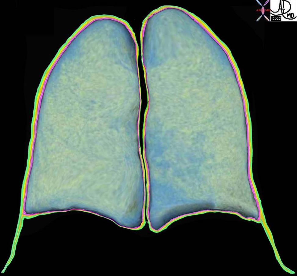
Pleura
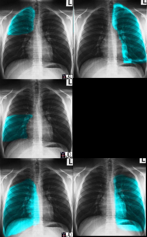
Lung Parts Relevant to the CXR
Heart Frontal View

If we were to “crack open” the chest of the chest X-ray, the structures that would dominate this bloody, black and white scene, would be the right sided chambers. The right ventricle (RV) would be the dominant anterior chamber, and would form the dominant interface with the diaphragm. The right atrium (RA) would form the border with the right lung. The RA would of course be slightly posterior to the RV. The left border would be formed by the left ventricle. Most the left ventricle is hidden posteriorly in this view. The left anterior descending artery would be visible from this anterior view. It marks the position of the interventricular septum.
Ashley Davidoff MD
SCALES
- 4 Review Spaces relating to 4 areas of major disease
- Pleura – PTX and Effusions
- Lungs – Pneumonia and Masses
- Hila – Masses and CHF
- Heart – Megalies and Failure
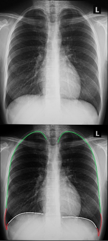
First Scale – Pleural Run
Ashley Davidoff MD
Pattern – start at the white lines to the right and left of the thoracic vertebra running along the diaphragms into the pleural recesses, up the lateral walls to the apices
2nd Scale – Lung Loops

Ashley Davidoff MD
- Pattern –
-
- Come down the trachea and and
- loop the upper lung fields, then the
- mid lung fields, and finally the
- lower lung fields
- looking for
- symmetry,
- masses,
- infiltrates,
- interstitial changes
-
3rd Scale – Skiing on the Moguls
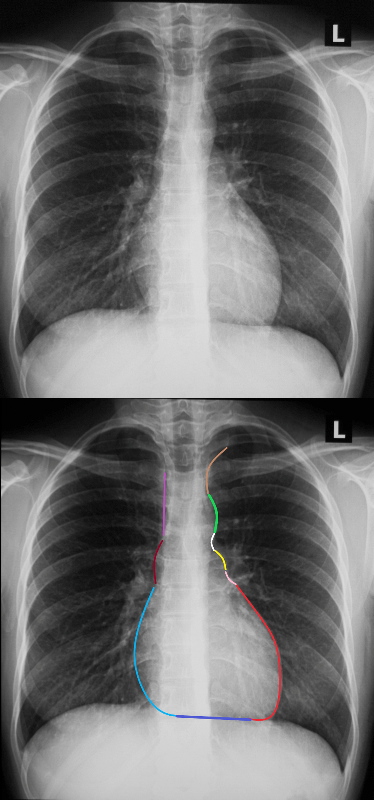
A methodical approach to evaluation of the cardiac silhouette is likened to skiing down a mogul laden ski slope and then taking a trip on the ski lift back to the top of the mountain. The ski slope starts at the left subclavian artery (light brown)
, followed by the mogul of the aortic knob (bright green) at the bottom of which is the A-P window (white) only to be presented with a second mogul of the main pulmonary artery(yellow), and then the bay of the left atrial appendage (pink)and finally free at last of moguls and an exciting and accelerating ski down the LV (red)
We then have to take a walk back to the ski lift. At the junction of the LV (red) and the RV (blue) , if we take a right ward look up the mountain we can spot the LAD on top of the interventricular septum.
The walk along the border of the RV is terminated at the junction of the RV (dark blue) and the right atrium (light blue). At this point we wait in line to get on to the ski lift. We ride up the right hand border of the right atrium (light blue) a little rough bump over the ascending aorta )maroon) and then straight to the top along the SVC (pink).
Ashley Davidoff MD
- Pattern of the Ski Run –
- Start at the left apex at the left subclavian artery,
- jump the aortic mogul and
- land in the AP window, and then
- jump the PA mogul,
- land in the LA appendiceal bay, and then
- ski all the way down the LV.
- Walk back to the ski lift via the inferior border of the RV and then the RA.
- At the bottom of the RA get on to the ski lift as the IVC enters the RA
- and then take the lift up the mountain via the RA ,
- portion of the ascending aorta and the
- the SVC
4th Scale – Hilar Hoops
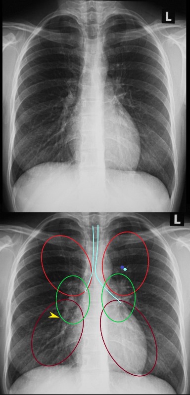
Down the airways for the carinal angle
Upper vs Lower Vessels
Artery vs Bronchus
Middle – Hilar Size and Shape
Lower – Descending Bronchovascular Bundle and specifically RPA yellow arrow
Ashley Davidoff MD
- Pattern of the Hilar Hoops –
-
- Trip down the trachea – Specifically to look at the carinal angle
- Upper hoop specifically to look at pa- bronchus ratio
- Middle Hoop – Specifically to look at hilar size and shape
- Lower Hoop – Specifically to look for redistribution and clarity of RPA
-
References and Links
 THE ART OF PERFECTING DANCE
Ashley Davidoff
TIGER WOODS AND TOM BRADY
NOTES SCALES AND MUSIC IN THE PURSUIT OF PERFECTION
Both images are in the public domain

