
NB
usually indistinguishable from symmetric HCM
can be asymmetric
LGE
mostly in the basal inferolateral LV wall
mostly mid myocardial (mesocardial)
subendocardium mostly spared
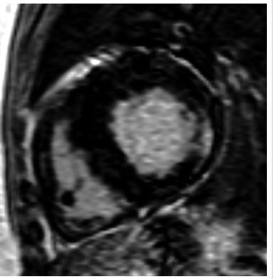
“Delayed enhanced images of 64-year-old heterozygotic woman with Fabry’s disease?related cardiac hypertrophy. Minimal thinning of basal segment of left ventricular inferolateral wall is associated with thick mesocardial striae of delayed enhancement shown on short-axis delayed enhanced images.”
De Cobelli et al Delayed-Enhanced Cardiac MRI for Differentiation of Fabry’s Disease from Symmetric Hypertrophic Cardiomyopathy
AJR Volume 192, Issue 3 2009
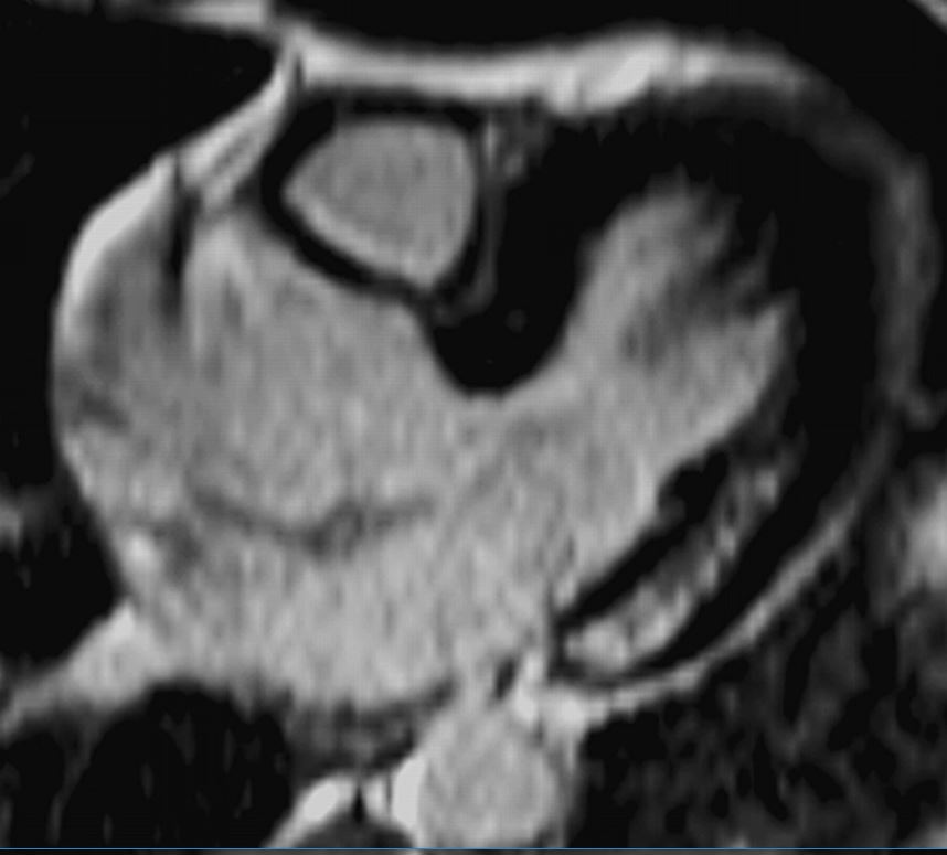
“40-year-old man with Fabry’s disease?related hypertrophy. Long-axis delayed enhanced images show typical pattern of delayed enhancement: thick striae involving inferolateral wall of basal segment of left ventricle in mesocardial distribution. Note sparing of subendocardial layer.
De Cobelli et al Delayed-Enhanced Cardiac MRI for Differentiation of Fabry’s Disease from Symmetric Hypertrophic Cardiomyopathy”
AJR Volume 192, Issue 3 2009
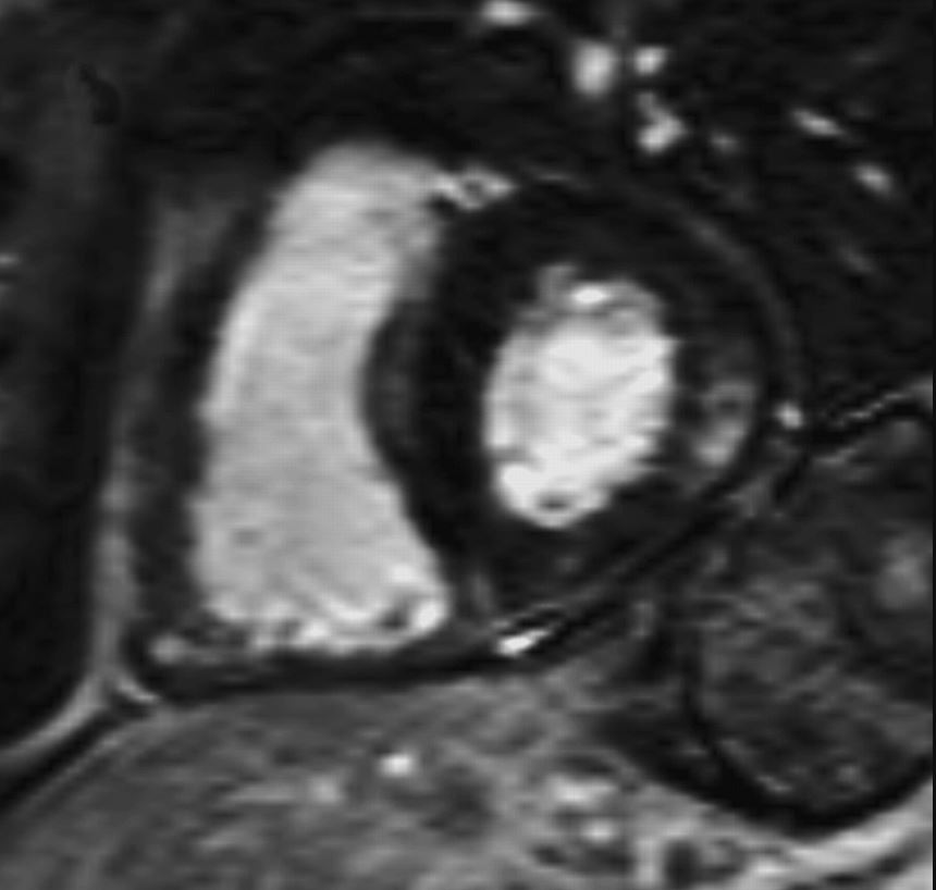
“40-year-old man with Fabry’s disease?related hypertrophy. Short -axis delayed enhanced images show typical pattern of delayed enhancement: thick striae involving inferolateral wall of basal segment of left ventricle in mesocardial distribution. Note sparing of subendocardial layer.”
De Cobelli et al Delayed-Enhanced Cardiac MRI for Differentiation of Fabry’s Disease from Symmetric Hypertrophic Cardiomyopathy”
AJR Volume 192, Issue 3 2009
need endomyocardial biopsy
Read More: https://www.ajronline.org/doi/10.2214/AJR.08.1201
References and Links
American Journal of Roentgenology Volume 192, Issue 3 2009
- TCV