SARCOID CARDIOMYOPATHY, HEART BLOCK
34-year-old male who was diagnosed with pulmonary sarcoidosis 1 year before, presents with progressive dyspnea over 6 months, previously able to run 5 miles per day and currently limited to walking short distances. Clinical evaluation revealed that he was in complete heart block with a heart rate of 35 /min. EKG showed a rate of 40 with AV dissociation with alternating junctional and ventricular escape rhythms.
CXR
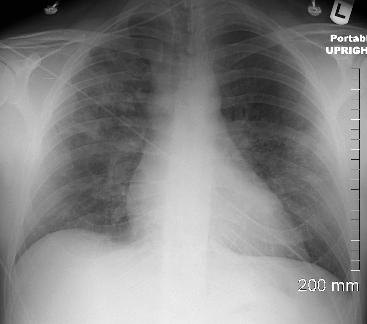
CXR shows upper lobe predominance interstitial changes, more prominent on the right side. There is evidence of CHF with enlargement of the LA (widening of the carinal angle) and cephalization of the vessels

CXR shows upper lobe predominance interstitial changes, slightly more prominent on the right side. There is evidence of CHF with enlargement of the LA (widening of the carinal angle) and cephalization of the vessels
Ashley Davidoff MD

CXR shows upper lobe predominance interstitial changes, slightly more prominent on the right side. There is evidence of CHF with enlargement of the LA (widening of the carinal angle) and cephalization of the vessels
Ashley Davidoff MD
An echocardiogram was normal with normal ejection fraction.
MRI
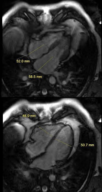
MRI revealed normal cardiac function, mild left atrial enlargement, and diffuse almost circumferential subendocardial enhancement of the LV, most prominent in the lateral wall. There was also a linear focus in the mid myocardium in the anterior region medially near the septum

MRI reveals normal cardiac function, mild left atrial enlargement, and diffuse almost circumferential subendocardial enhancement of the LV, most prominent in the lateral wall.
Ashley Davidoff MD

MRI reveals normal cardiac function, mild left atrial enlargement, and diffuse almost circumferential subendocardial enhancement of the LV, (red arrowheads) most prominent in the lateral wall. There was also a linear focus in the mid myocardium in the anterior region medially near the septum (green arrowheads)
Ashley Davidoff MD
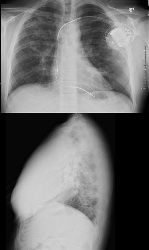
A dual lead pacemaker was placed and follow up CXR showed improved CHF an persistent interstitial findings dominant in the upper lobes consistent with sarcoidosis.

A dual lead pacemaker was placed and follow up CXR showed improved CHF and persistent interstitial findings dominant in the upper lobes consistent with sarcoidosis.
Ashley Davidoff MD
Follow up CXR 7 years later following shows a normal CXR. The pacemaker had been removed and the interstitial process had resolved

Follow up CXR 7 years later showed a normal CXR. The pacemaker had been removed and the interstitial process had resolved
Ashley Davidoff MD
