Normal thickness of the left ventricular myocardium is from 0.6 to 1.1 cm (as measured at the very end of diastole). If the myocardium is more than 1.1 cm thick, the diagnosis of LVH can be made.
The left ventricular free wall is thickest at the base and it
gradually thins towards the apex. At the tip of the apex
myocardium is 1?2 mm thick,
The interventricular septum increased from a median of 8.3 mm in the age group 20-29 to 11.2 mm in the group 60-70, whereas the posterior left ventricular wall increased from 7.5 mm to 9.8 mm.
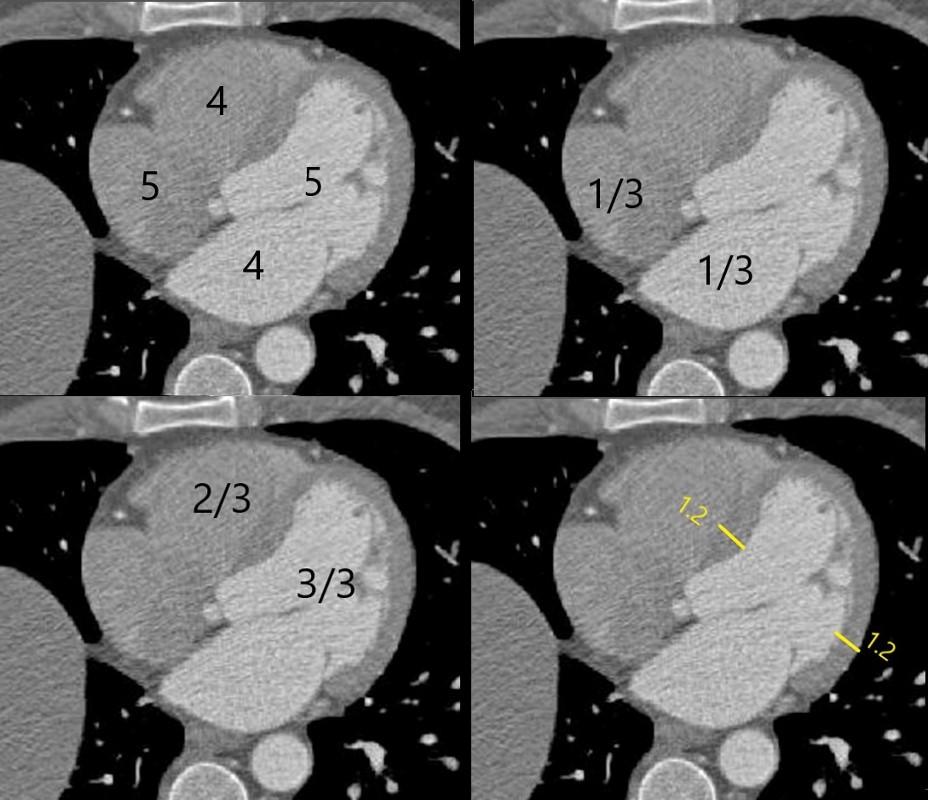
At the levelof the mitral valve which is also about mid septum, the chambers are relatively all best visualised. This is a place where approximate size can be evaluated
Ashley Davidoff MD
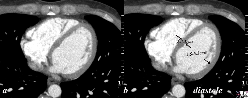
The gated CT scan was taken in diastole and shows the normal left ventricular dimensions which for the left ventricle wall is between .8 cms and 1.2 cms, and for the cavity is 4.5-5.5cms. the measurement should be taken at the level of the papillary muscles which is in mid ventricular cavity.
code
Courtesy Ashley Davidoff copyright 2018
34765c02.8s
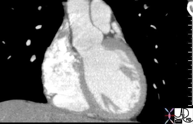
key words right atrium heart cardiac RA tricuspid valve TV left atrium LA MV mitralvalve RV right ventricle anterolateral papillary muscle interventricular septum left ventricle LV CTscan
Ashley Davidoff MD
34780
Applied Anatomy
Wall Thickness
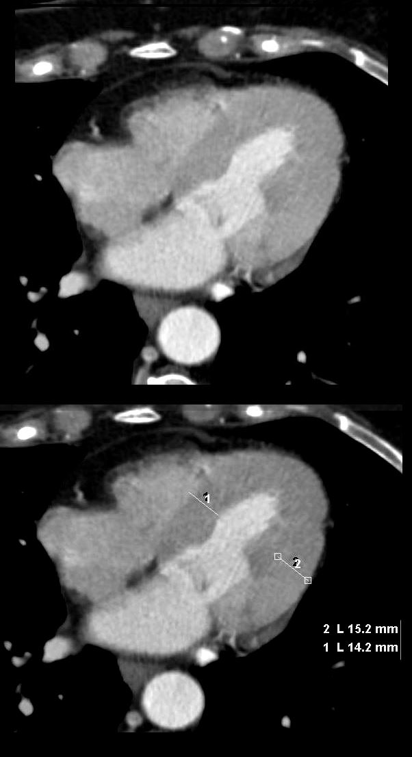
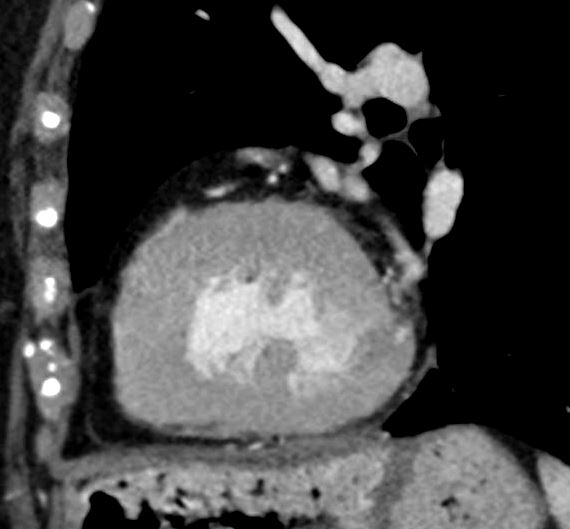
In diastole the septum measures 14mm and the free wall measures 15mm
(normal up to 12mm)
LVH-00b
tags concentric LVH
Ashley Davidoff MD
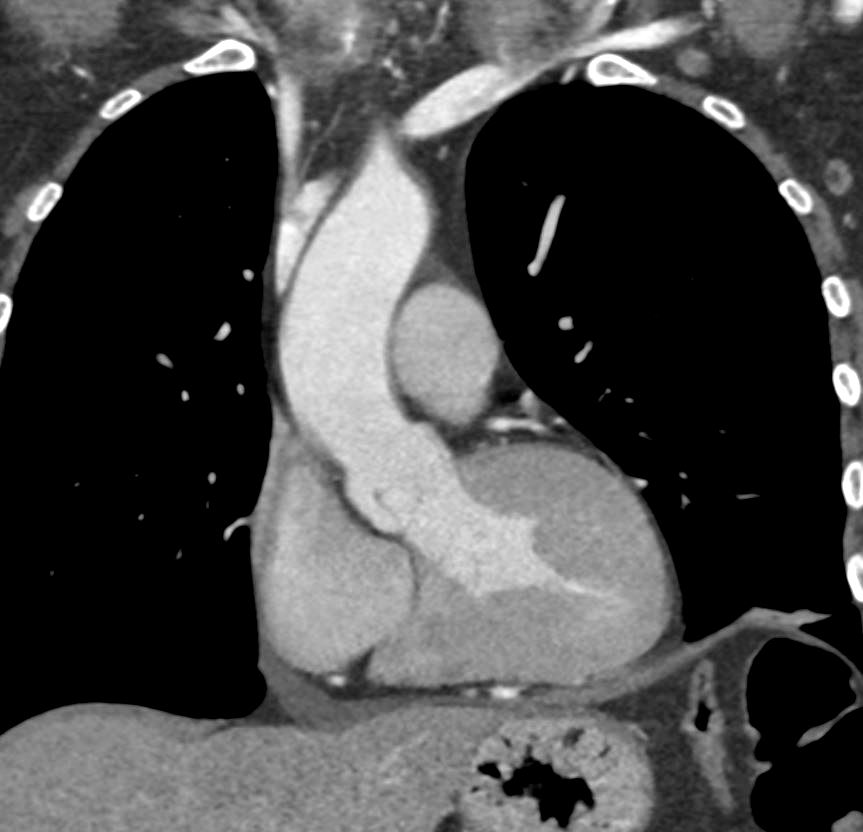
Coronal view in systole shows LVH and normal aortic valve
LVH-004b
Ashley Davidoff MD
LVH-003b
tags concentric hypertrophy LVH, hypertension
Ashley Davidoff MD
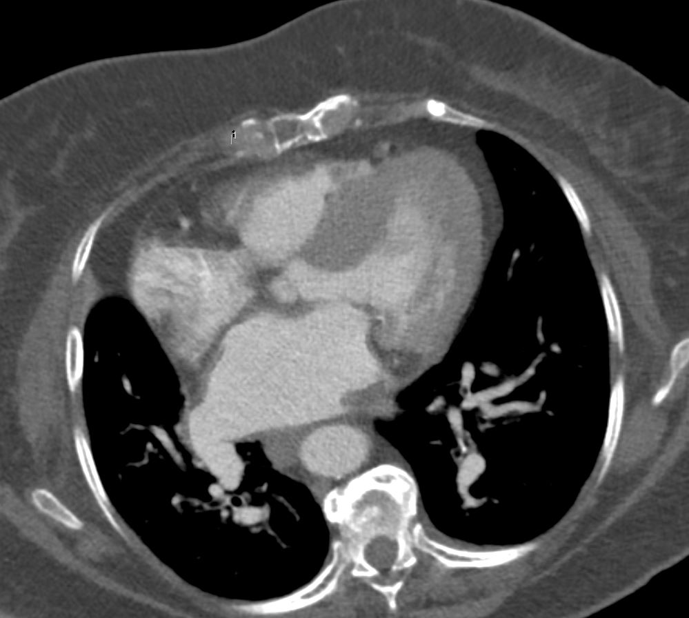
Asymmetric
Ashley Davidoff
tags asymmetric subaortic septum

Ashley Davidoff
tags asymmetric subaortic septum
96-LVH-002b
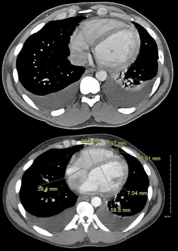
34-year-old male has a normal appearing CXR 1 year before presentation
At the time of his first presentation with dyspnea his CXR showed perihilar infiltrates.
A CT confirmed progressive alveolar edema, with bilateral effusions (right greater than left), mild left ventricular dilatation, Kerley B lines and centrilobular densities and small pericardial effusion.
Ashley Davidoff MD
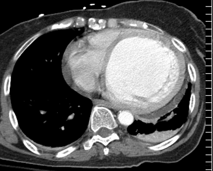
Recent MI
This elderly female recently sustained an acute MI and the resulting functional myocardial loss has resulted in a globular deformity of the heart as seen on this non gated CT through the chest. Note the malorientation of the posterior papillary muscle that prohibits normal function of the mitral valve and results in mitral regurgitation. A Small pericardial effusion is present.
Courtesy Ashley Davidoff MD copyright TCV 2008 75517
Canepa, M et al Distinguishing ventricular septal bulge versus hypertrophic cardiomyopathy in the elderly Heart. 2016 Jul 15; 102(14): 1087?1094.
