Venous Drainage
Copyright 2008
Ganesh Athappan MD Ashley Davidoff MD
Introduction
The nomenclature of the cardiac veins is confounding and it probably stems from the lack of sufficient attention to the anatomy of the cardiac veins in the past. Knowledge of the anatomy of the cardiac veins have become extremely important in recent years since it is imperative in the accurate and safe placement of pacing leads.Additional sources of confusion include the tendency to call the veins of the heart the coronary veins. ?Coronary veins? is infrequently used as a general term for the veins of the heart, but none of the veins is specifically named a coronary vein ie there is no such vein as the ?left coronary vein?for example. The use of the term ?coronary veins? for the heart is therefore discouraged, except for the use of coronary sinus which is universally accepted.
The coronary vein aka the left gastric vein, has also been a source of confusion since it has nothing to do with the heart and is named as such because it is shaped like a crown.
The Thebesian valve is a fenestrated membrane that is often present at the entrance of the coronary sinus into the right atrium
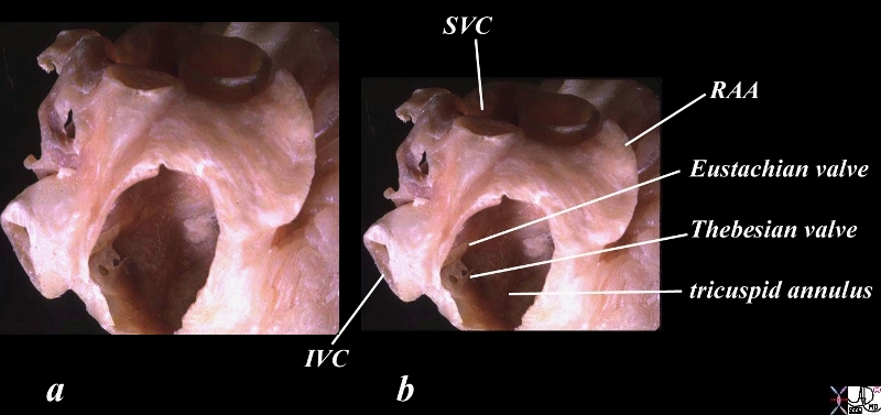
Thebesian Valves
The anterior wall has been removed In this waxed specimen created after distending the heart, allowing a window into the right atrial cavity and a focus on the fenestrated Thebesian valve which marks the entrance of the coronary sinus into the right atrium. The coronary sinus entrance lies just anterior and inferior to the Eustachian valve and the IVC, and just posterior to the tricuspid apparatus. right atrial appendage = RAA
Courtesy Ashley Davidoff TheCommonVein.net
06412cc02.81s
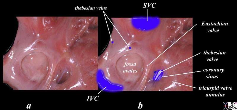
The right atrium has been opened in this normal heart in order to demonstrate the structure of the opening of the coronary sinus into the right atrium. The entrance of the SVC and IVC define superior and inferior and represent the right atrial side of the heart. The tricuspid annulus represents the anterior and leftward position of the entrance to the right ventricle (not shown). The coronary sinus is guarded by a fine mebrane called the thebesian valve and is bordered by the Eustachian valve of the IVC, the tricuspid annulus, and the A-V node (not shown. Two small thebesian veins enable venous return directly into the right atrium from heart muscle.
Courtesy Ashley Davidoff TheCommonVein.net
08452bb01c04b06.8s

The veins travel with the arteries and have been interpolated in this post mortem angiogram where the arteries are filled with barium. Images a and b represent an anterior view of the heart. The anterior interventricular vein drains into the great cardiac vein which runs with the circumflex coronary artery in the A-V groove. It collects the marginal veins of the LV including the left marginal vein. The great cardiac vein proceeds posteriorly in the A-V groove and enters the coronary sinus. (c,d)
Courtesy Ashley Davidoff TheCommonVein.net
15009d02c05b04.8s

The normal heart is displayed in the A-P projection (a,b) and posterior projection (c,d)
The anterior venous circulation of the right ventricle is small with many branches either draining into the small cardiac vein or into the Thebesian veins. In the diagram these veins are implied by overlay and cannot all be visualized.
The posterior view code heart (c,d) shows the entry of the small cardiac vein into the coronary sinus. It also shows the posterior tributaries of the right sided circulation including the middle cardiac vein and posterior left ventricular branches.
15009d02c11.81s Courtesy Ashley Davidoff TheCommonVein.net

The major veins of the heart have been overlaid and implied in this posterior view of a normal post mortem specimen arteriogram where barium has been instilled into the arteries. All the veins of the heart (except for those that drain directly into the myocardium) drain into the coronary sinus. There are three major veins. The great cardiac vein drains the territory of the LAD and circumflex, the middle cardiac vein drains the region of the PDA and other posterior ventricular vessels, and the small cardiac vein drains the anterior wall of the right ventricle and right atrium.
Courtesy Ashley Davidoff TheCommonVein.net
15009d02e04.8s
Coronary Sinus
The coronary sinus is the final common pathway of the veins of the heart, It empties into the right atrium at the entrance of the IVC into the heart.
The coronary sinus is formed by three major veins of the heart, the great, middle and small cardiac veins. It is approximately 2.5 cms in length and lies in the atrioventricular groove on the inferior wall of the heart, anterior to the atria, and posterior to the ventricles. It is formed behind the left atrium as a continuation of the great cardiac vein and proceeds right ward to enter the right atrium, covered by muscular fibers of the left atrium.
The Coronary Sinus
The coronary sinus enters the right atrium, anterior to the IVC, and just above and posterior to the septal leaflet of the tricuspid valve. Both the coronary sinus, a vein, and the circumflex coronary artery travel in the same direction in the AV groove ? a somewhat rare instance in the body.

Posterior and Inferior Aspect of the Heart
The heart specimen is viewed from its posterior aspect revealing the opened coronary sinus which lies in the A-V groove between the atria and ventricles. The blue arrow points to the coronary sinus and also to the direction of flow- from left to right, in the same direction as the distal left circumflex artery. Interestingly it is a rare instance where blood in the vein and artery of an organ are flowing in the same direction.
Courtesy Ashley Davidoff TheCommonVein.net
08423b02c03.8sb02
Relation of the
Coronary Sinus to the Right Atrium and Tricuspid Valve
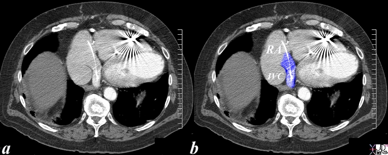
The axial CTscan demonstrates a pacing wire in the coronary sinus (blue overlay) The coronary sinus The coronary sinus is anterior and medial to the entrance of the IVC and posterior to the tricuspid annulus.
Courtesy Ashley Davidoff TheCommonVein.net 87579c02.8s
Normal Eustachian Valve in the Coronary Sinus

Linear filling defect in the coronary sinus reflecting the normal Eustachian valve
Ashley Davidoff MD TheCommonVein.net 136487
Interventricular Veins

This axial acquired CTscan of the heart shows a normal left main coronary artery dividing into a left anterior descending coronary artery and a smaller circumflex vessel. Note the accompanying great (anterior) cardiac vein that courses to the left of the left atrium as it heads towards the coronary sinus.
key words cardiac heart artery vein coronary normal anatomy LAD circumflex LM
Courtesy Ashley Davidoff TheCommonVein.net
39117
The Anterior Left Sided Venous Circulation

The axial images of two different patients reveal the left cardiac venous system including the anterior interventricular cardiac vein draining into the great cardiac vein (a,b). In images c and d the left marginal vein is seen to drain into the great cardiac vein which is now on the posterior margin of the A-V groove and which finally drains into the coronary sinus
47814c04.8s
Courtesy Ashley Davidoff TheCommonVein.net
Middle Cardiac vein
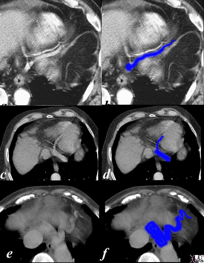
The axial images reflect the branches that drain the posterior venous circulation, though only the middle cardiac vein is shown. The small cardiac vein is difficult to visualize on axial images – possibly because it is too small or that most venous drainage for that territory drains directly into the right atrium and right ventricle. In images a and b the middle cardiac vein is seen traveling with the PDA. In images c and d the middle cardiac vein is seen draining into the coronary sinus. A hint of a small cardiac vein is noted but the distinction between the artery and vein is difficult. In images e and f, the middle cardiac vein is significantly enlarged due to right heart failure.
47814c07.8s Courtesy Ashley Davidoff TheCommonVein.net
Tributaries of the Coronary Sinus
The three major tributaries of the coronary sinus are;
a) great cardiac vein that follows the course of the LAD and circumflex territory
b) middle cardiac vein which follows the course of the PDA and its territory
c) small cardiac vein which drains the remaining portions of the RCA territory
The left sided arterial system has two main vessels (LAD and circumflex) and the right has only one named vessel ? the right coronary artery. Oddly in the venous system, the left only has one named major vessel ? the great cardiac vein and the right has two ? middle and small cardiac vein. Additionally bothte left and right systems drain into one common vein ? the coronary sinus
Left Sided Venous System of the Heart
The main left sided vein is the great cardiac vein which travels in the atrioventricular groove and it is formed by the anterior interventricular vein which travels with the LAD, and the left marginal vein which travels with the obtuse marginal vein (s)
Great Cardiac Vein
The great cardiac vein begins at the cardiac apex and ascends along the anterior interventricular groove progressing to the base of the ventricles as the anterior inter ventricular vein. At the coronary groove it turns to the left to form the base of the triangle of “Brocq and Mouchet ?with the two branches of the left coronary artery; the left anterior descending and the left circumflex arteries. The triangle is traversed by the diagonal branch of the LAD. The great cardiac vein runs along the coronary grove with the left circumflex to reach the posterior surface of the heart where it terminates by opening into the left extremity of the coronary sinus . (This is a rare occurrence of arterial and venous flow in the same direction ? in the coronary groove). The opening of the great cardiac vein is guarded by the valve of Vieussens.
The Right Sided Cardiac Veins
The right cardiac venous system drains into the coronary sinus via two major tributaries; the middle cardiac vein and the small cardiac vein. The middle cardiac vein drains the posterior interventricular system (PDA territory) and also the posterior left ventricular veins. The small cardiac vein drains the conal veins, anterior veins of the RV and the right marginal vein. Smaller tributaries of both the right ventricle and right atrium drain directly via Thebesian veins into the RA and RV cavity
Middle cardiac Vein (Posterior interventricular vein)
The middle cardiac vein begins at the cardiac apex and runs upwards in the posterior interventricular groove to reach the right extremity of the coronary sinus. The great cardiac vein and the middle cardiac vein are the two most consistent tributaries of the coronary sinus.
Small Cardiac Vein
The small cardiac vein runs in the coronary groove between the right atrium and ventricle. It drains the posterior surface of the right atrium and ventricle and opens into the right extremity of the coronary sinus. The middle cardiac and small cardiac vein drains most of the RCA territory. The right marginal vein ascends along the right margin of the heart and joins the small cardiac vein in the coronary sulcus, or opens directly into the right atrium.
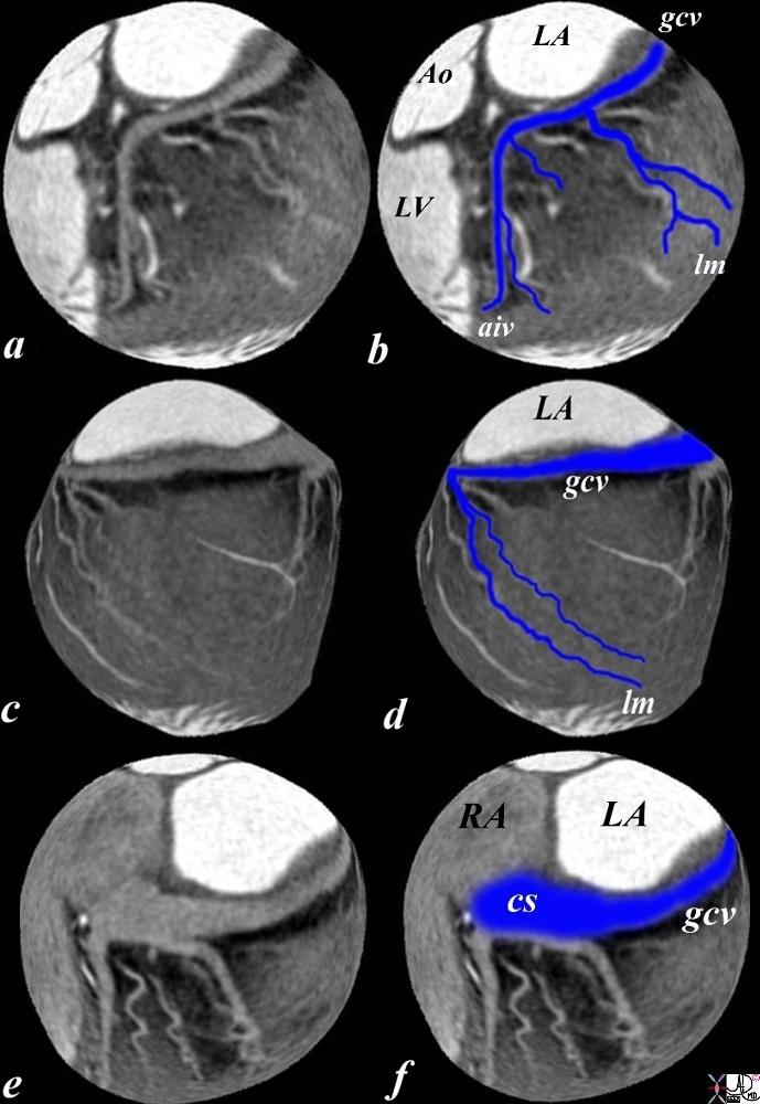
The CTA was tailored to evaluate the veins of the heart, and the following images were rendered in a sphere accounting for the distortion. This series represent the veins draining the LCA, and are viewed in frontal (a,b), lateral (c,d), and posterior views (e,f). Images a and b show the anterior interventricular vein which drains the septum anteriorly, together with the left marginal vein to form the great cardiac vein (gcv). Images c and d are a lateral view of the heart (anterior to your left) showing the left marginal vein joining the great cardiac vein in the atrioventricular groove. The last set of images show the great cardiac vein coursing on the posterior rim of the AV groove and joining the coronary sinus
47814c02.8s Courtesy Ashley Davidoff TheCommonVein.net

The posterior aspect of the heart has been oriented the way it is viewed in CT scan (as if you were looking from the feet) unlike the classicial anatomical orientation. This is by convention and is confusing. The continuation of the great cardiac vein in the left AV groove is shown as it proceeds posteriorly to enter the coronary sinus. (cs) It receives the middle cardiac vein which drains the interventricular septum from its inferior and posterior aspect. It also in this case receives posterior left ventricular (post LV) branches.
Courtesy Ashley Davidoff TheCommonVein.net 47809c06bb02b03.8s
The endocardium and innermost myocardium are drained by the least cardiac (Thebesian) veins which drain directly into the left atrium and left ventricle. Schuenke
Other Smaller Veins
?The Posterior Vein of the Left Ventricle courses along the diaphragmatic surface of the left ventricle to the coronary sinus, but may end in the great cardiac vein.
Anterior cardiac veins begin over the anterior surface of the right ventricle cross the coronary groove and end directly in the right atrium or by joining the small cardiac vein.
Smallest cardiac veins or The Thebesian veins are minute vessels that begin in capillary beds of the myocardium and open directly into the chambers of the heart .
The oblique vein of the left atrium (Marshall?s vein) descends obliquely on the back of the left atrium joins the coronary sinus at its left extremity. The vein represents the embryonic remnants of the left SVC.
Clinical considerations :
The coronary sinus enlarges with elevation of the right sided pressures and also with a persistent left sided superior vena cava which enters into the coronary sinus via a patent vein of Marshall
It has as mentioned significance to eletrophysiologists since it the rout that is used to place left ventricular epicardial leads for cardiac resynchronization therapy.
The coronary sinus (CS) has also been used in electrophysiological studies for ablation of the postero-septal and left-sided accessory pathways.
Clinical considerations :
coronary sinus is the site for left ventricular (LV) epicardial lead placement for cardiac resynchronization therapy.
The coronary sinus (CS) has been used in electrophysiological studies for ablation of the postero-septal and left-sided accessory pathways.
Dilatation of the MVA primarily affects its postero-lateral aspect, which is related to the coronary sinus.
The coronary venous anatomy as described has a high degree of variability which makes it unsuitable for electrophysiologists, mandating a segmental approach to coronary venous classification which will be subsequently described in later sections of the heart module .
“Initially described in 1706 by Vieussens and further elaborated by Thebesius in 1708, thebesian veins are small, valveless venous channels that provide direct connections between the coronary arteries or the coronary venous system and the chambers of the heart. They are thought to be more prevalent in the right atrium and the right ventricle. It has been proposed that thebesian veins provide an alternative channel of nutrition to the myocytes under certain conditions.”
References :
Maasarany SE, Ferret CG, Ferth A, Sheppard M, Henein MY. The coronary sinus conduit function . anatomical study . Europace 2005 7(5):475-481
http://education.yahoo.com/reference/gray/subjects/subject/166
Singh JP, Houser H, Heist EK, Ruskin JN. The Coronary Venous Anatomy A Segmental Approach to Aid Cardiac Resynchronization Therapy. JACC 2005;46(1):68-74
