CT scan is not used routinely for imaging of the heart because it is not real time, is expensive, requires gating for accurate measurements, requires contrast injection, and involves radiation exposure. On the other hand it is used routinely for examinations of the chest, and although this examination is not tailored for cardiac evaluation, evaluation of the size and an estimate of the thickness of component structures is possible.
Volume measurements and wall thickness measurements requires a diastolic frame in order to standardize the measurement and also because there is thickening of the myocardium during systole as it shortens. This requires a gated study in order to determine the phase of the heart
However the size of the atria and to some extent the ventricles can be fairly accurately and subjectively assessed often by reviewing the shape of the chambers.
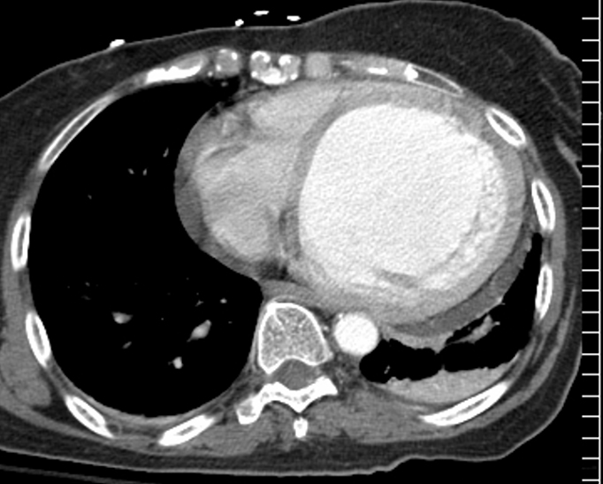
Comparing the Normal RV inflow with the Normal Left Ventricular Size
The axial gated CT scan through the right and left ventricle at end diastole shows the normal size and shape of the right ventricular inflow and left ventricle. The right ventricular inflow looks smaller than the LV in volume in this view since essentially it makes up for the volume in its second “floor” which sits more cranially as the right ventricular outflow tract. The left ventricle only has a single level or floor. Thus in this view the RV looks and measures smaller then the LV. Note also that the apex of the left ventricle protrude slightly more anteriorly than the RV even though it is the posterior ventricle, because it is the chamber that forms the apex of the heart. The septum also bulges toward the right ventricle due to the higher pressure in the left ventricle.
Courtesy Ashley DAvidoff MD copyright 2009 all rights reserved 37758b01c01.8s
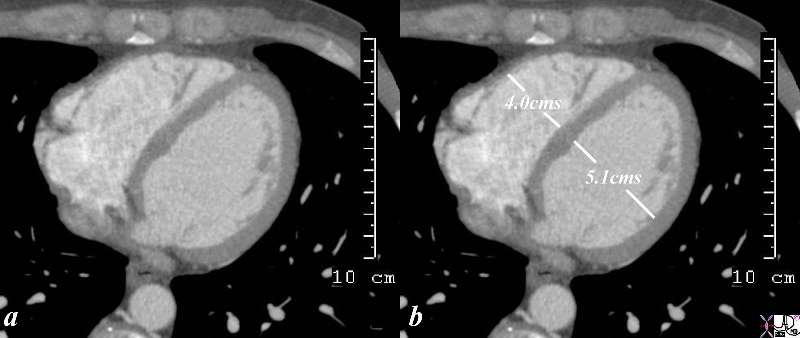
Dilated Cardiomyopathy
This elderly patient had sustained a cardiac arrest just 24 hours prior to the examination and the CT shows a bulbous and dilated left ventricle. Both the shape and the size of the LV cavity are clues to the enlargement of this heart. A small posterior pericardial effusion is present.
DOMElement Object
(
    [schemaTypeInfo] =>
    [tagName] => table
    [firstElementChild] => (object value omitted)
    [lastElementChild] => (object value omitted)
    [childElementCount] => 1
    [previousElementSibling] => (object value omitted)
    [nextElementSibling] => (object value omitted)
    [nodeName] => table
    [nodeValue] =>


Dilated Cardiomyopathy


This elderly patient had sustained a cardiac arrest just 24 hours prior to the examination and the  CT  shows a bulbous and dilated left ventricle.  Both the shape and the size of the LV cavity are clues to the enlargement of this heart.  A small posterior pericardial effusion is present.
Courtesy Ashley Davidoff MD copyright TCV 2008 75517


    [nodeType] => 1
    [parentNode] => (object value omitted)
    [childNodes] => (object value omitted)
    [firstChild] => (object value omitted)
    [lastChild] => (object value omitted)
    [previousSibling] => (object value omitted)
    [nextSibling] => (object value omitted)
    [attributes] => (object value omitted)
    [ownerDocument] => (object value omitted)
    [namespaceURI] =>
    [prefix] =>
    [localName] => table
    [baseURI] =>
    [textContent] =>


Dilated Cardiomyopathy


This elderly patient had sustained a cardiac arrest just 24 hours prior to the examination and the  CT  shows a bulbous and dilated left ventricle.  Both the shape and the size of the LV cavity are clues to the enlargement of this heart.  A small posterior pericardial effusion is present.
Courtesy Ashley Davidoff MD copyright TCV 2008 75517


)
DOMElement Object
(
    [schemaTypeInfo] =>
    [tagName] => td
    [firstElementChild] => (object value omitted)
    [lastElementChild] => (object value omitted)
    [childElementCount] => 2
    [previousElementSibling] =>
    [nextElementSibling] =>
    [nodeName] => td
    [nodeValue] =>
This elderly patient had sustained a cardiac arrest just 24 hours prior to the examination and the  CT  shows a bulbous and dilated left ventricle.  Both the shape and the size of the LV cavity are clues to the enlargement of this heart.  A small posterior pericardial effusion is present.
Courtesy Ashley Davidoff MD copyright TCV 2008 75517

    [nodeType] => 1
    [parentNode] => (object value omitted)
    [childNodes] => (object value omitted)
    [firstChild] => (object value omitted)
    [lastChild] => (object value omitted)
    [previousSibling] => (object value omitted)
    [nextSibling] => (object value omitted)
    [attributes] => (object value omitted)
    [ownerDocument] => (object value omitted)
    [namespaceURI] =>
    [prefix] =>
    [localName] => td
    [baseURI] =>
    [textContent] =>
This elderly patient had sustained a cardiac arrest just 24 hours prior to the examination and the  CT  shows a bulbous and dilated left ventricle.  Both the shape and the size of the LV cavity are clues to the enlargement of this heart.  A small posterior pericardial effusion is present.
Courtesy Ashley Davidoff MD copyright TCV 2008 75517

)
DOMElement Object
(
    [schemaTypeInfo] =>
    [tagName] => td
    [firstElementChild] => (object value omitted)
    [lastElementChild] => (object value omitted)
    [childElementCount] => 2
    [previousElementSibling] =>
    [nextElementSibling] =>
    [nodeName] => td
    [nodeValue] =>
Dilated Cardiomyopathy

    [nodeType] => 1
    [parentNode] => (object value omitted)
    [childNodes] => (object value omitted)
    [firstChild] => (object value omitted)
    [lastChild] => (object value omitted)
    [previousSibling] => (object value omitted)
    [nextSibling] => (object value omitted)
    [attributes] => (object value omitted)
    [ownerDocument] => (object value omitted)
    [namespaceURI] =>
    [prefix] =>
    [localName] => td
    [baseURI] =>
    [textContent] =>
Dilated Cardiomyopathy

)
DOMElement Object
(
    [schemaTypeInfo] =>
    [tagName] => table
    [firstElementChild] => (object value omitted)
    [lastElementChild] => (object value omitted)
    [childElementCount] => 1
    [previousElementSibling] => (object value omitted)
    [nextElementSibling] => (object value omitted)
    [nodeName] => table
    [nodeValue] =>


Comparing the Normal RV inflow with the Normal Left Ventricular Size


The axial gated CT scan through the right and left ventricle at end diastole shows the normal size and shape of the right ventricular inflow and left ventricle. The right ventricular inflow looks smaller than the LV in volume in this view since essentially it makes up for the volume in its second “floor” which sits more cranially as the right ventricular outflow tract. The left ventricle only has a single level or floor. Thus in this view the RV looks and measures smaller then the LV. Note also that the apex of the left ventricle protrude slightly more anteriorly than the RV even though it is the posterior ventricle, because it is the chamber that  forms the apex of the heart. The septum also bulges toward the right ventricle due to the higher pressure in the left ventricle.
Courtesy Ashley DAvidoff MD copyright 2009 all rights reserved 37758b01c01.8s


    [nodeType] => 1
    [parentNode] => (object value omitted)
    [childNodes] => (object value omitted)
    [firstChild] => (object value omitted)
    [lastChild] => (object value omitted)
    [previousSibling] => (object value omitted)
    [nextSibling] => (object value omitted)
    [attributes] => (object value omitted)
    [ownerDocument] => (object value omitted)
    [namespaceURI] =>
    [prefix] =>
    [localName] => table
    [baseURI] =>
    [textContent] =>


Comparing the Normal RV inflow with the Normal Left Ventricular Size


The axial gated CT scan through the right and left ventricle at end diastole shows the normal size and shape of the right ventricular inflow and left ventricle. The right ventricular inflow looks smaller than the LV in volume in this view since essentially it makes up for the volume in its second “floor” which sits more cranially as the right ventricular outflow tract. The left ventricle only has a single level or floor. Thus in this view the RV looks and measures smaller then the LV. Note also that the apex of the left ventricle protrude slightly more anteriorly than the RV even though it is the posterior ventricle, because it is the chamber that  forms the apex of the heart. The septum also bulges toward the right ventricle due to the higher pressure in the left ventricle.
Courtesy Ashley DAvidoff MD copyright 2009 all rights reserved 37758b01c01.8s


)
DOMElement Object
(
    [schemaTypeInfo] =>
    [tagName] => td
    [firstElementChild] => (object value omitted)
    [lastElementChild] => (object value omitted)
    [childElementCount] => 2
    [previousElementSibling] =>
    [nextElementSibling] =>
    [nodeName] => td
    [nodeValue] =>
The axial gated CT scan through the right and left ventricle at end diastole shows the normal size and shape of the right ventricular inflow and left ventricle. The right ventricular inflow looks smaller than the LV in volume in this view since essentially it makes up for the volume in its second “floor” which sits more cranially as the right ventricular outflow tract. The left ventricle only has a single level or floor. Thus in this view the RV looks and measures smaller then the LV. Note also that the apex of the left ventricle protrude slightly more anteriorly than the RV even though it is the posterior ventricle, because it is the chamber that  forms the apex of the heart. The septum also bulges toward the right ventricle due to the higher pressure in the left ventricle.
Courtesy Ashley DAvidoff MD copyright 2009 all rights reserved 37758b01c01.8s

    [nodeType] => 1
    [parentNode] => (object value omitted)
    [childNodes] => (object value omitted)
    [firstChild] => (object value omitted)
    [lastChild] => (object value omitted)
    [previousSibling] => (object value omitted)
    [nextSibling] => (object value omitted)
    [attributes] => (object value omitted)
    [ownerDocument] => (object value omitted)
    [namespaceURI] =>
    [prefix] =>
    [localName] => td
    [baseURI] =>
    [textContent] =>
The axial gated CT scan through the right and left ventricle at end diastole shows the normal size and shape of the right ventricular inflow and left ventricle. The right ventricular inflow looks smaller than the LV in volume in this view since essentially it makes up for the volume in its second “floor” which sits more cranially as the right ventricular outflow tract. The left ventricle only has a single level or floor. Thus in this view the RV looks and measures smaller then the LV. Note also that the apex of the left ventricle protrude slightly more anteriorly than the RV even though it is the posterior ventricle, because it is the chamber that  forms the apex of the heart. The septum also bulges toward the right ventricle due to the higher pressure in the left ventricle.
Courtesy Ashley DAvidoff MD copyright 2009 all rights reserved 37758b01c01.8s

)
DOMElement Object
(
    [schemaTypeInfo] =>
    [tagName] => td
    [firstElementChild] => (object value omitted)
    [lastElementChild] => (object value omitted)
    [childElementCount] => 2
    [previousElementSibling] =>
    [nextElementSibling] =>
    [nodeName] => td
    [nodeValue] =>

Comparing the Normal RV inflow with the Normal Left Ventricular Size
    [nodeType] => 1
    [parentNode] => (object value omitted)
    [childNodes] => (object value omitted)
    [firstChild] => (object value omitted)
    [lastChild] => (object value omitted)
    [previousSibling] => (object value omitted)
    [nextSibling] => (object value omitted)
    [attributes] => (object value omitted)
    [ownerDocument] => (object value omitted)
    [namespaceURI] =>
    [prefix] =>
    [localName] => td
    [baseURI] =>
    [textContent] =>

Comparing the Normal RV inflow with the Normal Left Ventricular Size
)
