Acknowledging lecture of professor Hany Shawky Nadin from Ain Shams U Cairo Egypt
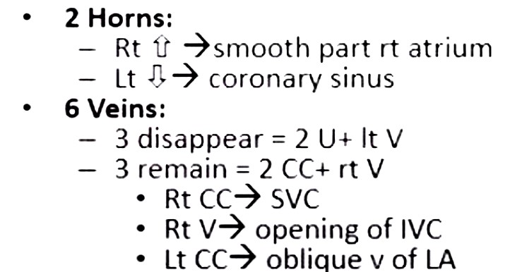
3 veins into common atrium
2 horns left horn becomes the sinus venosus and right horn becomes SVC and IVC
Common CArdinal Veins (anterior and post
Umbilical vein’from the placenta
Vitelline veins from yolk sac
The umbilical veins and left vitelline veins disappear
Acknowledging lecture of professor Hany Shawky Nadin from Ain Shams U Cairo Egypt
Acknowledging lecture of professor Hany Shawky Nadin from Ain Shams U Cairo Egypt
Oblique vein of Marshall
Acknowledging lecture of professor Hany Shawky Nadin from Ain Shams U Cairo Egypt
Cardinal Veins Vitelline Veins and Umbilical Veins

Cardianal System
2 systems connected by an oblique anastomosis
Vitelline System
Infra hepatic portion connected
Intrahepatic portion
left and right – will develop intosinusoid and connect with developing hepatocytes
Suprahepatic portion connect with both horns but will develop into suprahepatic IVC
Acknowledging lecture of professor Hany Shawky Nadin from Ain Shams U Cairo Egypt

2 systems connected by an oblique anastomosis
Left side resorbs and remains as the ligament of Marshall
Vitelline System
Infra hepatic evolves into SMV and splenic vein
Intrahepatic portion
left and right – will join and develop into sinusoids and connect with developing hepatocytes
Suprahepatic portion Left connects with right to form a common channel as the suprahepatic portion of the nepatic veins and join the IVC
Acknowledging lecture of professor Hany Shawky Nadin from Ain Shams U Cairo Egypt

2 systems connected by an oblique anastomosis
Left side resorbs and remains as the ligament of Marshall
Vitelline System
Infra hepatic evolves into SMV and splenic vein
Intrahepatic portion
left and right – will join and develop into sinusoids and connect with developing hepatocytes
Suprahepatic portion Left connects with right to form a common channel as the suprahepatic portion of the nepatic veins and join the IVC
Acknowledging lecture of professor Hany Shawky Nadin from Ain Shams U Cairo Egypt
