
The enlarged LV (a,b) is shaped like an oval and it is likened to a rugby ball or an American football placed on the field at kick off time. LVE on CXR is mostly assessed by an increased cardiothoracic ratio as well as the accentuation of the ovoid shape. (lower images c, d,e, f)
Ashley Davidoff MD
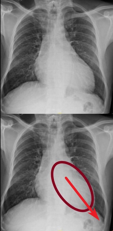
DOWN AND OUT
The left ventricle (LV) enlarges in a posterior, downward and lateral direction resulting in the characteristic changes of LVE on CXR
Ashley Davidoff MD

The enlarged LV (a,b) is shaped like an oval and it is likened to a rugby ball or an American football placed on the field at kick off time. LVE on CXR is mostly assessed by an increased cardiothoracic ratio as well as the accentuation of the ovoid shap. (lower images c, d,e, f)
Ashley Davidoff MD
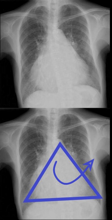
ANTERIOR LEFTWARD
The right ventricle (RV) enlarges with a clockwise rotation resulting in an upward turning of the apex and enlargement in a anterior and leftward lateral direction.
Ashley Davidoff MD
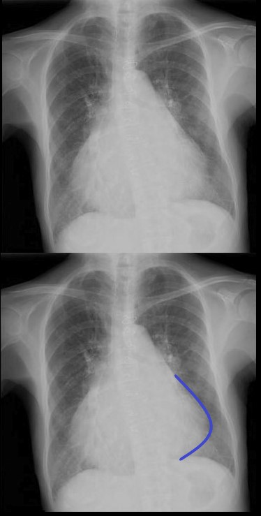
As the right ventricle (RV) enlarges with a clockwise rotation the apex of the small ventricle succumbs to the larger silhouette of the RV which pints upward and to the left and has been called the “proud breast” appearance.
Ashley Davidoff MD

Ashley Davidoff MD
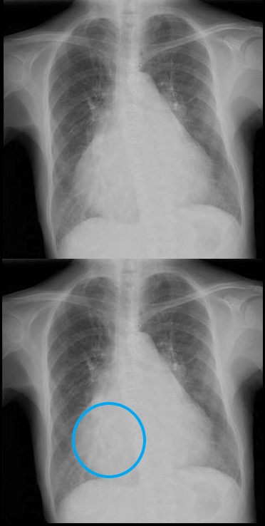
The right atrium is the most difficult chamber to assess unless it is very large in which case it will present on the frontal CXR with a very large right paravertebral border. This is a 71 year old female person with rheumatic heart disease with pulmonary hypertension and tricuspid regurgitation hence resulting in a large right atrium (RAE)
Ashley Davidoff MD
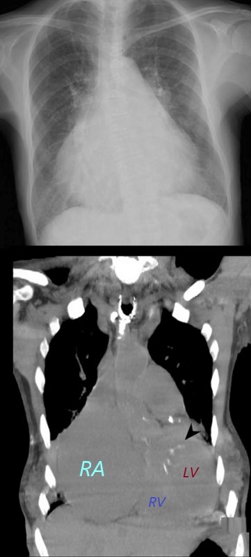
The right atrium is the most difficult chamber to assess unless it is very large in which case it will present on the frontal CXR with a very large right paravertebral border. The frontal CXR and coronal CT through the RA is from a 71 year old female with rheumatic heart disease with pulmonary hypertension and tricuspid regurgitation resulting in a giant right atrium (RAE). The RA accounts for the large bulge of the right border of the cardiac silhouette. The black arrowhead in the loer image points to the calcified mitral valve.
Ashley Davidoff MD
