Ashley Davidoff MD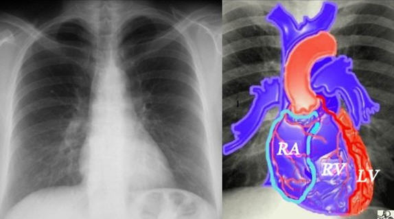
Ashley Davidoff MD
Ashley Davidoff MD
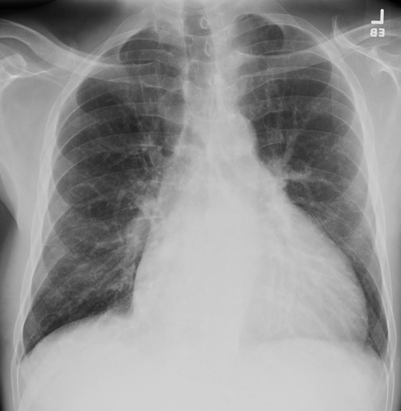
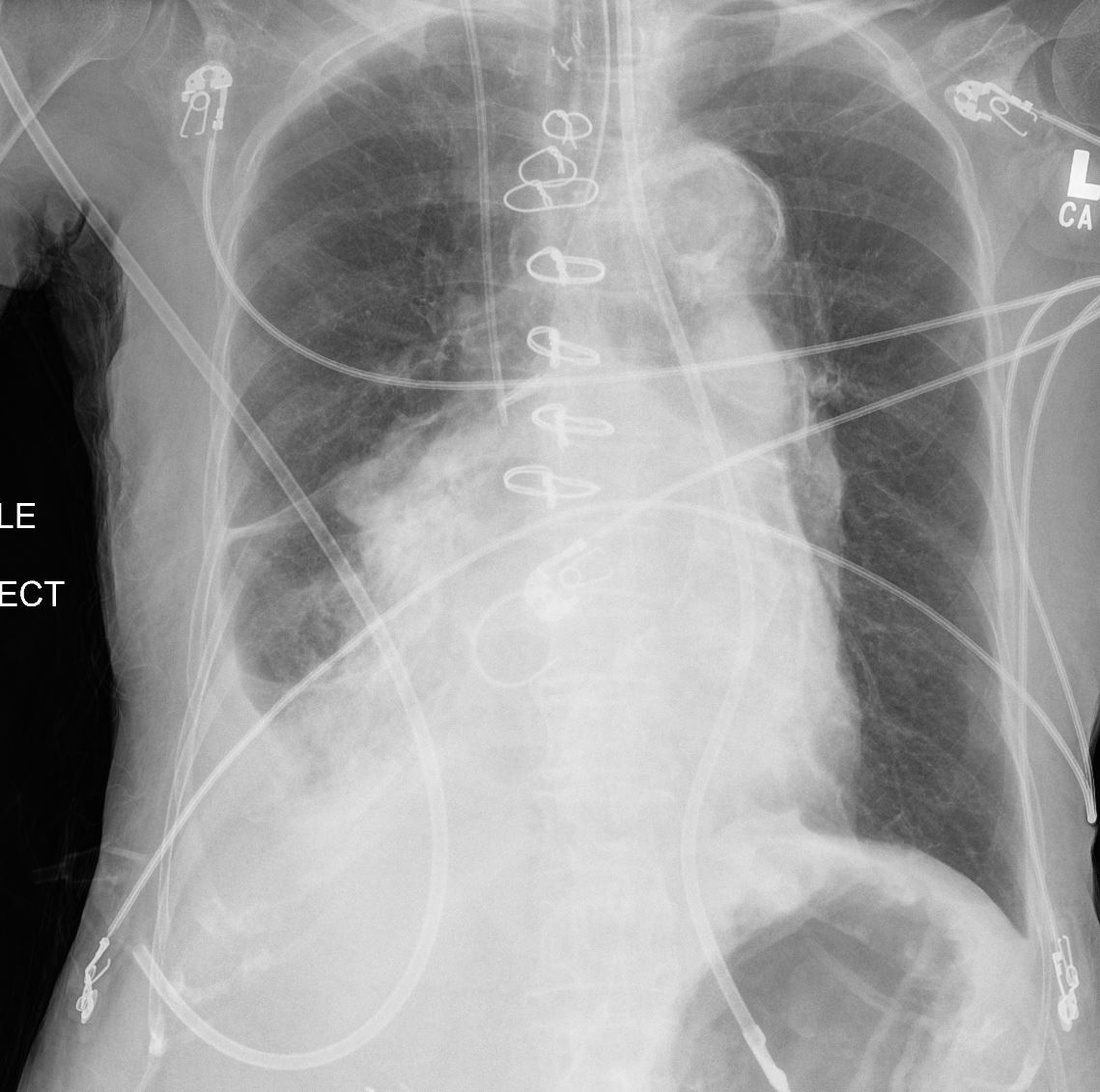
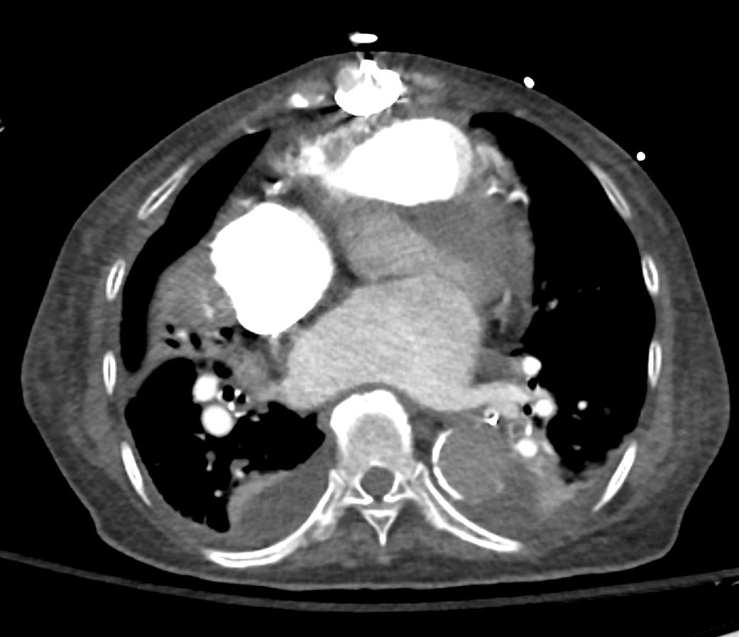
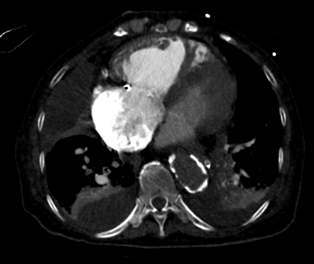
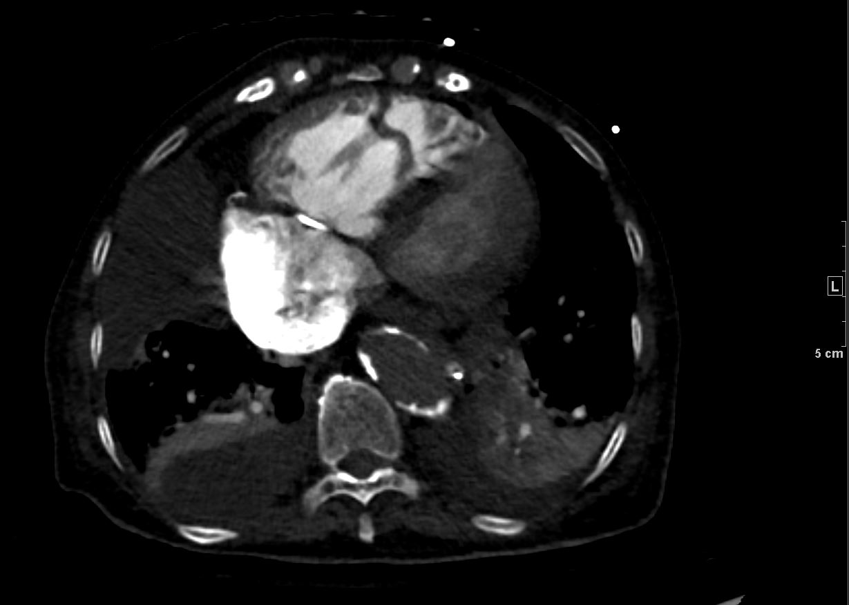
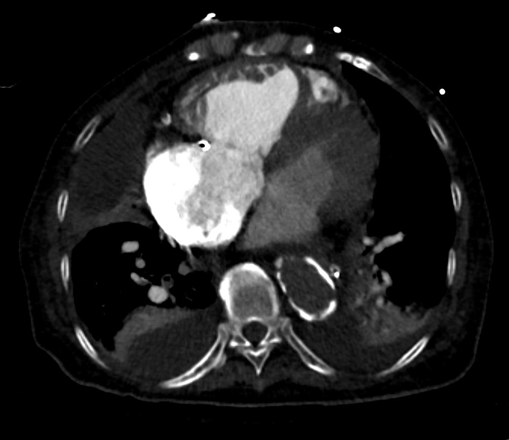
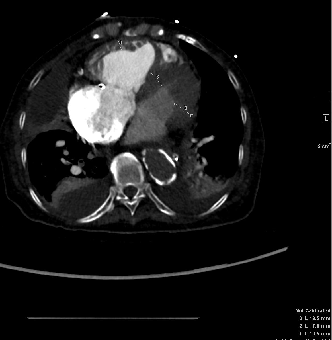
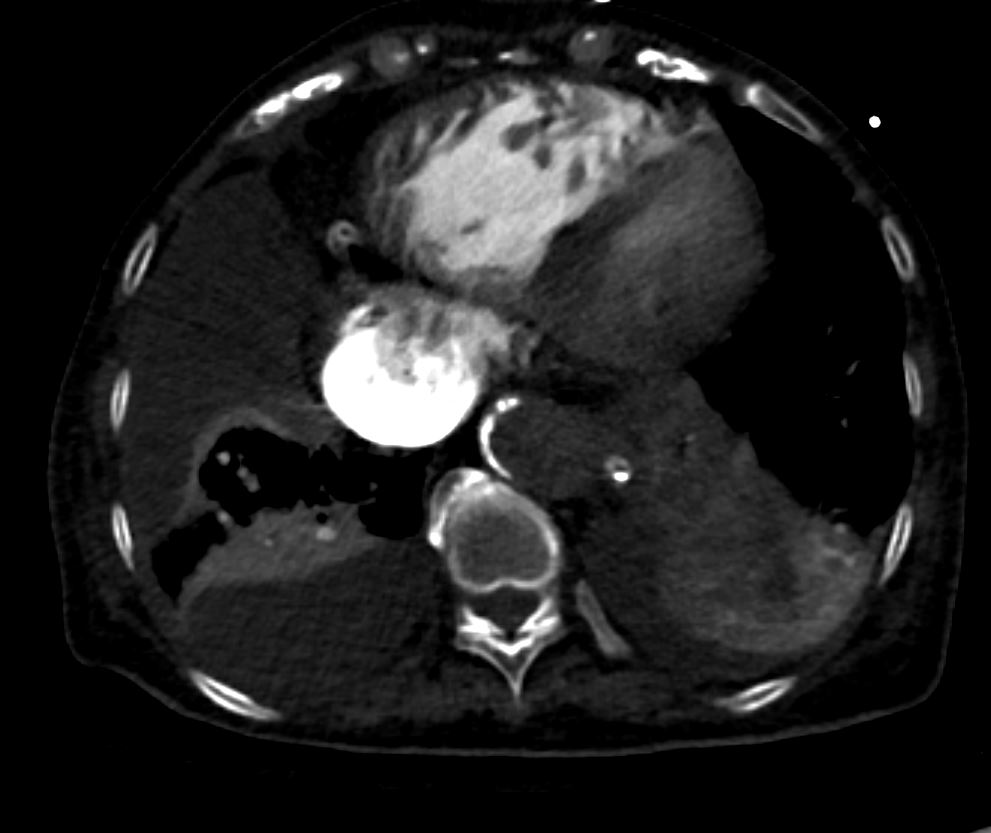
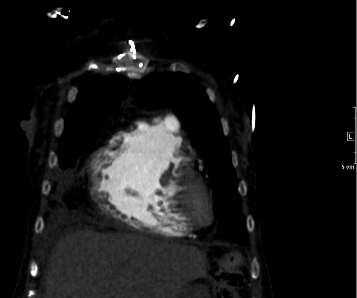
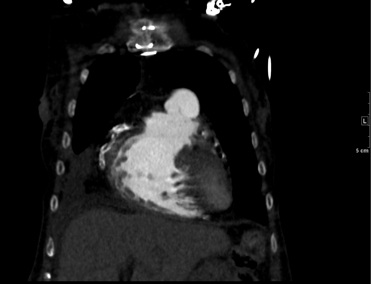
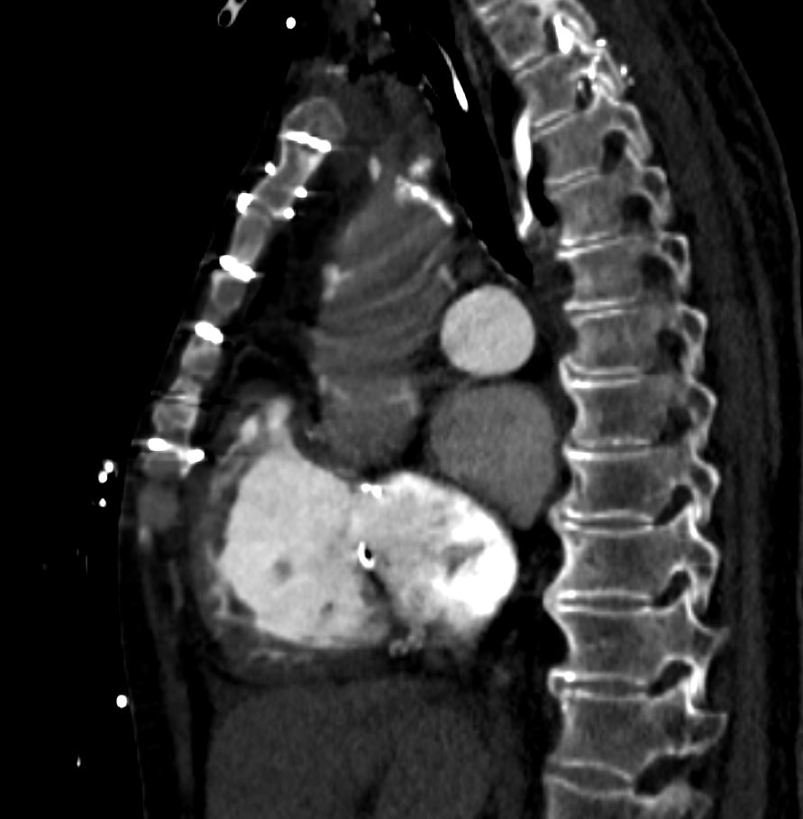
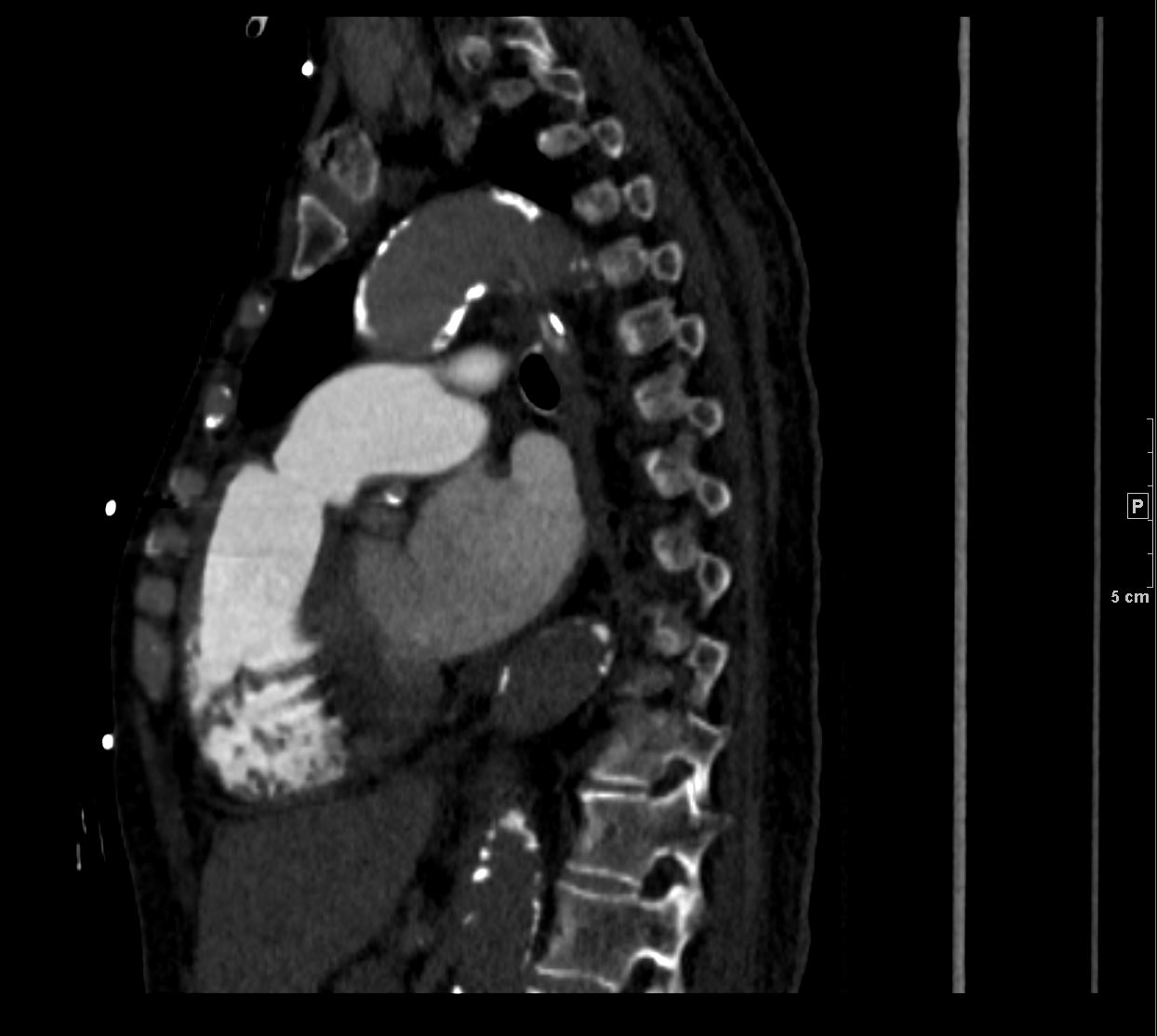
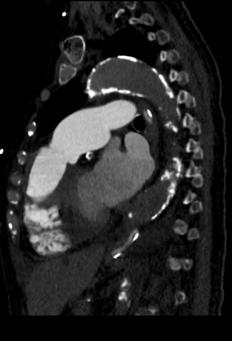
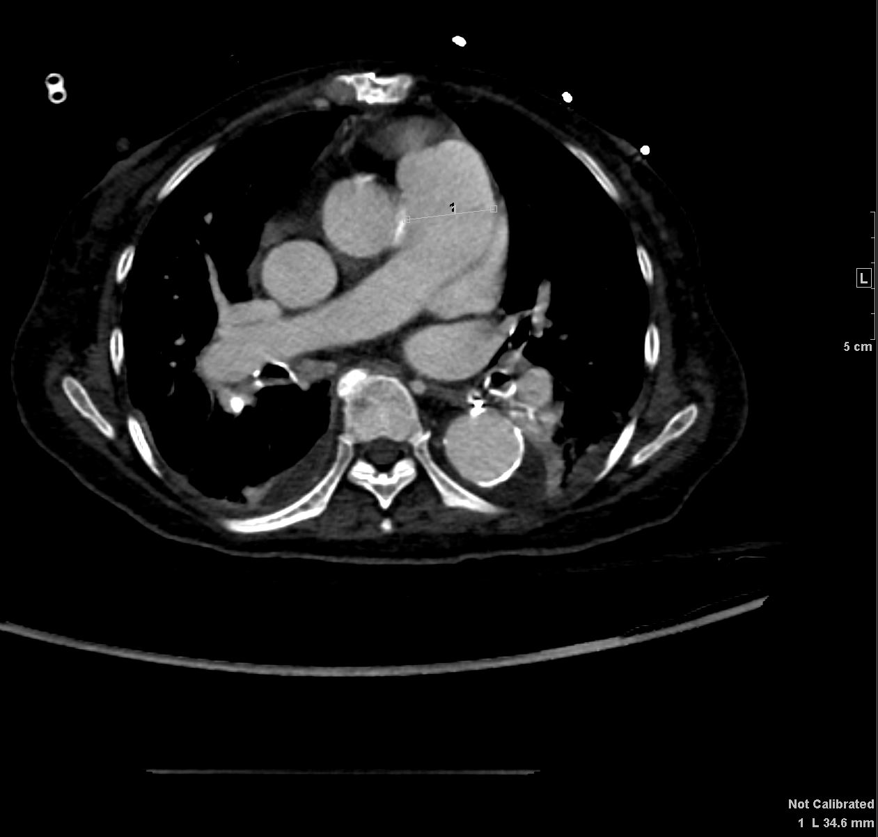
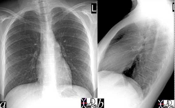
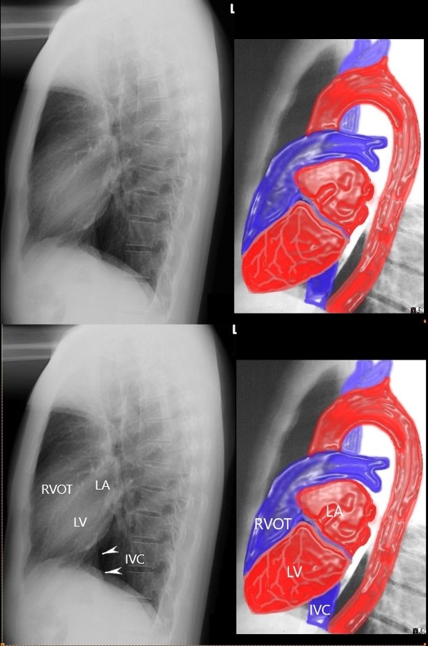
57-year-old female with 4 chamber enlargement consistent with a dilated cardiomyopathy.
CXR showed cardiomegaly with a triangular shaped heart
CT confirmed 4 chamber cardiomegaly with pulmonary hypertension. The left atrium (LA) measured 5cms, the right atrium was 8.2cms, right ventricle 4.9cms, left ventricle 5.7cms, and PA 3.5cms. LV thickness was 1.4cms
On the coronal view the triangular shaped RV dominated the shape of the heart. There was mild tricuspid regurgitation
Ashley Davidoff MD
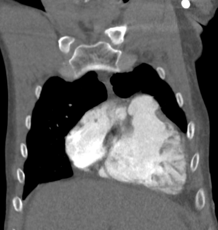
DILATED CARDIOMYOPATHY WITH 4 CHAMBER ENLARGEMENT and TRIANGULAR HEART ON CXR
Ashley Davidoff MD
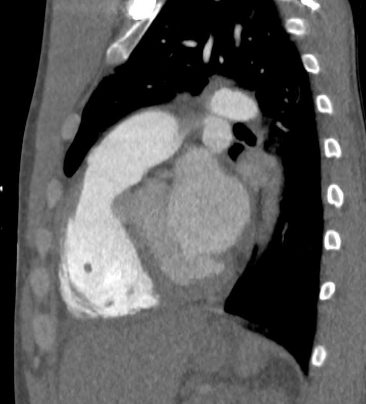
DILATED CARDIOMYOPATHY WITH 4 CHAMBER ENLARGEMENT and TRIANGULAR HEART ON CXR
Ashley Davidoff MD
87-M-mitral-stenosis-PHT-001.jpg 87-M-mitral-stenosis-PHT-003.jpg 87-M-mitral-stenosis-PHT-010.jpg 87-M-mitral-stenosis-PHT-012.jpg
87-M-mitral-stenosis-PHT-013.jpg 87-M-mitral-stenosis-PHT-004.jpg 87-M-mitral-stenosis-PHT-005.jpg 87-M-mitral-stenosis-PHT-006.jpg 87-M-mitral-stenosis-PHT-007.jpg 87-M-mitral-stenosis-PHT-008.jpg 87-M-mitral-stenosis-PHT-009.jpg 87-M-mitral-stenosis-PHT-014.jpg
RVH
By MRI
Ashley Davidoff MD
key words
normal
anatomy
right atrium and right ventricle (RV)
130630.8
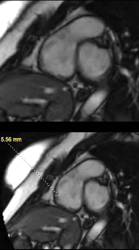
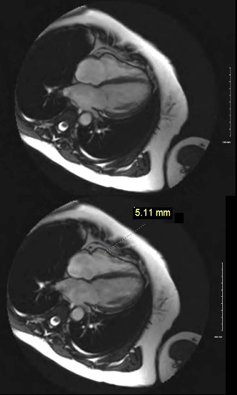
Short axis “white blood ” MRI sequence through the normal right ventricle (RV) sinus and right ventricular outflow tract (RVOT) reveal normal myocardial thickness in the 5mm range of the anterior wall
Ashley Davidoff MD
key words
normal
anatomy
right atrium and right ventricle (RV)
130627.8
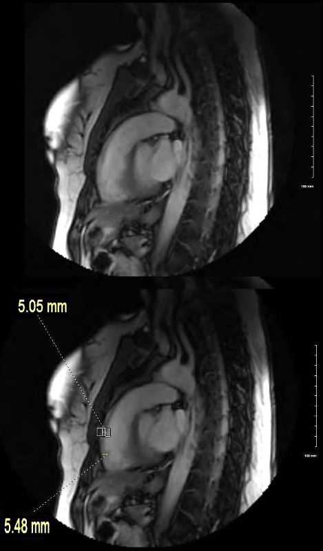
Sagittal “white blood ” MRI sequence through the normal right ventricular (RV) inflow and right ventricular outflow tract (RVOT) reveal myocardial thickness in the 5mm range of the anterior wall of the RV sinus and RVOT..
Ashley Davidoff MD
key words
normal
anatomy
right atrium and right ventricle (RV)
130631.8
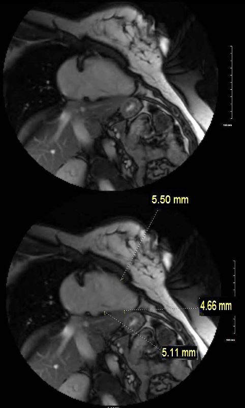
Parasagittal “white blood ” MRI sequence throuGh the normal reveal myocardial thickness in the 5mm range of the inferior and anterior wall
Ashley Davidoff MD
key words
normal
anatomy
right atrium and right ventricle (RV)
130629.8

35 year old patient with severe mitral stenosis . with left atrial enlargement (LAE) characterized by elevation of the left mainstem bronchus, straightening of the left heart border, and prominence of the upper 1/3 of the posterior border of the heart. The right atrium is also enlarged characterized by a rotund right heart border. There is right ventricular enlargement characterized by filling in of the retrosternal airspace.
Ashley Davidoff MD
References and Links

