Nutshells and buzz words
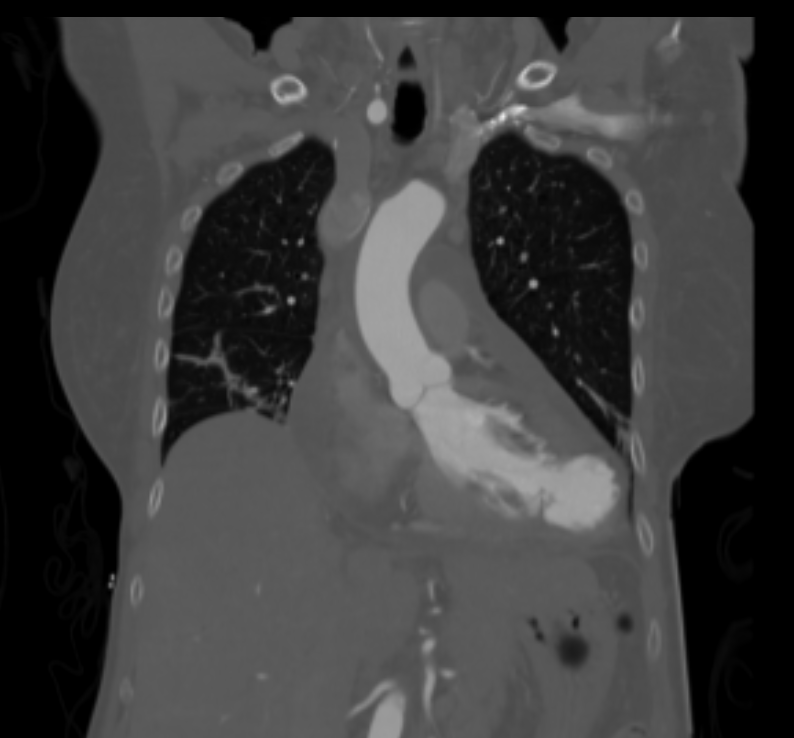
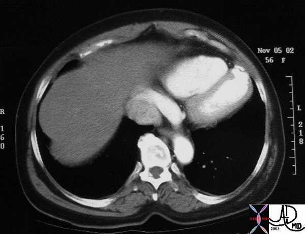
posterior and basal


1280px-Heart_pseudoaneurysm_a4c.jpg
Pseudoaneurysm of the left ventricle, four-chamber echocardiography view
Nutshells and buzz words
posterior and basal


1280px-Heart_pseudoaneurysm_a4c.jpg
Pseudoaneurysm of the left ventricle, four-chamber echocardiography view