Imaging
The Common Vein Copyright 2008
Introduction
The goal of medical imaging is to reflect the true structure and function of a biological unit in order to evaluate whether it is in a state of order or disorder. We have come to a time when the imaging modalities can reflect the macroscopic structures of the body with great accuracy so that the evaluation regarding size shape and position are fairly straightforward, while the characterisation of the tissue remains the challenge.
Structure
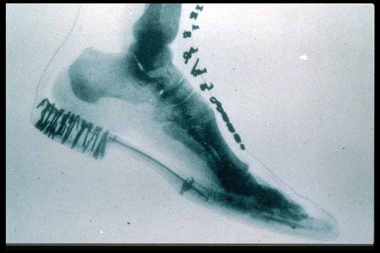
Evaluation of structure has advanced significantly as depicted in the two forms of X-ray use as outlined below
54464 |
| This X-ray of a woman’s foot and shoe was taken in 1896 by W. H. McElwain and Company. It is one of the first X-ray photographs made in the United States. From the Smithsonian National Museum of American History 54464 |

Structure of the Foot – CTscan |
| 49549c01 foot bone #D normal anatomy ligaments talus navicular cuboid tarsals metatarsal phalanges 3D volume rendering surface rendering CTscan Courtesy Ashley Davidoff MD |
Function
Function has been mostly in the domain of Echocardiography and nuclear medicine.
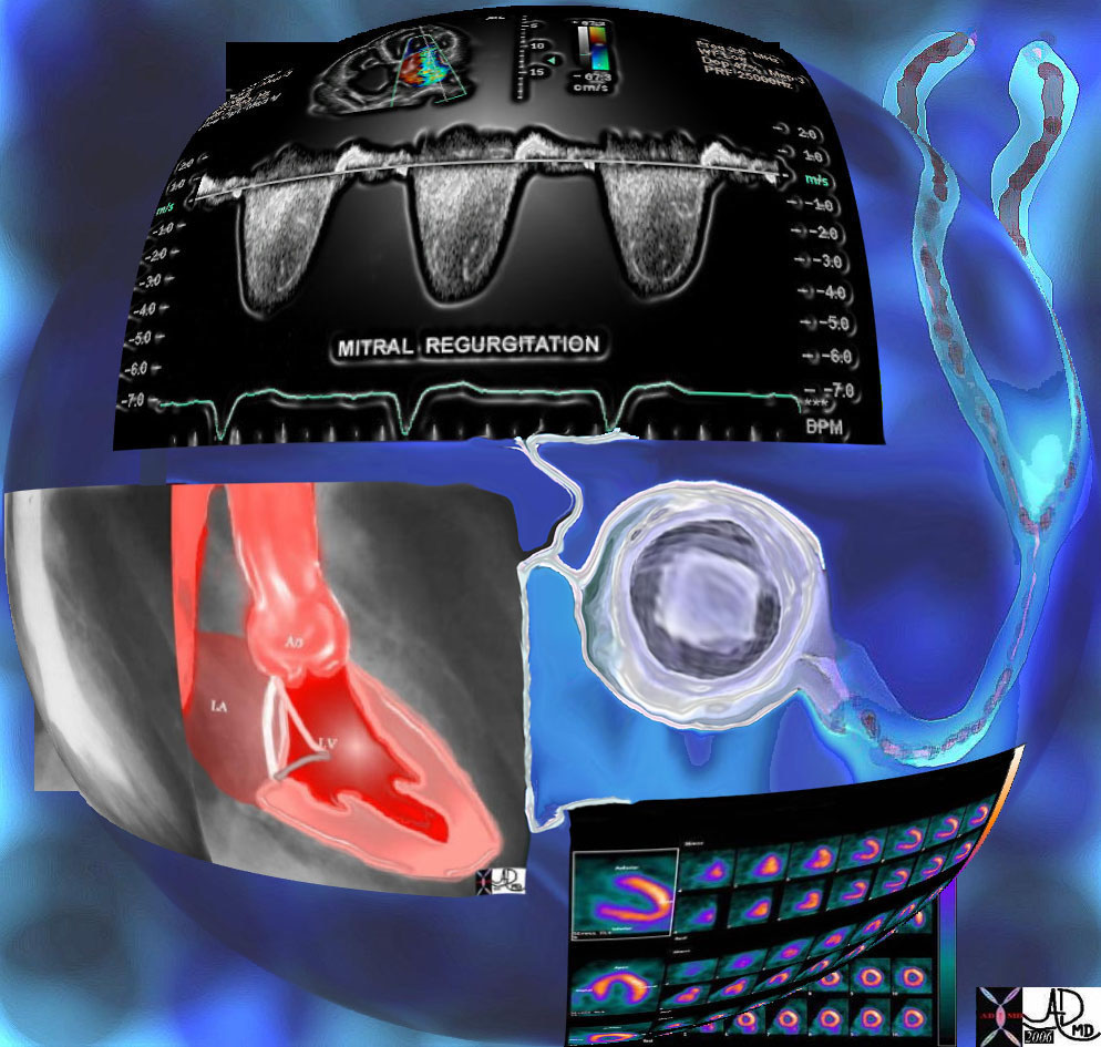
The Technologies of the Heart |
| 32647d06b01 Courtesy Ashley Davidoff MD. accessory interesting Imaging Strategies Chest stethoscope heart cardiac chest echocardiography echcardiogram color doppler pulsed doppler dx MR mitral regurgitation angiogram angiography ventriculogram LV gram radionucliide study MIBI Davidoff art |
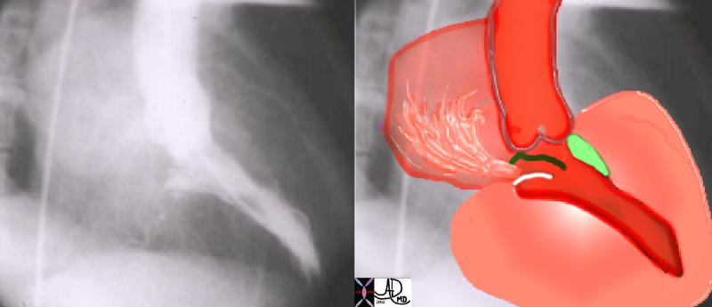
Mitral Regurgitation |
| This angiogram in RAO projection shows a hypercontractile left ventricle that has a ballet shoe appearance, with mitral regurgitation filling the left atrium. The drawing shows the significant LVH small cavity of the LV, the area of subaortic muscle bundle (green) and the mitral regurgitation caused by the systolic anterior motion of the mitral valve. Courtesy Ashley Davidoff 34805 cardiac heart MV interventriclar septum LVH IHSS SAM MR imaging radiology angiography disease overlay |
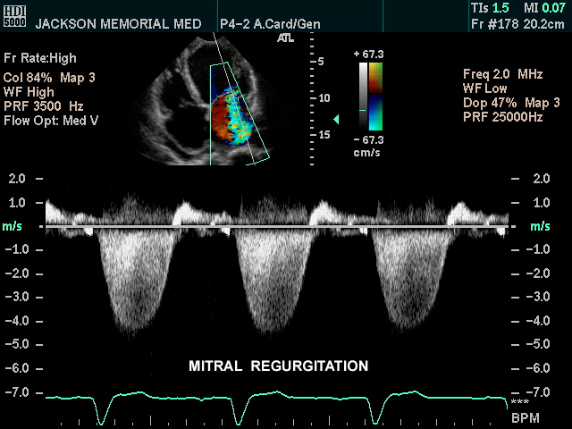 Mitral Regurgitation Mitral Regurgitation |
| 4 chamber view of the heart using gray scale echo, pulse doppler, as well as color flow interrogated at the left atrial (LA) level. The image demonstrates an example of mitral regurgitation Courtesy Philips Medical Systems 33119 cardiac heart echo mitral valve regurgitation MV MR imaging cardiac echo |
Disease
Specific disease entities including inflammation infection and malignancy
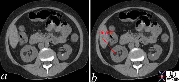 Lipoma Lipoma |
| The low density lesion in the right kidney measures -58 HU units – a finding characteristic of a fat containing lesion in the kidney. The differential is virtually limited to a diagnosis of angiomyolipoma. The nephrolithiasis of the right kidney is also obvious. Courtesy Ashley Davidoff MD 38006c01 code kidney mass fat angiomyolipoma density calcification |
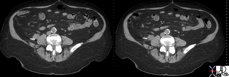 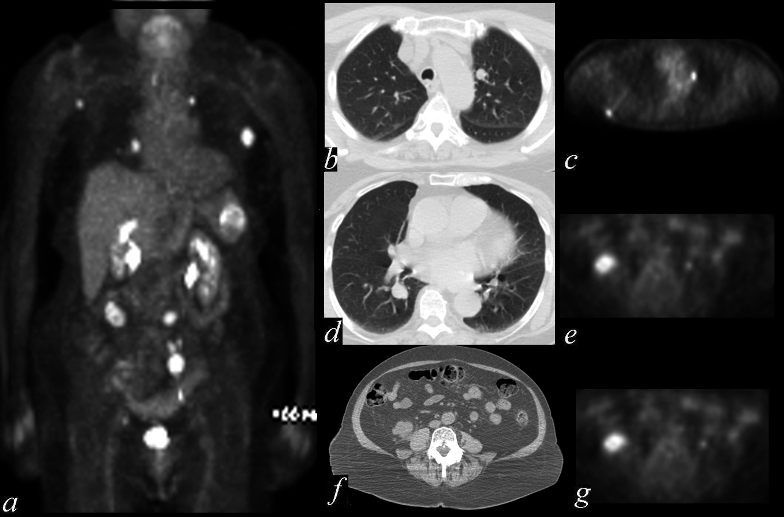
Diagnosis of Malignancy |
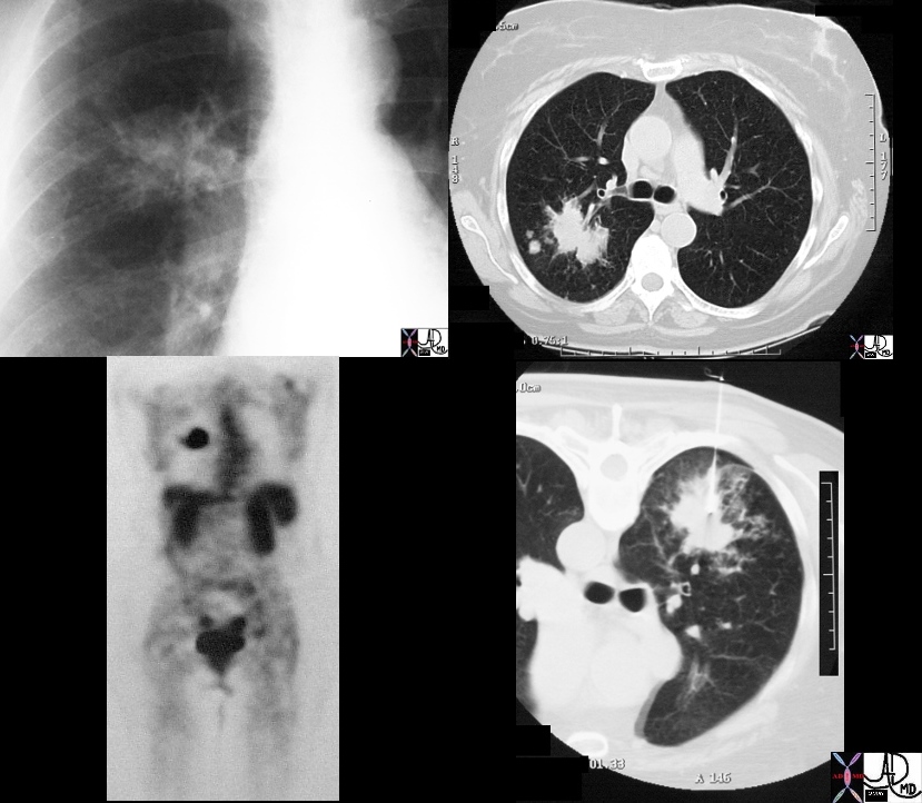
| 49726c01.800 nodules lungs peritoneal cavity retropritoneum CT scan PET scan dx metastattic melanoma metastases Courtesy Ashley DAvidoff MD 49725.800 mets |
Diagnosis
The diagnosis of a disease process is not a black or white answer. Knowledge of the cause, effect, severity, and complcations are needed to be understood in their entirety in order to identify the approriate treatment.
Treatment
Treatments guided by imaging techniques include embolisation, stent placement, thrombolytic therapy., and radiofrequency ablation among many others that are evolving
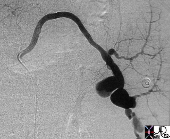  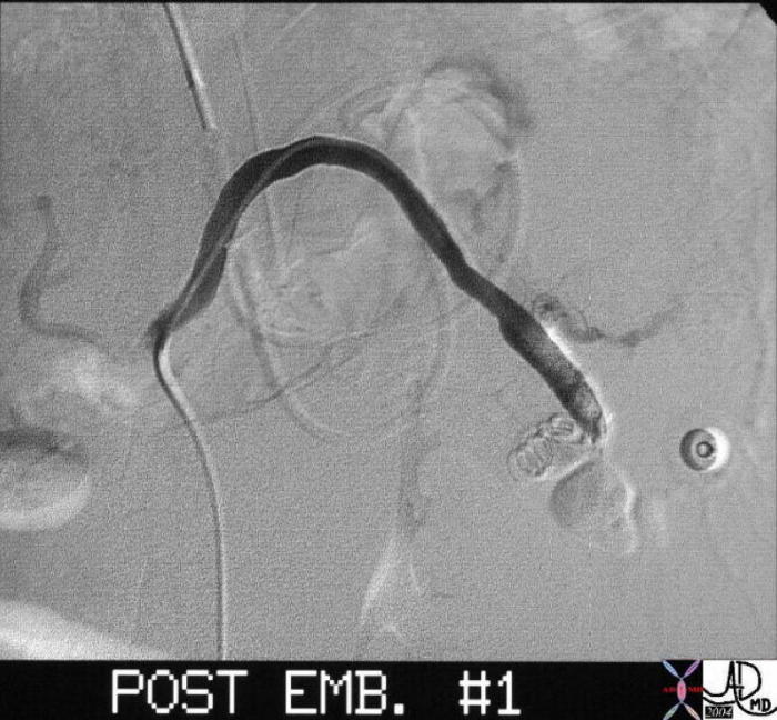 Bleeding Splenic Artery Embolized Bleeding Splenic Artery Embolized |
| 22521 Courtesy Ashley Davidoff MD code spleen + artery + dx ruptured splenic artery aneurysm pseudoaneurysm + cystic fibrosis biliary cirrhosis portal hypertension angiography emergency |
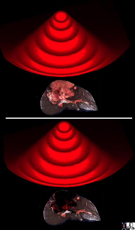 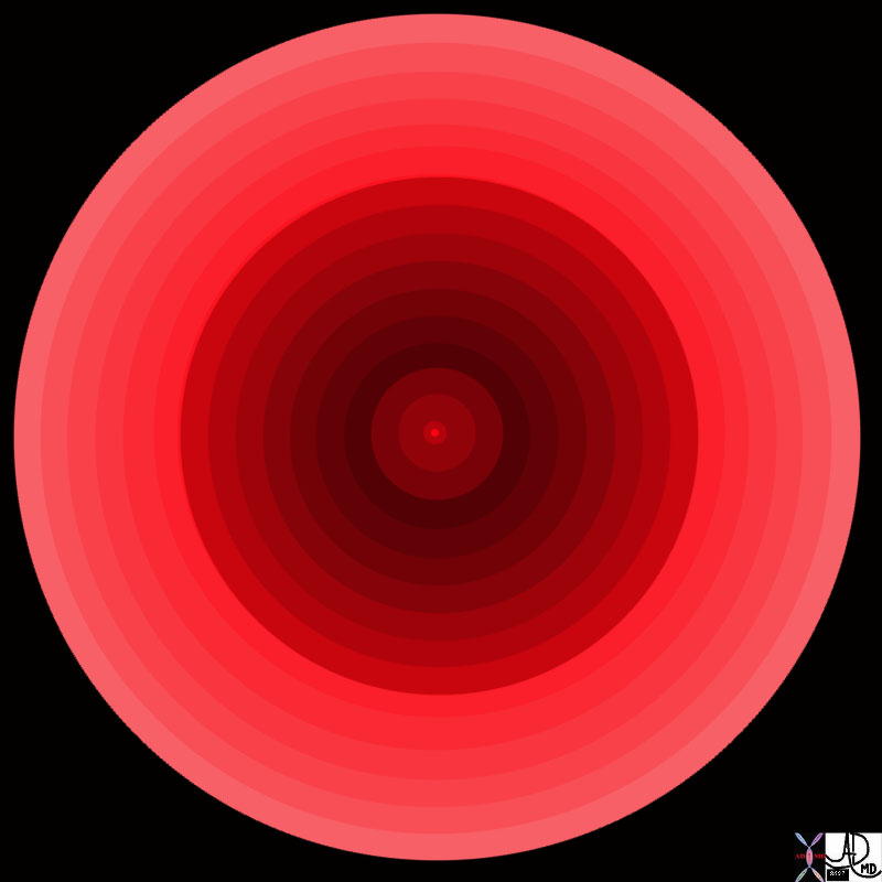 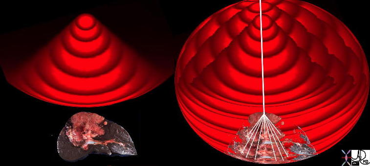 Radiofrequecy Ablation Radiofrequecy Ablation |
| 47678c03 47678c02 47678c01.800 47678f06.800 concemtric rings pulsatile waves heat RFA radiofrequency ablation principles HCC RFA charred tissue Davidoff Art Courtesy Ashley Davidoff MD |

Radiofrequency Ablation of Renal Cell Carcinoma in a 90Year Old Woman |
| 22365c02 90 year old female kidney mass hypervascularity hypervascular mass dx RCC renal cell carcinoma CTscan CTguided RFA radiofrequency ablation of a malignant tumor CTscan Courtesy Ashley Davidoff MD |
Our tools generate an energy source and direct the energy source toward the tissues to ascertain the difference between two side by side structures. X-rays for example, generated by an X-ray tube, penetrate the body and interact with the tissues in different ways depending on the tissue. When they interact with bone for example the X-rays are reflected off the bone, and their interaction with air on the other hand is limited andt they can pass right through an air filled structure without impedence. The ways in which X-rays, gamma rays, ultrasound, lightwaves behave when they come into contact with the tissues is recorded and reflected in the image so that the size, shape, position, and character of the structure can be evaluated.
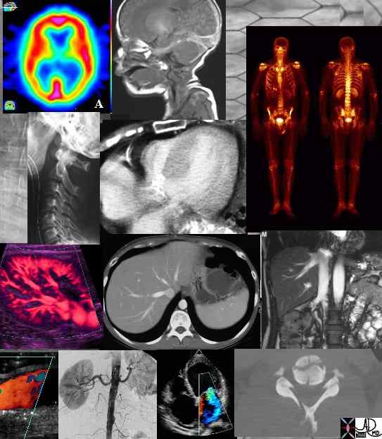
Imaging modalities |
| (Image courtesy of Ashley Davidoff M.D.) 45829 |
X-RAYS:
X-rays are generated producing a photon that is able to penetrate the body. The photons have a relatively high energy with a wavelength ranging between 0.01 to 10 nanometers. These photons interact with the tissues and are reflected, or transmitted to variable degree. The net pattern of their interactions with tissue, is reflected on photosensitive ” film” positioned on the exiting side of the body. Bone will reflect a large portion of the X-ray, and appear as white, while air will allow the X-rays tpo pass right through appearing as black or lucent on the film. Soft tissues and fat will reflect and absorb the x-rays to different degrees with fat absorbing less and therefore appearing a little blacker than soft tissue.
CT scan uses the same principles except because its a digital technique and it allows gray zone area, ie. the gray zone to be expanded so that there are a whole lot more levels of black and white or gray in the gray zone. Digital method allows us therefore to distinguish very subtle differences in the degree of transmission and deflection allowing us, for example, to distinguish between gray and white matter. The CT scan still uses an x-ray method but added to it incorporates a digitalization of that signal. This allows us to expand the gray scale.
Ultrasound
Ultrasound in very similar fashion is ultrasound waves that are thrown to the body structures these are transmitted or deflected. If they are totally transmitted then we can characterize water because water always almost totally transmits the sound waves. If there is anything other than water in the path of the ultrasound wave we get deflection to varying degrees. This deflection allows for a white spot. The degree of multiplicity of defection allows us to get some sense of the collagenous nature of a structure the area that we work. If something is very echogenic other than bone and air and calcium then one can imply that it contains a lot of collagen. If there is a shadow this implies that there is absence through transmission and most sound waves can get through usually implying calcium or air. I imagine that most of you understand these principles but they have a simplicity about them that is applicable and understandable.
Nuclear Medicine
Nuclear medicine is an imaging technique that use radioactive substances to evaluate structure, function and disease and it is also used in the treatment of disease.
MRI
MRI on the other hand is difficult for most people to understand. For instance, its ruled by the same principles as I introduced. Things interact with things in the given environment that is constant at a moment in time that result in a change. The things we are lured to are the protons in the body. Protons in all structures of the body. These act as little magnets they interact with other things which are the electromagnetic waves that are pulsed into the body and they interact in a given environment. That environment is in the magnetic environment. The magnetic environment brings all the players in the body, i.e. all the protons in the body into the playing field so they all start off at relatively the same position and starting rules so that this is how we do it. We place the patient in the magnet, all the protons get aligned according to the axis of the magnetic field which is along the axis of the body, i.e. the long axis of the body. So imagine a whole lot of musical strings going from head to toe in the body all in disarray from the beginning and then these then get all aligned in the same direction once the patient gets put in the magnet. Which means they are all taught and stretched and ready to go. You then plunk in radiofrequency waves that will make them all stand to attention in a different direction most of the time at 90 degrees long axis. And then you switch the radiofrequency wave and you see how they behave, so this is an interaction between protons and electromagnetic waves in a given environment which is magnetic field with a subsequent change. This change for given tissue is going to be very characteristic. In the same way as if you pulled on an elastic band and it returned to its normal position once you let it go you give it a character called elastic – this is what MR does – investigates a tissue and sees how it behaves. If it behaves like water – then its water – if it behaves like bone – then its bone. If it behaves like spleen – then its spleen. So the art to MRI is to know how things behave when subjected to electromagnetic waves. It becomes more complicated because we don?t only look at its behavior with one pulse sequence. Most of the time tissue are characterized by MR but at least two pulse sequences as we move onto to complexity we investigate the character of the tissue by multiple pulse sequences. So from the body point of view, i.e. from the abdomen what you have to realize is that MR is best at characterizing tissue not necessarily at detecting abnormalities.
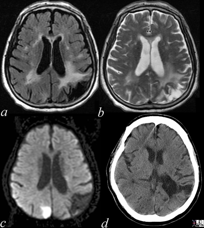 Acute and Chronic Infarction Acute and Chronic Infarction |
| 49685C01 brain DWI occipital lobe fx vague hypodensity right occipital lobe with encephalomalacia and ex vacuo changes in the left occipital and posterior parietal region dx acute infarction right occipital lobe chronic infarction left occipItoparietal lobe a IR white matter disease vague increase in right b T2 gliosis left parietal c DWI bright acute right occipital d CT vague hypodensity right occipital old infarct left dx acute infarction right occipital lobe chronic infarction left parietal lobe MRI diffusion weighted imaging CTscan Courtesy Ashley Davidoff MD |
I like to equate the MR experiment with the playing of a string instrument. The strings of the protons in your body, your fingers – plucking fingers are the electromagnetic waves that are going to be put in by the radiofrequency wave and the magnetic field is the tuned strings, i.e. the strings get put at a fixed tension but they are ready to be played. This is like the magnetic field. So before you start the experiment you tune your piano, you tune your guitar, the strings are at a given tension, protons are given direction in position. You pluck a string and you listen – the same way you put patient in the MR – the magnetic field aligns the protons – you put in the pulse wave a radiofrequency pulse wave, a electromagnetic pulse wave and the protons begin to sing and to sound. And you listen for two components of this sound – you listen for the T1 component and you listen for the T2 component. All tissues have both T1 and T2 characteristics and by virtue of the pulse sequence you can bring out this character. Either the T1 or T2 if you are investigating T1 behavior – time between plucking the instrument, ie. the time between putting a radiofrequency wave into the body is relatively short in the order of 600 mlsec and the time between listening is also relatively short. When you want to bring out the T2 characteristics of a tissue, the time between plucking is very long usually greater than 2000 mlsec and in the order of 6000 mlsec. When the TR is long, which is the repetition rate or the time between plucking is long we bring out the characteristic of the T2 characteristics of the tissue. When its short we bring out the T1 characteristics.
So from a positive point of view if your asked whether something is a T1 or a T2 weighted image, i.e. is it bringing out T1 characteristics or T2 characteristics are reasonable but not always consistent pattern because there are some other variables of determining this – is to look and see whether the TR is short. If it is short – its T1, if its long its T2. From a practical
standpoint if you look at the CSF and it is white then it is a T2 weighted image because water is white on T2 and if it is black its a T1 weighted sequence. In general if the fat is white its a T1 weighted sequence. Other practical rules: fat is white on T1, bone and iron is black on all sequences i.e. cortical bone. Spleen is wet – its a large sponge containing a lot of blood, therefore it tends to be brighter than the liver on T2. Tumor is wet – due to an absence of lymphatic drainage and in efficient venous drainage. The wetness of tumor is similar to the wetness of the spleen, and therefore, tumor has similar characteristics to the spleen. Brightness on T1 is unusual. It is normal and usual for fat. If something is not fat and its bright on T1 then it may be acute blood, melanin, mucin or some other material that is high in protein.
Molecular Imaging
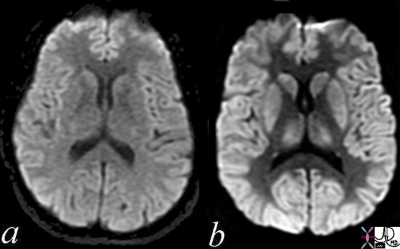 Normal and Global Ischemia after Cardiac Arrest Normal and Global Ischemia after Cardiac Arrest |
| The two images represent a diffusion weighted MRI image which measures Brownian motion of molecules. In acute infarction there is restricted Bronian motion of the affected area and the image can be manipulated to present this a s a bright region. In this case the acute infarction or ischemia (b) is relatively bright compared to the white namatter and compared to the gray matterof the normal (a)
49433c02.800 brain cerebral cerebrum white matter gray matter basal ganglia fx increase intensity in gray matter relative decrease in intensity in white matter dx global ischemia question brain death probable irreversible brain death cerebral infarction s/p arrest MRI DWI diffusion weighted imaging normal and abnormal Courtesy Ashley Davidoff MD |
References
Links from India 5star
DOMElement Object
(
    [schemaTypeInfo] =>
    [tagName] => table
    [firstElementChild] => (object value omitted)
    [lastElementChild] => (object value omitted)
    [childElementCount] => 1
    [previousElementSibling] => (object value omitted)
    [nextElementSibling] => (object value omitted)
    [nodeName] => table
    [nodeValue] =>


Normal and Global Ischemia after Cardiac Arrest


The two images represent a diffusion weighted MRI image which measures Brownian motion of molecules.  In acute infarction there is restricted Bronian motion of the affected area and the image can be manipulated to present this a s a bright region.  In this case the acute infarction or ischemia (b) is relatively bright compared to the white namatter and compared to the gray matterof the normal (a)
49433c02.800 brain cerebral cerebrum white matter gray matter basal ganglia fx increase intensity in gray matter relative decrease in intensity in white matter dx global ischemia question brain death probable irreversible brain death cerebral infarction s/p arrest MRI DWI diffusion weighted imaging normal and abnormal Courtesy Ashley Davidoff MD


    [nodeType] => 1
    [parentNode] => (object value omitted)
    [childNodes] => (object value omitted)
    [firstChild] => (object value omitted)
    [lastChild] => (object value omitted)
    [previousSibling] => (object value omitted)
    [nextSibling] => (object value omitted)
    [attributes] => (object value omitted)
    [ownerDocument] => (object value omitted)
    [namespaceURI] =>
    [prefix] =>
    [localName] => table
    [baseURI] =>
    [textContent] =>


Normal and Global Ischemia after Cardiac Arrest


The two images represent a diffusion weighted MRI image which measures Brownian motion of molecules.  In acute infarction there is restricted Bronian motion of the affected area and the image can be manipulated to present this a s a bright region.  In this case the acute infarction or ischemia (b) is relatively bright compared to the white namatter and compared to the gray matterof the normal (a)
49433c02.800 brain cerebral cerebrum white matter gray matter basal ganglia fx increase intensity in gray matter relative decrease in intensity in white matter dx global ischemia question brain death probable irreversible brain death cerebral infarction s/p arrest MRI DWI diffusion weighted imaging normal and abnormal Courtesy Ashley Davidoff MD


)
DOMElement Object
(
    [schemaTypeInfo] =>
    [tagName] => td
    [firstElementChild] => (object value omitted)
    [lastElementChild] => (object value omitted)
    [childElementCount] => 1
    [previousElementSibling] =>
    [nextElementSibling] =>
    [nodeName] => td
    [nodeValue] => The two images represent a diffusion weighted MRI image which measures Brownian motion of molecules.  In acute infarction there is restricted Bronian motion of the affected area and the image can be manipulated to present this a s a bright region.  In this case the acute infarction or ischemia (b) is relatively bright compared to the white namatter and compared to the gray matterof the normal (a)
49433c02.800 brain cerebral cerebrum white matter gray matter basal ganglia fx increase intensity in gray matter relative decrease in intensity in white matter dx global ischemia question brain death probable irreversible brain death cerebral infarction s/p arrest MRI DWI diffusion weighted imaging normal and abnormal Courtesy Ashley Davidoff MD
    [nodeType] => 1
    [parentNode] => (object value omitted)
    [childNodes] => (object value omitted)
    [firstChild] => (object value omitted)
    [lastChild] => (object value omitted)
    [previousSibling] => (object value omitted)
    [nextSibling] => (object value omitted)
    [attributes] => (object value omitted)
    [ownerDocument] => (object value omitted)
    [namespaceURI] =>
    [prefix] =>
    [localName] => td
    [baseURI] =>
    [textContent] => The two images represent a diffusion weighted MRI image which measures Brownian motion of molecules.  In acute infarction there is restricted Bronian motion of the affected area and the image can be manipulated to present this a s a bright region.  In this case the acute infarction or ischemia (b) is relatively bright compared to the white namatter and compared to the gray matterof the normal (a)
49433c02.800 brain cerebral cerebrum white matter gray matter basal ganglia fx increase intensity in gray matter relative decrease in intensity in white matter dx global ischemia question brain death probable irreversible brain death cerebral infarction s/p arrest MRI DWI diffusion weighted imaging normal and abnormal Courtesy Ashley Davidoff MD
)
DOMElement Object
(
    [schemaTypeInfo] =>
    [tagName] => td
    [firstElementChild] => (object value omitted)
    [lastElementChild] => (object value omitted)
    [childElementCount] => 1
    [previousElementSibling] =>
    [nextElementSibling] =>
    [nodeName] => td
    [nodeValue] => Normal and Global Ischemia after Cardiac Arrest
    [nodeType] => 1
    [parentNode] => (object value omitted)
    [childNodes] => (object value omitted)
    [firstChild] => (object value omitted)
    [lastChild] => (object value omitted)
    [previousSibling] => (object value omitted)
    [nextSibling] => (object value omitted)
    [attributes] => (object value omitted)
    [ownerDocument] => (object value omitted)
    [namespaceURI] =>
    [prefix] =>
    [localName] => td
    [baseURI] =>
    [textContent] => Normal and Global Ischemia after Cardiac Arrest
)
DOMElement Object
(
    [schemaTypeInfo] =>
    [tagName] => table
    [firstElementChild] => (object value omitted)
    [lastElementChild] => (object value omitted)
    [childElementCount] => 1
    [previousElementSibling] => (object value omitted)
    [nextElementSibling] => (object value omitted)
    [nodeName] => table
    [nodeValue] =>


Acute and Chronic Infarction


49685C01 brain DWI occipital lobe fx vague hypodensity right occipital lobe with encephalomalacia and ex vacuo changes in the left occipital and posterior parietal region dx acute infarction right occipital lobe chronic infarction left occipItoparietal lobe a IR white matter disease vague increase in right b T2 gliosis left parietal c DWI bright acute right occipital d CT vague hypodensity right occipital old infarct left dx acute infarction right occipital lobe chronic infarction left parietal lobe MRI diffusion weighted imaging CTscan Courtesy Ashley Davidoff MD


    [nodeType] => 1
    [parentNode] => (object value omitted)
    [childNodes] => (object value omitted)
    [firstChild] => (object value omitted)
    [lastChild] => (object value omitted)
    [previousSibling] => (object value omitted)
    [nextSibling] => (object value omitted)
    [attributes] => (object value omitted)
    [ownerDocument] => (object value omitted)
    [namespaceURI] =>
    [prefix] =>
    [localName] => table
    [baseURI] =>
    [textContent] =>


Acute and Chronic Infarction


49685C01 brain DWI occipital lobe fx vague hypodensity right occipital lobe with encephalomalacia and ex vacuo changes in the left occipital and posterior parietal region dx acute infarction right occipital lobe chronic infarction left occipItoparietal lobe a IR white matter disease vague increase in right b T2 gliosis left parietal c DWI bright acute right occipital d CT vague hypodensity right occipital old infarct left dx acute infarction right occipital lobe chronic infarction left parietal lobe MRI diffusion weighted imaging CTscan Courtesy Ashley Davidoff MD


)
DOMElement Object
(
    [schemaTypeInfo] =>
    [tagName] => td
    [firstElementChild] =>
    [lastElementChild] =>
    [childElementCount] => 0
    [previousElementSibling] =>
    [nextElementSibling] =>
    [nodeName] => td
    [nodeValue] => 49685C01 brain DWI occipital lobe fx vague hypodensity right occipital lobe with encephalomalacia and ex vacuo changes in the left occipital and posterior parietal region dx acute infarction right occipital lobe chronic infarction left occipItoparietal lobe a IR white matter disease vague increase in right b T2 gliosis left parietal c DWI bright acute right occipital d CT vague hypodensity right occipital old infarct left dx acute infarction right occipital lobe chronic infarction left parietal lobe MRI diffusion weighted imaging CTscan Courtesy Ashley Davidoff MD
    [nodeType] => 1
    [parentNode] => (object value omitted)
    [childNodes] => (object value omitted)
    [firstChild] => (object value omitted)
    [lastChild] => (object value omitted)
    [previousSibling] => (object value omitted)
    [nextSibling] => (object value omitted)
    [attributes] => (object value omitted)
    [ownerDocument] => (object value omitted)
    [namespaceURI] =>
    [prefix] =>
    [localName] => td
    [baseURI] =>
    [textContent] => 49685C01 brain DWI occipital lobe fx vague hypodensity right occipital lobe with encephalomalacia and ex vacuo changes in the left occipital and posterior parietal region dx acute infarction right occipital lobe chronic infarction left occipItoparietal lobe a IR white matter disease vague increase in right b T2 gliosis left parietal c DWI bright acute right occipital d CT vague hypodensity right occipital old infarct left dx acute infarction right occipital lobe chronic infarction left parietal lobe MRI diffusion weighted imaging CTscan Courtesy Ashley Davidoff MD
)
DOMElement Object
(
    [schemaTypeInfo] =>
    [tagName] => td
    [firstElementChild] => (object value omitted)
    [lastElementChild] => (object value omitted)
    [childElementCount] => 1
    [previousElementSibling] =>
    [nextElementSibling] =>
    [nodeName] => td
    [nodeValue] => Acute and Chronic Infarction
    [nodeType] => 1
    [parentNode] => (object value omitted)
    [childNodes] => (object value omitted)
    [firstChild] => (object value omitted)
    [lastChild] => (object value omitted)
    [previousSibling] => (object value omitted)
    [nextSibling] => (object value omitted)
    [attributes] => (object value omitted)
    [ownerDocument] => (object value omitted)
    [namespaceURI] =>
    [prefix] =>
    [localName] => td
    [baseURI] =>
    [textContent] => Acute and Chronic Infarction
)
DOMElement Object
(
    [schemaTypeInfo] =>
    [tagName] => table
    [firstElementChild] => (object value omitted)
    [lastElementChild] => (object value omitted)
    [childElementCount] => 1
    [previousElementSibling] => (object value omitted)
    [nextElementSibling] => (object value omitted)
    [nodeName] => table
    [nodeValue] =>


Imaging modalities


 (Image courtesy of Ashley Davidoff M.D.) 45829


    [nodeType] => 1
    [parentNode] => (object value omitted)
    [childNodes] => (object value omitted)
    [firstChild] => (object value omitted)
    [lastChild] => (object value omitted)
    [previousSibling] => (object value omitted)
    [nextSibling] => (object value omitted)
    [attributes] => (object value omitted)
    [ownerDocument] => (object value omitted)
    [namespaceURI] =>
    [prefix] =>
    [localName] => table
    [baseURI] =>
    [textContent] =>


Imaging modalities


 (Image courtesy of Ashley Davidoff M.D.) 45829


)
DOMElement Object
(
    [schemaTypeInfo] =>
    [tagName] => td
    [firstElementChild] =>
    [lastElementChild] =>
    [childElementCount] => 0
    [previousElementSibling] =>
    [nextElementSibling] =>
    [nodeName] => td
    [nodeValue] =>  (Image courtesy of Ashley Davidoff M.D.) 45829
    [nodeType] => 1
    [parentNode] => (object value omitted)
    [childNodes] => (object value omitted)
    [firstChild] => (object value omitted)
    [lastChild] => (object value omitted)
    [previousSibling] => (object value omitted)
    [nextSibling] => (object value omitted)
    [attributes] => (object value omitted)
    [ownerDocument] => (object value omitted)
    [namespaceURI] =>
    [prefix] =>
    [localName] => td
    [baseURI] =>
    [textContent] =>  (Image courtesy of Ashley Davidoff M.D.) 45829
)
DOMElement Object
(
    [schemaTypeInfo] =>
    [tagName] => td
    [firstElementChild] => (object value omitted)
    [lastElementChild] => (object value omitted)
    [childElementCount] => 2
    [previousElementSibling] =>
    [nextElementSibling] =>
    [nodeName] => td
    [nodeValue] =>
Imaging modalities
    [nodeType] => 1
    [parentNode] => (object value omitted)
    [childNodes] => (object value omitted)
    [firstChild] => (object value omitted)
    [lastChild] => (object value omitted)
    [previousSibling] => (object value omitted)
    [nextSibling] => (object value omitted)
    [attributes] => (object value omitted)
    [ownerDocument] => (object value omitted)
    [namespaceURI] =>
    [prefix] =>
    [localName] => td
    [baseURI] =>
    [textContent] =>
Imaging modalities
)
DOMElement Object
(
    [schemaTypeInfo] =>
    [tagName] => table
    [firstElementChild] => (object value omitted)
    [lastElementChild] => (object value omitted)
    [childElementCount] => 1
    [previousElementSibling] => (object value omitted)
    [nextElementSibling] => (object value omitted)
    [nodeName] => table
    [nodeValue] =>


Radiofrequency Ablation  of Renal Cell Carcinoma in a 90Year Old Woman


22365c02 90 year old female kidney mass hypervascularity hypervascular mass dx RCC renal cell carcinoma CTscan CTguided RFA radiofrequency ablation of a malignant tumor CTscan Courtesy Ashley Davidoff MD


    [nodeType] => 1
    [parentNode] => (object value omitted)
    [childNodes] => (object value omitted)
    [firstChild] => (object value omitted)
    [lastChild] => (object value omitted)
    [previousSibling] => (object value omitted)
    [nextSibling] => (object value omitted)
    [attributes] => (object value omitted)
    [ownerDocument] => (object value omitted)
    [namespaceURI] =>
    [prefix] =>
    [localName] => table
    [baseURI] =>
    [textContent] =>


Radiofrequency Ablation  of Renal Cell Carcinoma in a 90Year Old Woman


22365c02 90 year old female kidney mass hypervascularity hypervascular mass dx RCC renal cell carcinoma CTscan CTguided RFA radiofrequency ablation of a malignant tumor CTscan Courtesy Ashley Davidoff MD


)
DOMElement Object
(
    [schemaTypeInfo] =>
    [tagName] => td
    [firstElementChild] =>
    [lastElementChild] =>
    [childElementCount] => 0
    [previousElementSibling] =>
    [nextElementSibling] =>
    [nodeName] => td
    [nodeValue] => 22365c02 90 year old female kidney mass hypervascularity hypervascular mass dx RCC renal cell carcinoma CTscan CTguided RFA radiofrequency ablation of a malignant tumor CTscan Courtesy Ashley Davidoff MD
    [nodeType] => 1
    [parentNode] => (object value omitted)
    [childNodes] => (object value omitted)
    [firstChild] => (object value omitted)
    [lastChild] => (object value omitted)
    [previousSibling] => (object value omitted)
    [nextSibling] => (object value omitted)
    [attributes] => (object value omitted)
    [ownerDocument] => (object value omitted)
    [namespaceURI] =>
    [prefix] =>
    [localName] => td
    [baseURI] =>
    [textContent] => 22365c02 90 year old female kidney mass hypervascularity hypervascular mass dx RCC renal cell carcinoma CTscan CTguided RFA radiofrequency ablation of a malignant tumor CTscan Courtesy Ashley Davidoff MD
)
DOMElement Object
(
    [schemaTypeInfo] =>
    [tagName] => td
    [firstElementChild] => (object value omitted)
    [lastElementChild] => (object value omitted)
    [childElementCount] => 2
    [previousElementSibling] =>
    [nextElementSibling] =>
    [nodeName] => td
    [nodeValue] =>
Radiofrequency Ablation  of Renal Cell Carcinoma in a 90Year Old Woman
    [nodeType] => 1
    [parentNode] => (object value omitted)
    [childNodes] => (object value omitted)
    [firstChild] => (object value omitted)
    [lastChild] => (object value omitted)
    [previousSibling] => (object value omitted)
    [nextSibling] => (object value omitted)
    [attributes] => (object value omitted)
    [ownerDocument] => (object value omitted)
    [namespaceURI] =>
    [prefix] =>
    [localName] => td
    [baseURI] =>
    [textContent] =>
Radiofrequency Ablation  of Renal Cell Carcinoma in a 90Year Old Woman
)
DOMElement Object
(
    [schemaTypeInfo] =>
    [tagName] => table
    [firstElementChild] => (object value omitted)
    [lastElementChild] => (object value omitted)
    [childElementCount] => 1
    [previousElementSibling] => (object value omitted)
    [nextElementSibling] => (object value omitted)
    [nodeName] => table
    [nodeValue] =>


Radiofrequecy Ablation


47678c03 47678c02 47678c01.800 47678f06.800 concemtric rings pulsatile waves heat RFA radiofrequency ablation principles HCC RFA charred tissue Davidoff Art Courtesy Ashley Davidoff MD


    [nodeType] => 1
    [parentNode] => (object value omitted)
    [childNodes] => (object value omitted)
    [firstChild] => (object value omitted)
    [lastChild] => (object value omitted)
    [previousSibling] => (object value omitted)
    [nextSibling] => (object value omitted)
    [attributes] => (object value omitted)
    [ownerDocument] => (object value omitted)
    [namespaceURI] =>
    [prefix] =>
    [localName] => table
    [baseURI] =>
    [textContent] =>


Radiofrequecy Ablation


47678c03 47678c02 47678c01.800 47678f06.800 concemtric rings pulsatile waves heat RFA radiofrequency ablation principles HCC RFA charred tissue Davidoff Art Courtesy Ashley Davidoff MD


)
DOMElement Object
(
    [schemaTypeInfo] =>
    [tagName] => td
    [firstElementChild] =>
    [lastElementChild] =>
    [childElementCount] => 0
    [previousElementSibling] =>
    [nextElementSibling] =>
    [nodeName] => td
    [nodeValue] => 47678c03 47678c02 47678c01.800 47678f06.800 concemtric rings pulsatile waves heat RFA radiofrequency ablation principles HCC RFA charred tissue Davidoff Art Courtesy Ashley Davidoff MD
    [nodeType] => 1
    [parentNode] => (object value omitted)
    [childNodes] => (object value omitted)
    [firstChild] => (object value omitted)
    [lastChild] => (object value omitted)
    [previousSibling] => (object value omitted)
    [nextSibling] => (object value omitted)
    [attributes] => (object value omitted)
    [ownerDocument] => (object value omitted)
    [namespaceURI] =>
    [prefix] =>
    [localName] => td
    [baseURI] =>
    [textContent] => 47678c03 47678c02 47678c01.800 47678f06.800 concemtric rings pulsatile waves heat RFA radiofrequency ablation principles HCC RFA charred tissue Davidoff Art Courtesy Ashley Davidoff MD
)
DOMElement Object
(
    [schemaTypeInfo] =>
    [tagName] => td
    [firstElementChild] => (object value omitted)
    [lastElementChild] => (object value omitted)
    [childElementCount] => 1
    [previousElementSibling] =>
    [nextElementSibling] =>
    [nodeName] => td
    [nodeValue] => Radiofrequecy Ablation
    [nodeType] => 1
    [parentNode] => (object value omitted)
    [childNodes] => (object value omitted)
    [firstChild] => (object value omitted)
    [lastChild] => (object value omitted)
    [previousSibling] => (object value omitted)
    [nextSibling] => (object value omitted)
    [attributes] => (object value omitted)
    [ownerDocument] => (object value omitted)
    [namespaceURI] =>
    [prefix] =>
    [localName] => td
    [baseURI] =>
    [textContent] => Radiofrequecy Ablation
)
https://beta.thecommonvein.net/wp-content/uploads/2023/06/47678f06.800.jpg https://beta.thecommonvein.net/wp-content/uploads/2023/06/47678c01.800.jpg https://beta.thecommonvein.net/wp-content/uploads/2023/06/47678c03.jpg
http://thecommonvein.net/media/47678c03.jpg
DOMElement Object
(
    [schemaTypeInfo] =>
    [tagName] => table
    [firstElementChild] => (object value omitted)
    [lastElementChild] => (object value omitted)
    [childElementCount] => 1
    [previousElementSibling] => (object value omitted)
    [nextElementSibling] => (object value omitted)
    [nodeName] => table
    [nodeValue] =>


Bleeding Splenic Artery Embolized


22521 Courtesy Ashley Davidoff MD code spleen + artery + dx ruptured splenic artery aneurysm pseudoaneurysm + cystic fibrosis biliary cirrhosis portal hypertension angiography emergency


    [nodeType] => 1
    [parentNode] => (object value omitted)
    [childNodes] => (object value omitted)
    [firstChild] => (object value omitted)
    [lastChild] => (object value omitted)
    [previousSibling] => (object value omitted)
    [nextSibling] => (object value omitted)
    [attributes] => (object value omitted)
    [ownerDocument] => (object value omitted)
    [namespaceURI] =>
    [prefix] =>
    [localName] => table
    [baseURI] =>
    [textContent] =>


Bleeding Splenic Artery Embolized


22521 Courtesy Ashley Davidoff MD code spleen + artery + dx ruptured splenic artery aneurysm pseudoaneurysm + cystic fibrosis biliary cirrhosis portal hypertension angiography emergency


)
DOMElement Object
(
    [schemaTypeInfo] =>
    [tagName] => td
    [firstElementChild] =>
    [lastElementChild] =>
    [childElementCount] => 0
    [previousElementSibling] =>
    [nextElementSibling] =>
    [nodeName] => td
    [nodeValue] => 22521 Courtesy Ashley Davidoff MD code spleen + artery + dx ruptured splenic artery aneurysm pseudoaneurysm + cystic fibrosis biliary cirrhosis portal hypertension angiography emergency
    [nodeType] => 1
    [parentNode] => (object value omitted)
    [childNodes] => (object value omitted)
    [firstChild] => (object value omitted)
    [lastChild] => (object value omitted)
    [previousSibling] => (object value omitted)
    [nextSibling] => (object value omitted)
    [attributes] => (object value omitted)
    [ownerDocument] => (object value omitted)
    [namespaceURI] =>
    [prefix] =>
    [localName] => td
    [baseURI] =>
    [textContent] => 22521 Courtesy Ashley Davidoff MD code spleen + artery + dx ruptured splenic artery aneurysm pseudoaneurysm + cystic fibrosis biliary cirrhosis portal hypertension angiography emergency
)
DOMElement Object
(
    [schemaTypeInfo] =>
    [tagName] => td
    [firstElementChild] => (object value omitted)
    [lastElementChild] => (object value omitted)
    [childElementCount] => 2
    [previousElementSibling] =>
    [nextElementSibling] =>
    [nodeName] => td
    [nodeValue] => Bleeding Splenic Artery Embolized
    [nodeType] => 1
    [parentNode] => (object value omitted)
    [childNodes] => (object value omitted)
    [firstChild] => (object value omitted)
    [lastChild] => (object value omitted)
    [previousSibling] => (object value omitted)
    [nextSibling] => (object value omitted)
    [attributes] => (object value omitted)
    [ownerDocument] => (object value omitted)
    [namespaceURI] =>
    [prefix] =>
    [localName] => td
    [baseURI] =>
    [textContent] => Bleeding Splenic Artery Embolized
)
https://beta.thecommonvein.net/wp-content/uploads/2023/05/22521.jpg
http://thecommonvein.net/media/22521.JPG http://thecommonvein.net/media/22522.JPG
DOMElement Object
(
    [schemaTypeInfo] =>
    [tagName] => table
    [firstElementChild] => (object value omitted)
    [lastElementChild] => (object value omitted)
    [childElementCount] => 1
    [previousElementSibling] => (object value omitted)
    [nextElementSibling] => (object value omitted)
    [nodeName] => table
    [nodeValue] =>


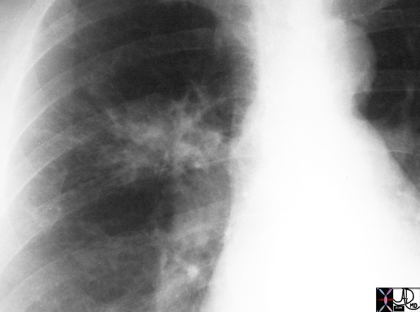
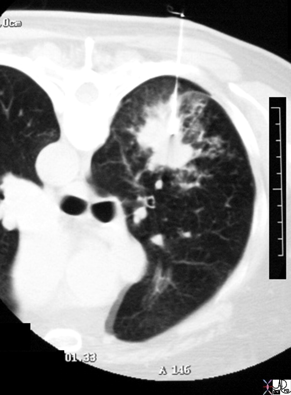
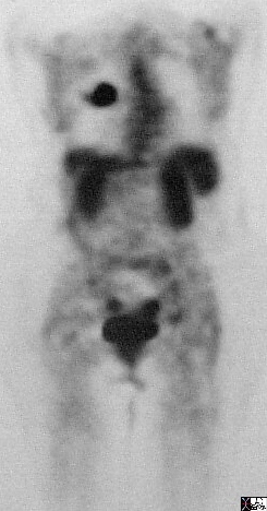
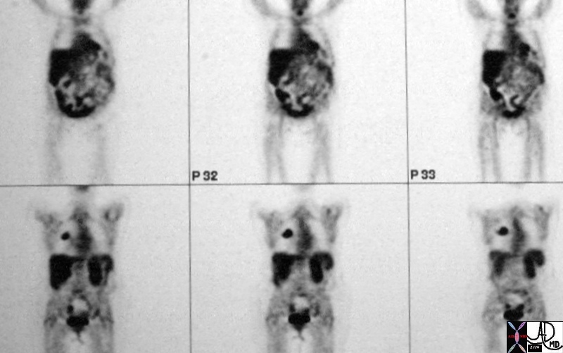
Lung Cancer – From the plain Film, CTscan, PET scan and CTguided biopsy


28986c01 hx 66F lung spiculated mass biopsy satellite nodules malignant carcinoma PETscan CTscan plain CXR Davidoff MD 28979 28980 28981 28984 28985 28986c01


    [nodeType] => 1
    [parentNode] => (object value omitted)
    [childNodes] => (object value omitted)
    [firstChild] => (object value omitted)
    [lastChild] => (object value omitted)
    [previousSibling] => (object value omitted)
    [nextSibling] => (object value omitted)
    [attributes] => (object value omitted)
    [ownerDocument] => (object value omitted)
    [namespaceURI] =>
    [prefix] =>
    [localName] => table
    [baseURI] =>
    [textContent] =>


Lung Cancer – From the plain Film, CTscan, PET scan and CTguided biopsy


28986c01 hx 66F lung spiculated mass biopsy satellite nodules malignant carcinoma PETscan CTscan plain CXR Davidoff MD 28979 28980 28981 28984 28985 28986c01


)
DOMElement Object
(
    [schemaTypeInfo] =>
    [tagName] => td
    [firstElementChild] =>
    [lastElementChild] =>
    [childElementCount] => 0
    [previousElementSibling] =>
    [nextElementSibling] =>
    [nodeName] => td
    [nodeValue] => 28986c01 hx 66F lung spiculated mass biopsy satellite nodules malignant carcinoma PETscan CTscan plain CXR Davidoff MD 28979 28980 28981 28984 28985 28986c01
    [nodeType] => 1
    [parentNode] => (object value omitted)
    [childNodes] => (object value omitted)
    [firstChild] => (object value omitted)
    [lastChild] => (object value omitted)
    [previousSibling] => (object value omitted)
    [nextSibling] => (object value omitted)
    [attributes] => (object value omitted)
    [ownerDocument] => (object value omitted)
    [namespaceURI] =>
    [prefix] =>
    [localName] => td
    [baseURI] =>
    [textContent] => 28986c01 hx 66F lung spiculated mass biopsy satellite nodules malignant carcinoma PETscan CTscan plain CXR Davidoff MD 28979 28980 28981 28984 28985 28986c01
)
DOMElement Object
(
    [schemaTypeInfo] =>
    [tagName] => td
    [firstElementChild] => (object value omitted)
    [lastElementChild] => (object value omitted)
    [childElementCount] => 2
    [previousElementSibling] =>
    [nextElementSibling] =>
    [nodeName] => td
    [nodeValue] =>
Lung Cancer – From the plain Film, CTscan, PET scan and CTguided biopsy
    [nodeType] => 1
    [parentNode] => (object value omitted)
    [childNodes] => (object value omitted)
    [firstChild] => (object value omitted)
    [lastChild] => (object value omitted)
    [previousSibling] => (object value omitted)
    [nextSibling] => (object value omitted)
    [attributes] => (object value omitted)
    [ownerDocument] => (object value omitted)
    [namespaceURI] =>
    [prefix] =>
    [localName] => td
    [baseURI] =>
    [textContent] =>
Lung Cancer – From the plain Film, CTscan, PET scan and CTguided biopsy
)
https://beta.thecommonvein.net/wp-content/uploads/2023/05/28986c01.jpg https://beta.thecommonvein.net/wp-content/uploads/2023/05/28985.jpg https://beta.thecommonvein.net/wp-content/uploads/2023/05/28984.jpg https://beta.thecommonvein.net/wp-content/uploads/2023/05/28981.jpg https://beta.thecommonvein.net/wp-content/uploads/2023/05/28980.jpg https://beta.thecommonvein.net/wp-content/uploads/2023/05/28979.jpg
http://thecommonvein.net/media/28986c01.jpg
DOMElement Object
(
    [schemaTypeInfo] =>
    [tagName] => table
    [firstElementChild] => (object value omitted)
    [lastElementChild] => (object value omitted)
    [childElementCount] => 1
    [previousElementSibling] => (object value omitted)
    [nextElementSibling] => (object value omitted)
    [nodeName] => table
    [nodeValue] =>


Diagnosis of Malignancy


49726c01.800 nodules lungs peritoneal cavity retropritoneum CT scan PET scan dx metastattic melanoma metastases Courtesy Ashley DAvidoff MD 49725.800 mets


    [nodeType] => 1
    [parentNode] => (object value omitted)
    [childNodes] => (object value omitted)
    [firstChild] => (object value omitted)
    [lastChild] => (object value omitted)
    [previousSibling] => (object value omitted)
    [nextSibling] => (object value omitted)
    [attributes] => (object value omitted)
    [ownerDocument] => (object value omitted)
    [namespaceURI] =>
    [prefix] =>
    [localName] => table
    [baseURI] =>
    [textContent] =>


Diagnosis of Malignancy


49726c01.800 nodules lungs peritoneal cavity retropritoneum CT scan PET scan dx metastattic melanoma metastases Courtesy Ashley DAvidoff MD 49725.800 mets


)
DOMElement Object
(
    [schemaTypeInfo] =>
    [tagName] => td
    [firstElementChild] =>
    [lastElementChild] =>
    [childElementCount] => 0
    [previousElementSibling] =>
    [nextElementSibling] =>
    [nodeName] => td
    [nodeValue] => 49726c01.800 nodules lungs peritoneal cavity retropritoneum CT scan PET scan dx metastattic melanoma metastases Courtesy Ashley DAvidoff MD 49725.800 mets
    [nodeType] => 1
    [parentNode] => (object value omitted)
    [childNodes] => (object value omitted)
    [firstChild] => (object value omitted)
    [lastChild] => (object value omitted)
    [previousSibling] => (object value omitted)
    [nextSibling] => (object value omitted)
    [attributes] => (object value omitted)
    [ownerDocument] => (object value omitted)
    [namespaceURI] =>
    [prefix] =>
    [localName] => td
    [baseURI] =>
    [textContent] => 49726c01.800 nodules lungs peritoneal cavity retropritoneum CT scan PET scan dx metastattic melanoma metastases Courtesy Ashley DAvidoff MD 49725.800 mets
)
DOMElement Object
(
    [schemaTypeInfo] =>
    [tagName] => td
    [firstElementChild] => (object value omitted)
    [lastElementChild] => (object value omitted)
    [childElementCount] => 2
    [previousElementSibling] =>
    [nextElementSibling] =>
    [nodeName] => td
    [nodeValue] =>
Diagnosis of Malignancy
    [nodeType] => 1
    [parentNode] => (object value omitted)
    [childNodes] => (object value omitted)
    [firstChild] => (object value omitted)
    [lastChild] => (object value omitted)
    [previousSibling] => (object value omitted)
    [nextSibling] => (object value omitted)
    [attributes] => (object value omitted)
    [ownerDocument] => (object value omitted)
    [namespaceURI] =>
    [prefix] =>
    [localName] => td
    [baseURI] =>
    [textContent] =>
Diagnosis of Malignancy
)
https://beta.thecommonvein.net/wp-content/uploads/2023/06/49726c01.800.jpg https://beta.thecommonvein.net/wp-content/uploads/2023/06/49725.800.jpg
http://thecommonvein.net/media/49726c01.800.jpg
DOMElement Object
(
    [schemaTypeInfo] =>
    [tagName] => table
    [firstElementChild] => (object value omitted)
    [lastElementChild] => (object value omitted)
    [childElementCount] => 1
    [previousElementSibling] => (object value omitted)
    [nextElementSibling] => (object value omitted)
    [nodeName] => table
    [nodeValue] =>


Lipoma


The low density lesion in the right kidney measures -58 HU units – a finding characteristic of a fat containing lesion in the kidney. The differential is virtually limited to a diagnosis of angiomyolipoma. The nephrolithiasis of the right kidney is also obvious. Courtesy Ashley Davidoff MD 38006c01 code kidney mass fat angiomyolipoma density calcification


    [nodeType] => 1
    [parentNode] => (object value omitted)
    [childNodes] => (object value omitted)
    [firstChild] => (object value omitted)
    [lastChild] => (object value omitted)
    [previousSibling] => (object value omitted)
    [nextSibling] => (object value omitted)
    [attributes] => (object value omitted)
    [ownerDocument] => (object value omitted)
    [namespaceURI] =>
    [prefix] =>
    [localName] => table
    [baseURI] =>
    [textContent] =>


Lipoma


The low density lesion in the right kidney measures -58 HU units – a finding characteristic of a fat containing lesion in the kidney. The differential is virtually limited to a diagnosis of angiomyolipoma. The nephrolithiasis of the right kidney is also obvious. Courtesy Ashley Davidoff MD 38006c01 code kidney mass fat angiomyolipoma density calcification


)
DOMElement Object
(
    [schemaTypeInfo] =>
    [tagName] => td
    [firstElementChild] =>
    [lastElementChild] =>
    [childElementCount] => 0
    [previousElementSibling] =>
    [nextElementSibling] =>
    [nodeName] => td
    [nodeValue] => The low density lesion in the right kidney measures -58 HU units – a finding characteristic of a fat containing lesion in the kidney. The differential is virtually limited to a diagnosis of angiomyolipoma. The nephrolithiasis of the right kidney is also obvious. Courtesy Ashley Davidoff MD 38006c01 code kidney mass fat angiomyolipoma density calcification
    [nodeType] => 1
    [parentNode] => (object value omitted)
    [childNodes] => (object value omitted)
    [firstChild] => (object value omitted)
    [lastChild] => (object value omitted)
    [previousSibling] => (object value omitted)
    [nextSibling] => (object value omitted)
    [attributes] => (object value omitted)
    [ownerDocument] => (object value omitted)
    [namespaceURI] =>
    [prefix] =>
    [localName] => td
    [baseURI] =>
    [textContent] => The low density lesion in the right kidney measures -58 HU units – a finding characteristic of a fat containing lesion in the kidney. The differential is virtually limited to a diagnosis of angiomyolipoma. The nephrolithiasis of the right kidney is also obvious. Courtesy Ashley Davidoff MD 38006c01 code kidney mass fat angiomyolipoma density calcification
)
DOMElement Object
(
    [schemaTypeInfo] =>
    [tagName] => td
    [firstElementChild] => (object value omitted)
    [lastElementChild] => (object value omitted)
    [childElementCount] => 1
    [previousElementSibling] =>
    [nextElementSibling] =>
    [nodeName] => td
    [nodeValue] => Lipoma
    [nodeType] => 1
    [parentNode] => (object value omitted)
    [childNodes] => (object value omitted)
    [firstChild] => (object value omitted)
    [lastChild] => (object value omitted)
    [previousSibling] => (object value omitted)
    [nextSibling] => (object value omitted)
    [attributes] => (object value omitted)
    [ownerDocument] => (object value omitted)
    [namespaceURI] =>
    [prefix] =>
    [localName] => td
    [baseURI] =>
    [textContent] => Lipoma
)
DOMElement Object
(
    [schemaTypeInfo] =>
    [tagName] => table
    [firstElementChild] => (object value omitted)
    [lastElementChild] => (object value omitted)
    [childElementCount] => 1
    [previousElementSibling] => (object value omitted)
    [nextElementSibling] => (object value omitted)
    [nodeName] => table
    [nodeValue] =>


Mitral Regurgitation


4 chamber view of the heart using gray scale echo, pulse doppler, as well as color flow interrogated at the left atrial (LA) level. The image demonstrates an example of mitral regurgitation Courtesy Philips Medical Systems 33119 cardiac heart echo mitral valve regurgitation MV MR imaging cardiac echo


    [nodeType] => 1
    [parentNode] => (object value omitted)
    [childNodes] => (object value omitted)
    [firstChild] => (object value omitted)
    [lastChild] => (object value omitted)
    [previousSibling] => (object value omitted)
    [nextSibling] => (object value omitted)
    [attributes] => (object value omitted)
    [ownerDocument] => (object value omitted)
    [namespaceURI] =>
    [prefix] =>
    [localName] => table
    [baseURI] =>
    [textContent] =>


Mitral Regurgitation


4 chamber view of the heart using gray scale echo, pulse doppler, as well as color flow interrogated at the left atrial (LA) level. The image demonstrates an example of mitral regurgitation Courtesy Philips Medical Systems 33119 cardiac heart echo mitral valve regurgitation MV MR imaging cardiac echo


)
DOMElement Object
(
    [schemaTypeInfo] =>
    [tagName] => td
    [firstElementChild] =>
    [lastElementChild] =>
    [childElementCount] => 0
    [previousElementSibling] =>
    [nextElementSibling] =>
    [nodeName] => td
    [nodeValue] => 4 chamber view of the heart using gray scale echo, pulse doppler, as well as color flow interrogated at the left atrial (LA) level. The image demonstrates an example of mitral regurgitation Courtesy Philips Medical Systems 33119 cardiac heart echo mitral valve regurgitation MV MR imaging cardiac echo
    [nodeType] => 1
    [parentNode] => (object value omitted)
    [childNodes] => (object value omitted)
    [firstChild] => (object value omitted)
    [lastChild] => (object value omitted)
    [previousSibling] => (object value omitted)
    [nextSibling] => (object value omitted)
    [attributes] => (object value omitted)
    [ownerDocument] => (object value omitted)
    [namespaceURI] =>
    [prefix] =>
    [localName] => td
    [baseURI] =>
    [textContent] => 4 chamber view of the heart using gray scale echo, pulse doppler, as well as color flow interrogated at the left atrial (LA) level. The image demonstrates an example of mitral regurgitation Courtesy Philips Medical Systems 33119 cardiac heart echo mitral valve regurgitation MV MR imaging cardiac echo
)
DOMElement Object
(
    [schemaTypeInfo] =>
    [tagName] => td
    [firstElementChild] =>
    [lastElementChild] =>
    [childElementCount] => 0
    [previousElementSibling] =>
    [nextElementSibling] =>
    [nodeName] => td
    [nodeValue] => Mitral Regurgitation
    [nodeType] => 1
    [parentNode] => (object value omitted)
    [childNodes] => (object value omitted)
    [firstChild] => (object value omitted)
    [lastChild] => (object value omitted)
    [previousSibling] => (object value omitted)
    [nextSibling] => (object value omitted)
    [attributes] => (object value omitted)
    [ownerDocument] => (object value omitted)
    [namespaceURI] =>
    [prefix] =>
    [localName] => td
    [baseURI] =>
    [textContent] => Mitral Regurgitation
)
https://beta.thecommonvein.net/wp-content/uploads/2023/05/33119.jpg
DOMElement Object
(
    [schemaTypeInfo] =>
    [tagName] => table
    [firstElementChild] => (object value omitted)
    [lastElementChild] => (object value omitted)
    [childElementCount] => 1
    [previousElementSibling] => (object value omitted)
    [nextElementSibling] => (object value omitted)
    [nodeName] => table
    [nodeValue] =>


Mitral Regurgitation


This angiogram in RAO projection shows a hypercontractile left ventricle that has a ballet shoe appearance, with mitral regurgitation filling the left atrium. The drawing shows the significant LVH small cavity of the LV, the area of subaortic muscle bundle (green) and the mitral regurgitation caused by the systolic anterior motion of the mitral valve. Courtesy Ashley Davidoff 34805 cardiac heart MV interventriclar septum LVH IHSS SAM MR imaging radiology angiography disease overlay


    [nodeType] => 1
    [parentNode] => (object value omitted)
    [childNodes] => (object value omitted)
    [firstChild] => (object value omitted)
    [lastChild] => (object value omitted)
    [previousSibling] => (object value omitted)
    [nextSibling] => (object value omitted)
    [attributes] => (object value omitted)
    [ownerDocument] => (object value omitted)
    [namespaceURI] =>
    [prefix] =>
    [localName] => table
    [baseURI] =>
    [textContent] =>


Mitral Regurgitation


This angiogram in RAO projection shows a hypercontractile left ventricle that has a ballet shoe appearance, with mitral regurgitation filling the left atrium. The drawing shows the significant LVH small cavity of the LV, the area of subaortic muscle bundle (green) and the mitral regurgitation caused by the systolic anterior motion of the mitral valve. Courtesy Ashley Davidoff 34805 cardiac heart MV interventriclar septum LVH IHSS SAM MR imaging radiology angiography disease overlay


)
DOMElement Object
(
    [schemaTypeInfo] =>
    [tagName] => td
    [firstElementChild] =>
    [lastElementChild] =>
    [childElementCount] => 0
    [previousElementSibling] =>
    [nextElementSibling] =>
    [nodeName] => td
    [nodeValue] => This angiogram in RAO projection shows a hypercontractile left ventricle that has a ballet shoe appearance, with mitral regurgitation filling the left atrium. The drawing shows the significant LVH small cavity of the LV, the area of subaortic muscle bundle (green) and the mitral regurgitation caused by the systolic anterior motion of the mitral valve. Courtesy Ashley Davidoff 34805 cardiac heart MV interventriclar septum LVH IHSS SAM MR imaging radiology angiography disease overlay
    [nodeType] => 1
    [parentNode] => (object value omitted)
    [childNodes] => (object value omitted)
    [firstChild] => (object value omitted)
    [lastChild] => (object value omitted)
    [previousSibling] => (object value omitted)
    [nextSibling] => (object value omitted)
    [attributes] => (object value omitted)
    [ownerDocument] => (object value omitted)
    [namespaceURI] =>
    [prefix] =>
    [localName] => td
    [baseURI] =>
    [textContent] => This angiogram in RAO projection shows a hypercontractile left ventricle that has a ballet shoe appearance, with mitral regurgitation filling the left atrium. The drawing shows the significant LVH small cavity of the LV, the area of subaortic muscle bundle (green) and the mitral regurgitation caused by the systolic anterior motion of the mitral valve. Courtesy Ashley Davidoff 34805 cardiac heart MV interventriclar septum LVH IHSS SAM MR imaging radiology angiography disease overlay
)
DOMElement Object
(
    [schemaTypeInfo] =>
    [tagName] => td
    [firstElementChild] => (object value omitted)
    [lastElementChild] => (object value omitted)
    [childElementCount] => 2
    [previousElementSibling] =>
    [nextElementSibling] =>
    [nodeName] => td
    [nodeValue] =>
Mitral Regurgitation
    [nodeType] => 1
    [parentNode] => (object value omitted)
    [childNodes] => (object value omitted)
    [firstChild] => (object value omitted)
    [lastChild] => (object value omitted)
    [previousSibling] => (object value omitted)
    [nextSibling] => (object value omitted)
    [attributes] => (object value omitted)
    [ownerDocument] => (object value omitted)
    [namespaceURI] =>
    [prefix] =>
    [localName] => td
    [baseURI] =>
    [textContent] =>
Mitral Regurgitation
)
DOMElement Object
(
    [schemaTypeInfo] =>
    [tagName] => table
    [firstElementChild] => (object value omitted)
    [lastElementChild] => (object value omitted)
    [childElementCount] => 1
    [previousElementSibling] => (object value omitted)
    [nextElementSibling] => (object value omitted)
    [nodeName] => table
    [nodeValue] =>


The Technologies of the Heart


32647d06b01 Courtesy Ashley Davidoff MD. accessory interesting Imaging Strategies Chest stethoscope heart cardiac chest echocardiography echcardiogram color doppler pulsed doppler dx MR mitral regurgitation angiogram angiography ventriculogram LV gram radionucliide study MIBI Davidoff art


    [nodeType] => 1
    [parentNode] => (object value omitted)
    [childNodes] => (object value omitted)
    [firstChild] => (object value omitted)
    [lastChild] => (object value omitted)
    [previousSibling] => (object value omitted)
    [nextSibling] => (object value omitted)
    [attributes] => (object value omitted)
    [ownerDocument] => (object value omitted)
    [namespaceURI] =>
    [prefix] =>
    [localName] => table
    [baseURI] =>
    [textContent] =>


The Technologies of the Heart


32647d06b01 Courtesy Ashley Davidoff MD. accessory interesting Imaging Strategies Chest stethoscope heart cardiac chest echocardiography echcardiogram color doppler pulsed doppler dx MR mitral regurgitation angiogram angiography ventriculogram LV gram radionucliide study MIBI Davidoff art


)
DOMElement Object
(
    [schemaTypeInfo] =>
    [tagName] => td
    [firstElementChild] =>
    [lastElementChild] =>
    [childElementCount] => 0
    [previousElementSibling] =>
    [nextElementSibling] =>
    [nodeName] => td
    [nodeValue] => 32647d06b01 Courtesy Ashley Davidoff MD. accessory interesting Imaging Strategies Chest stethoscope heart cardiac chest echocardiography echcardiogram color doppler pulsed doppler dx MR mitral regurgitation angiogram angiography ventriculogram LV gram radionucliide study MIBI Davidoff art
    [nodeType] => 1
    [parentNode] => (object value omitted)
    [childNodes] => (object value omitted)
    [firstChild] => (object value omitted)
    [lastChild] => (object value omitted)
    [previousSibling] => (object value omitted)
    [nextSibling] => (object value omitted)
    [attributes] => (object value omitted)
    [ownerDocument] => (object value omitted)
    [namespaceURI] =>
    [prefix] =>
    [localName] => td
    [baseURI] =>
    [textContent] => 32647d06b01 Courtesy Ashley Davidoff MD. accessory interesting Imaging Strategies Chest stethoscope heart cardiac chest echocardiography echcardiogram color doppler pulsed doppler dx MR mitral regurgitation angiogram angiography ventriculogram LV gram radionucliide study MIBI Davidoff art
)
DOMElement Object
(
    [schemaTypeInfo] =>
    [tagName] => td
    [firstElementChild] => (object value omitted)
    [lastElementChild] => (object value omitted)
    [childElementCount] => 2
    [previousElementSibling] =>
    [nextElementSibling] =>
    [nodeName] => td
    [nodeValue] =>
The Technologies of the Heart
    [nodeType] => 1
    [parentNode] => (object value omitted)
    [childNodes] => (object value omitted)
    [firstChild] => (object value omitted)
    [lastChild] => (object value omitted)
    [previousSibling] => (object value omitted)
    [nextSibling] => (object value omitted)
    [attributes] => (object value omitted)
    [ownerDocument] => (object value omitted)
    [namespaceURI] =>
    [prefix] =>
    [localName] => td
    [baseURI] =>
    [textContent] =>
The Technologies of the Heart
)
DOMElement Object
(
    [schemaTypeInfo] =>
    [tagName] => table
    [firstElementChild] => (object value omitted)
    [lastElementChild] => (object value omitted)
    [childElementCount] => 1
    [previousElementSibling] => (object value omitted)
    [nextElementSibling] => (object value omitted)
    [nodeName] => table
    [nodeValue] =>


Structure of the Foot – CTscan


49549c01 foot bone #D normal anatomy ligaments talus navicular cuboid tarsals metatarsal phalanges 3D volume rendering surface rendering CTscan Courtesy Ashley Davidoff MD


    [nodeType] => 1
    [parentNode] => (object value omitted)
    [childNodes] => (object value omitted)
    [firstChild] => (object value omitted)
    [lastChild] => (object value omitted)
    [previousSibling] => (object value omitted)
    [nextSibling] => (object value omitted)
    [attributes] => (object value omitted)
    [ownerDocument] => (object value omitted)
    [namespaceURI] =>
    [prefix] =>
    [localName] => table
    [baseURI] =>
    [textContent] =>


Structure of the Foot – CTscan


49549c01 foot bone #D normal anatomy ligaments talus navicular cuboid tarsals metatarsal phalanges 3D volume rendering surface rendering CTscan Courtesy Ashley Davidoff MD


)
DOMElement Object
(
    [schemaTypeInfo] =>
    [tagName] => td
    [firstElementChild] =>
    [lastElementChild] =>
    [childElementCount] => 0
    [previousElementSibling] =>
    [nextElementSibling] =>
    [nodeName] => td
    [nodeValue] => 49549c01 foot bone #D normal anatomy ligaments talus navicular cuboid tarsals metatarsal phalanges 3D volume rendering surface rendering CTscan Courtesy Ashley Davidoff MD
    [nodeType] => 1
    [parentNode] => (object value omitted)
    [childNodes] => (object value omitted)
    [firstChild] => (object value omitted)
    [lastChild] => (object value omitted)
    [previousSibling] => (object value omitted)
    [nextSibling] => (object value omitted)
    [attributes] => (object value omitted)
    [ownerDocument] => (object value omitted)
    [namespaceURI] =>
    [prefix] =>
    [localName] => td
    [baseURI] =>
    [textContent] => 49549c01 foot bone #D normal anatomy ligaments talus navicular cuboid tarsals metatarsal phalanges 3D volume rendering surface rendering CTscan Courtesy Ashley Davidoff MD
)
DOMElement Object
(
    [schemaTypeInfo] =>
    [tagName] => td
    [firstElementChild] => (object value omitted)
    [lastElementChild] => (object value omitted)
    [childElementCount] => 2
    [previousElementSibling] =>
    [nextElementSibling] =>
    [nodeName] => td
    [nodeValue] =>
Structure of the Foot – CTscan
    [nodeType] => 1
    [parentNode] => (object value omitted)
    [childNodes] => (object value omitted)
    [firstChild] => (object value omitted)
    [lastChild] => (object value omitted)
    [previousSibling] => (object value omitted)
    [nextSibling] => (object value omitted)
    [attributes] => (object value omitted)
    [ownerDocument] => (object value omitted)
    [namespaceURI] =>
    [prefix] =>
    [localName] => td
    [baseURI] =>
    [textContent] =>
Structure of the Foot – CTscan
)
DOMElement Object
(
    [schemaTypeInfo] =>
    [tagName] => table
    [firstElementChild] => (object value omitted)
    [lastElementChild] => (object value omitted)
    [childElementCount] => 1
    [previousElementSibling] => (object value omitted)
    [nextElementSibling] => (object value omitted)
    [nodeName] => table
    [nodeValue] =>


54464


This X-ray of a woman’s foot and shoe was taken in 1896 by W. H. McElwain and Company. It is one of the first X-ray photographs made in the United States. From the Smithsonian National Museum of American History 54464


    [nodeType] => 1
    [parentNode] => (object value omitted)
    [childNodes] => (object value omitted)
    [firstChild] => (object value omitted)
    [lastChild] => (object value omitted)
    [previousSibling] => (object value omitted)
    [nextSibling] => (object value omitted)
    [attributes] => (object value omitted)
    [ownerDocument] => (object value omitted)
    [namespaceURI] =>
    [prefix] =>
    [localName] => table
    [baseURI] =>
    [textContent] =>


54464


This X-ray of a woman’s foot and shoe was taken in 1896 by W. H. McElwain and Company. It is one of the first X-ray photographs made in the United States. From the Smithsonian National Museum of American History 54464


)
DOMElement Object
(
    [schemaTypeInfo] =>
    [tagName] => td
    [firstElementChild] =>
    [lastElementChild] =>
    [childElementCount] => 0
    [previousElementSibling] =>
    [nextElementSibling] =>
    [nodeName] => td
    [nodeValue] => This X-ray of a woman’s foot and shoe was taken in 1896 by W. H. McElwain and Company. It is one of the first X-ray photographs made in the United States. From the Smithsonian National Museum of American History 54464
    [nodeType] => 1
    [parentNode] => (object value omitted)
    [childNodes] => (object value omitted)
    [firstChild] => (object value omitted)
    [lastChild] => (object value omitted)
    [previousSibling] => (object value omitted)
    [nextSibling] => (object value omitted)
    [attributes] => (object value omitted)
    [ownerDocument] => (object value omitted)
    [namespaceURI] =>
    [prefix] =>
    [localName] => td
    [baseURI] =>
    [textContent] => This X-ray of a woman’s foot and shoe was taken in 1896 by W. H. McElwain and Company. It is one of the first X-ray photographs made in the United States. From the Smithsonian National Museum of American History 54464
)
DOMElement Object
(
    [schemaTypeInfo] =>
    [tagName] => td
    [firstElementChild] => (object value omitted)
    [lastElementChild] => (object value omitted)
    [childElementCount] => 2
    [previousElementSibling] =>
    [nextElementSibling] =>
    [nodeName] => td
    [nodeValue] =>
54464
    [nodeType] => 1
    [parentNode] => (object value omitted)
    [childNodes] => (object value omitted)
    [firstChild] => (object value omitted)
    [lastChild] => (object value omitted)
    [previousSibling] => (object value omitted)
    [nextSibling] => (object value omitted)
    [attributes] => (object value omitted)
    [ownerDocument] => (object value omitted)
    [namespaceURI] =>
    [prefix] =>
    [localName] => td
    [baseURI] =>
    [textContent] =>
54464
)


