The Common Vein Copyright 2008
Definition
Pneumonia is a n infection of the lung
characterized by a consolidation of peribronchial tissue, susegments, segments or lobes of the lung, but may also be interstitial in nature.
Pneumonia can be caused by bacteria, viruses, fungi, and by atypical bacteria
resulting in fever, cough, sometimes productive, elevated white count, malaise, and shortness of breath
Sometimes complicated bylung abscesss and empyema.
Diagnosis is suspected clinically and confirmed bya chest X-ray
Imaging includes the use of CXR and occasionally CTscan is necessary.
Treatment options depend on the cause but since most consolidative pneumonias are bacterial in origin antibiotics are utilized.
Patients with pneumonia can also present with chest pain, classicilly due to involvement of the pleural surface with the inflammatory process. Thus the pain would be similar to that described above with pleuritic PE pain, somatic and sharp, aggravated by deep inspiration and coughing and lessened by shallow breathing. Patients usually present with shortness of breath, a productive cough and fever as well.

Sharp Intense Pleuritic Pain on Inspiration Due to Peripheral Based Pneumonia |
| The pleuritic pain of pulmonary embolus, pneumonia or pleurisy is not distinct in itself, but the associated findings relating to the specific disease will help differentiate the variety of causes of pleuritic chest pain.
In this instance a periphral based pneumonia abuts the pleural surface and parapneumonic pleural inflammation involves both the visceral pleura and the parietal pleura. When the lung moves with respiration the rubbing of inflammed structures and particulalrly the pleura brings about the sharp focal pleuritic pain localized over the pneumonia.
42540c02a01 lung inspiration expiration pain on inspiration focal severe pain pleuritic pain PE pulmonary embolus Davidoff art Courtesy Ashley Davidoff MD |

Right Upper Lobe Pneumonia |
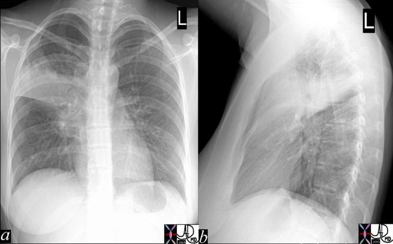
| The AP and lateral CXR shows a right upper lobe pneumonia actually localized to the right upper lobe on the PA view (a) while on the lateral view it appears that the posterior segment of the right upper lobe is involved. In this patient the pleuritic pain may be felt in the back.
41800c Courtesy Ashley Davidoff MD medical students code chest infiltrate lung pneumonia right upper lobe RUL |

Hemorrhagic Pneumonia |
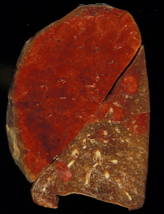
| The gross pathology specimen shows a hemorrhagic lobar pneumonia (red hepatisation) in the left upper lobe with patchy subsegmental bronchopneumonic disease in the lower lobe. The relationship to the surface pleura may result in pleuritic pain.
Courtesy Jeffrey Pierce and Ashley Davidoff MD 32320 |
An infection of the trachea or bronchi, tracheobronchitis, may present with sternal chest pain in a patient. The patient may also experience some discomfort in breathing and a cough that can worsen the pain.
DOMElement Object
(
    [schemaTypeInfo] =>
    [tagName] => table
    [firstElementChild] => (object value omitted)
    [lastElementChild] => (object value omitted)
    [childElementCount] => 1
    [previousElementSibling] => (object value omitted)
    [nextElementSibling] => (object value omitted)
    [nodeName] => table
    [nodeValue] =>


Hemorrhagic Pneumonia


The  gross pathology specimen shows a hemorrhagic lobar pneumonia (red hepatisation)  in the left upper lobe with patchy subsegmental bronchopneumonic disease in the lower lobe.  The relationship to the surface pleura may result in pleuritic pain.
Courtesy Jeffrey Pierce and Ashley Davidoff MD 32320


    [nodeType] => 1
    [parentNode] => (object value omitted)
    [childNodes] => (object value omitted)
    [firstChild] => (object value omitted)
    [lastChild] => (object value omitted)
    [previousSibling] => (object value omitted)
    [nextSibling] => (object value omitted)
    [attributes] => (object value omitted)
    [ownerDocument] => (object value omitted)
    [namespaceURI] =>
    [prefix] =>
    [localName] => table
    [baseURI] =>
    [textContent] =>


Hemorrhagic Pneumonia


The  gross pathology specimen shows a hemorrhagic lobar pneumonia (red hepatisation)  in the left upper lobe with patchy subsegmental bronchopneumonic disease in the lower lobe.  The relationship to the surface pleura may result in pleuritic pain.
Courtesy Jeffrey Pierce and Ashley Davidoff MD 32320


)
DOMElement Object
(
    [schemaTypeInfo] =>
    [tagName] => td
    [firstElementChild] => (object value omitted)
    [lastElementChild] => (object value omitted)
    [childElementCount] => 1
    [previousElementSibling] =>
    [nextElementSibling] =>
    [nodeName] => td
    [nodeValue] => The  gross pathology specimen shows a hemorrhagic lobar pneumonia (red hepatisation)  in the left upper lobe with patchy subsegmental bronchopneumonic disease in the lower lobe.  The relationship to the surface pleura may result in pleuritic pain.
Courtesy Jeffrey Pierce and Ashley Davidoff MD 32320
    [nodeType] => 1
    [parentNode] => (object value omitted)
    [childNodes] => (object value omitted)
    [firstChild] => (object value omitted)
    [lastChild] => (object value omitted)
    [previousSibling] => (object value omitted)
    [nextSibling] => (object value omitted)
    [attributes] => (object value omitted)
    [ownerDocument] => (object value omitted)
    [namespaceURI] =>
    [prefix] =>
    [localName] => td
    [baseURI] =>
    [textContent] => The  gross pathology specimen shows a hemorrhagic lobar pneumonia (red hepatisation)  in the left upper lobe with patchy subsegmental bronchopneumonic disease in the lower lobe.  The relationship to the surface pleura may result in pleuritic pain.
Courtesy Jeffrey Pierce and Ashley Davidoff MD 32320
)
DOMElement Object
(
    [schemaTypeInfo] =>
    [tagName] => td
    [firstElementChild] => (object value omitted)
    [lastElementChild] => (object value omitted)
    [childElementCount] => 2
    [previousElementSibling] =>
    [nextElementSibling] =>
    [nodeName] => td
    [nodeValue] =>
Hemorrhagic Pneumonia
    [nodeType] => 1
    [parentNode] => (object value omitted)
    [childNodes] => (object value omitted)
    [firstChild] => (object value omitted)
    [lastChild] => (object value omitted)
    [previousSibling] => (object value omitted)
    [nextSibling] => (object value omitted)
    [attributes] => (object value omitted)
    [ownerDocument] => (object value omitted)
    [namespaceURI] =>
    [prefix] =>
    [localName] => td
    [baseURI] =>
    [textContent] =>
Hemorrhagic Pneumonia
)
DOMElement Object
(
    [schemaTypeInfo] =>
    [tagName] => table
    [firstElementChild] => (object value omitted)
    [lastElementChild] => (object value omitted)
    [childElementCount] => 1
    [previousElementSibling] => (object value omitted)
    [nextElementSibling] => (object value omitted)
    [nodeName] => table
    [nodeValue] =>


Right Upper Lobe Pneumonia


The AP and lateral CXR shows a right upper lobe pneumonia actually localized to the right upper lobe on the PA view (a) while on the lateral view it appears that the posterior segment of the right upper lobe is involved.  In this patient the pleuritic pain may be felt in the back.
41800c Courtesy Ashley Davidoff MD medical students code chest infiltrate lung pneumonia right upper lobe RUL


    [nodeType] => 1
    [parentNode] => (object value omitted)
    [childNodes] => (object value omitted)
    [firstChild] => (object value omitted)
    [lastChild] => (object value omitted)
    [previousSibling] => (object value omitted)
    [nextSibling] => (object value omitted)
    [attributes] => (object value omitted)
    [ownerDocument] => (object value omitted)
    [namespaceURI] =>
    [prefix] =>
    [localName] => table
    [baseURI] =>
    [textContent] =>


Right Upper Lobe Pneumonia


The AP and lateral CXR shows a right upper lobe pneumonia actually localized to the right upper lobe on the PA view (a) while on the lateral view it appears that the posterior segment of the right upper lobe is involved.  In this patient the pleuritic pain may be felt in the back.
41800c Courtesy Ashley Davidoff MD medical students code chest infiltrate lung pneumonia right upper lobe RUL


)
DOMElement Object
(
    [schemaTypeInfo] =>
    [tagName] => td
    [firstElementChild] => (object value omitted)
    [lastElementChild] => (object value omitted)
    [childElementCount] => 1
    [previousElementSibling] =>
    [nextElementSibling] =>
    [nodeName] => td
    [nodeValue] => The AP and lateral CXR shows a right upper lobe pneumonia actually localized to the right upper lobe on the PA view (a) while on the lateral view it appears that the posterior segment of the right upper lobe is involved.  In this patient the pleuritic pain may be felt in the back.
41800c Courtesy Ashley Davidoff MD medical students code chest infiltrate lung pneumonia right upper lobe RUL
    [nodeType] => 1
    [parentNode] => (object value omitted)
    [childNodes] => (object value omitted)
    [firstChild] => (object value omitted)
    [lastChild] => (object value omitted)
    [previousSibling] => (object value omitted)
    [nextSibling] => (object value omitted)
    [attributes] => (object value omitted)
    [ownerDocument] => (object value omitted)
    [namespaceURI] =>
    [prefix] =>
    [localName] => td
    [baseURI] =>
    [textContent] => The AP and lateral CXR shows a right upper lobe pneumonia actually localized to the right upper lobe on the PA view (a) while on the lateral view it appears that the posterior segment of the right upper lobe is involved.  In this patient the pleuritic pain may be felt in the back.
41800c Courtesy Ashley Davidoff MD medical students code chest infiltrate lung pneumonia right upper lobe RUL
)
DOMElement Object
(
    [schemaTypeInfo] =>
    [tagName] => td
    [firstElementChild] => (object value omitted)
    [lastElementChild] => (object value omitted)
    [childElementCount] => 2
    [previousElementSibling] =>
    [nextElementSibling] =>
    [nodeName] => td
    [nodeValue] =>
Right Upper Lobe Pneumonia
    [nodeType] => 1
    [parentNode] => (object value omitted)
    [childNodes] => (object value omitted)
    [firstChild] => (object value omitted)
    [lastChild] => (object value omitted)
    [previousSibling] => (object value omitted)
    [nextSibling] => (object value omitted)
    [attributes] => (object value omitted)
    [ownerDocument] => (object value omitted)
    [namespaceURI] =>
    [prefix] =>
    [localName] => td
    [baseURI] =>
    [textContent] =>
Right Upper Lobe Pneumonia
)
DOMElement Object
(
    [schemaTypeInfo] =>
    [tagName] => table
    [firstElementChild] => (object value omitted)
    [lastElementChild] => (object value omitted)
    [childElementCount] => 1
    [previousElementSibling] => (object value omitted)
    [nextElementSibling] => (object value omitted)
    [nodeName] => table
    [nodeValue] =>


Sharp Intense Pleuritic Pain on Inspiration Due to Peripheral Based Pneumonia


The pleuritic pain of pulmonary embolus, pneumonia or pleurisy is not distinct in itself, but the associated findings relating to the specific disease will help differentiate the variety of causes of pleuritic chest pain.
In this instance a periphral based pneumonia abuts the pleural surface and parapneumonic pleural inflammation involves both the visceral pleura and the parietal pleura.  When the lung moves with respiration the rubbing of inflammed structures and particulalrly the pleura brings about the sharp focal pleuritic pain localized over the pneumonia.
42540c02a01  lung inspiration expiration pain on inspiration focal severe pain pleuritic pain PE pulmonary embolus Davidoff art Courtesy Ashley Davidoff MD


    [nodeType] => 1
    [parentNode] => (object value omitted)
    [childNodes] => (object value omitted)
    [firstChild] => (object value omitted)
    [lastChild] => (object value omitted)
    [previousSibling] => (object value omitted)
    [nextSibling] => (object value omitted)
    [attributes] => (object value omitted)
    [ownerDocument] => (object value omitted)
    [namespaceURI] =>
    [prefix] =>
    [localName] => table
    [baseURI] =>
    [textContent] =>


Sharp Intense Pleuritic Pain on Inspiration Due to Peripheral Based Pneumonia


The pleuritic pain of pulmonary embolus, pneumonia or pleurisy is not distinct in itself, but the associated findings relating to the specific disease will help differentiate the variety of causes of pleuritic chest pain.
In this instance a periphral based pneumonia abuts the pleural surface and parapneumonic pleural inflammation involves both the visceral pleura and the parietal pleura.  When the lung moves with respiration the rubbing of inflammed structures and particulalrly the pleura brings about the sharp focal pleuritic pain localized over the pneumonia.
42540c02a01  lung inspiration expiration pain on inspiration focal severe pain pleuritic pain PE pulmonary embolus Davidoff art Courtesy Ashley Davidoff MD


)
DOMElement Object
(
    [schemaTypeInfo] =>
    [tagName] => td
    [firstElementChild] => (object value omitted)
    [lastElementChild] => (object value omitted)
    [childElementCount] => 2
    [previousElementSibling] =>
    [nextElementSibling] =>
    [nodeName] => td
    [nodeValue] => The pleuritic pain of pulmonary embolus, pneumonia or pleurisy is not distinct in itself, but the associated findings relating to the specific disease will help differentiate the variety of causes of pleuritic chest pain.
In this instance a periphral based pneumonia abuts the pleural surface and parapneumonic pleural inflammation involves both the visceral pleura and the parietal pleura.  When the lung moves with respiration the rubbing of inflammed structures and particulalrly the pleura brings about the sharp focal pleuritic pain localized over the pneumonia.
42540c02a01  lung inspiration expiration pain on inspiration focal severe pain pleuritic pain PE pulmonary embolus Davidoff art Courtesy Ashley Davidoff MD
    [nodeType] => 1
    [parentNode] => (object value omitted)
    [childNodes] => (object value omitted)
    [firstChild] => (object value omitted)
    [lastChild] => (object value omitted)
    [previousSibling] => (object value omitted)
    [nextSibling] => (object value omitted)
    [attributes] => (object value omitted)
    [ownerDocument] => (object value omitted)
    [namespaceURI] =>
    [prefix] =>
    [localName] => td
    [baseURI] =>
    [textContent] => The pleuritic pain of pulmonary embolus, pneumonia or pleurisy is not distinct in itself, but the associated findings relating to the specific disease will help differentiate the variety of causes of pleuritic chest pain.
In this instance a periphral based pneumonia abuts the pleural surface and parapneumonic pleural inflammation involves both the visceral pleura and the parietal pleura.  When the lung moves with respiration the rubbing of inflammed structures and particulalrly the pleura brings about the sharp focal pleuritic pain localized over the pneumonia.
42540c02a01  lung inspiration expiration pain on inspiration focal severe pain pleuritic pain PE pulmonary embolus Davidoff art Courtesy Ashley Davidoff MD
)
DOMElement Object
(
    [schemaTypeInfo] =>
    [tagName] => td
    [firstElementChild] => (object value omitted)
    [lastElementChild] => (object value omitted)
    [childElementCount] => 2
    [previousElementSibling] =>
    [nextElementSibling] =>
    [nodeName] => td
    [nodeValue] =>
Sharp Intense Pleuritic Pain on Inspiration Due to Peripheral Based Pneumonia
    [nodeType] => 1
    [parentNode] => (object value omitted)
    [childNodes] => (object value omitted)
    [firstChild] => (object value omitted)
    [lastChild] => (object value omitted)
    [previousSibling] => (object value omitted)
    [nextSibling] => (object value omitted)
    [attributes] => (object value omitted)
    [ownerDocument] => (object value omitted)
    [namespaceURI] =>
    [prefix] =>
    [localName] => td
    [baseURI] =>
    [textContent] =>
Sharp Intense Pleuritic Pain on Inspiration Due to Peripheral Based Pneumonia
)


