Author Ashley Davidoff

Pulmonary Hypertension – Scleroderma
|
|
This unfortunate 32 year old female with scleroderma has many causes for chest pain but the cause under discussion is a relatively uncommon cause for chest pain – pulmonary hypertension. This is best diagnosed with echocardiogram or cardiac catherization but in this CT the large pulmonary artery (blue – which should be the same size of the red aorta normally) is indicative of pulmonary hypertension. Her heart is enlarged (a) and she has a pericardial effusion, and bilateral pleural effusions (d) both of which can cause pain. In addition she has an enlarged patulous esophagus (seen as a black slit behind the heart (d) characteristic of scleroderma, and associated with reflux which is another cause for chest pain.
30464c12 32 female lungs pleura heart cardiac RA RV right ventricle right atrium pericardium esophagus ILD basal interstitial lung disease pericardial effusion pleural effusion cardiomegaly enlarged esophagus patulous esophagus gallbladder wall edema congestive cardiac failure RHF right heart failure rifht ventricular enlargement RVE RAE right atrial enlargement pulmonary hypertension cor pulmonale dx scleroderma Courtesy Ashley Davidoff MD |
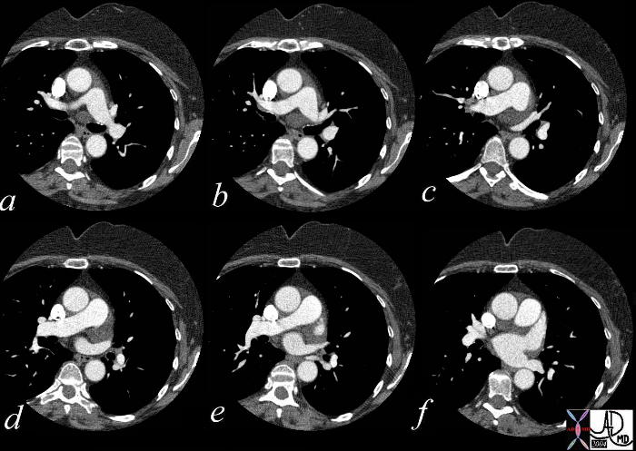
This series of axial CTscans shows the antomy of the superior recesses of the pericardium. Note that fluid can be seen anterior to the pulmonary artery, (a-f) to the left of the MPA posterior to the bifurcation of MPA, posterior and to the left of the ascending aorta (a-f) and superior to the LAA. (d,e) Courtesy Ashley Davidoff MD.
00164.jpg
The mediastinal windows of the CTscan of this patient who had a sharp penetrating trauma shows pericardial fluid which has to be considered a hemorrhagic pericardial effusion and monitored very carefully. Courtesy Ashley Davidoff MD. 00164 code cardiac accessory interesting pericardium heart trauma imaging radiology CTscan
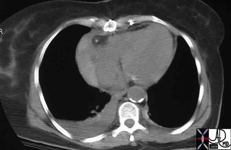
28761b01 s/p cardiac surgery heart pericardium fx high density accumulation right cardiac dx hemorhagic pericardial effusion dx hemopericardium imaging radiology CTscan Courtesy Ashley Davidoff MD
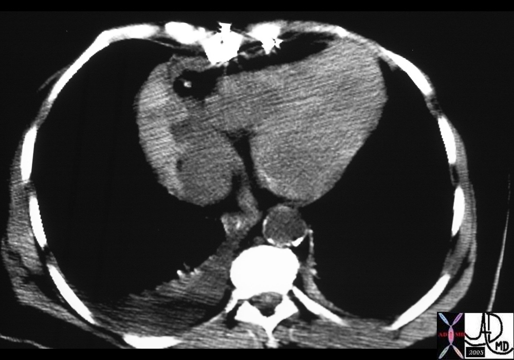
28761 s/p cardiac surgery heart pericardium fx high density accumulation right cardiac dx hemorhagic pericardial effusion dx hemopericardium imaging radiology CTscan Courtesy Ashley Davidoff MD

A low density mass is seen in intimate association with the pericardium which itself demonstrates a small amount of fluid. The water density measured is reminiscent of a pericardial cyst. Courtesy Ashley Davidoff MD. 15647
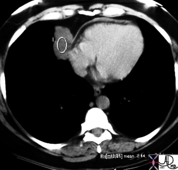
A low density mass is seen in intimate association with the pericardium which itself demonstrates a small amount of fluid. The water density measured at 8.4 HU is reminiscent of a pericardial cyst. Courtesy Ashley Davidoff MD. 15648
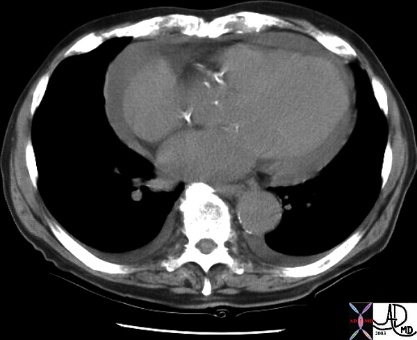
This CT shows a moderate pericardial effusion. Courtesy Ashley Davidoff MD. 16796
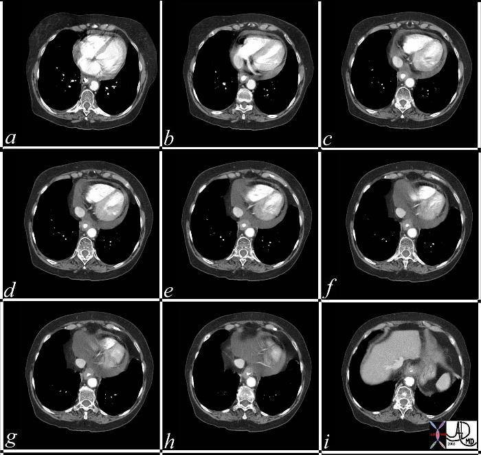
This combination of CT images through the heart show a pericardial effusion associated wih a thick walled and “stiff” esophagus in this patient with known carcinoma of the distal esophagus. The effusion is most likely from direct extension of the malignat process. Courtesy Ashley Davidoff MD 37284c

This combination of images are from a patient with mesothelioma associated with asbestos related disease. Note the pleural plaques in images 3,4,5, the contracted hemithorax (1), the pleural origin of the density (2) characterised by blunted costophrenic angle, and the rind of soft tissue around the heart (3) and in the posterior costophrenic spavce. (3,4). The pathology shows a combination of malignant proliferation of stromal and glandular elements (7) abutting relatively normal lung (6) Courtesy Ashley Davidoff MD. 32215c
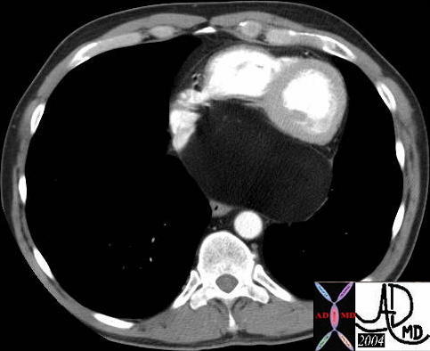
This axial CTscan from an asymptomatic patient reveals a psoterior mediastinal fatty mass displacing right ventricle and left ventricle forward. The mass appears to arise from the pericardium and a diagnosis of lipoma of the pericardium is most likely. Courtesy Ashley Davidoff MD and Gerard Hayes MD. 38433
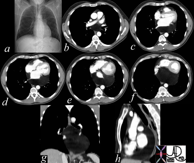
This series of plain film (a) and CTscan axial (b,c,d,e,f) coronal (g) and sagittal (h) images, from an asymptomatic patient reveal a posterior mediastinal fatty mass displacing the left bronchus upward (a) and pushing the left atrium , right ventricle and left ventricle forward. The mass appears to arise from the pericadium and a diagnosis of lipoma of the pericardium is most likely. Courtesy Ashley Davidoff MD and Gerard Hayes MD. 38436c
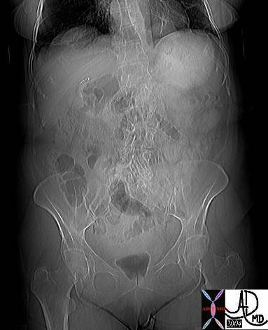
What is the finding for you detectives with sharp eyes? The scout film from a CTscan shows a left retrocardiac density. What is your diagnosis? Courtesy Ashley Davidoff MD Bochdalek hernia 39226i
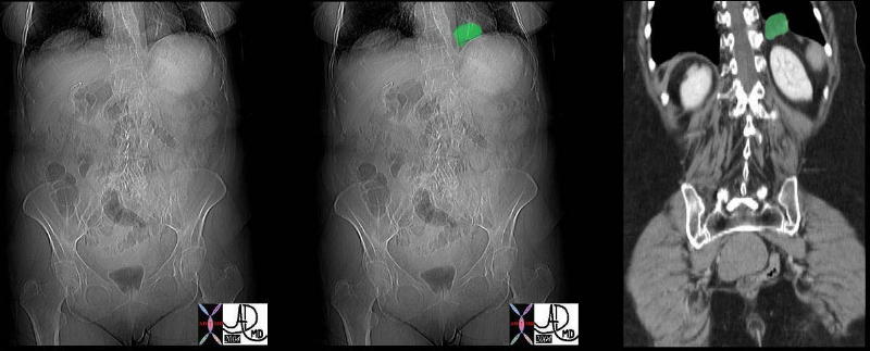
The scout film shows a left retrocardiac density that is of lower density (a) highlighted in green in b, and seen on the coronal CTscan as a herniation of fat through the diaphragm in c. The finding is characteristic of a Bochdalek hernia Courtesy Ashley Davidoff MD 39235c
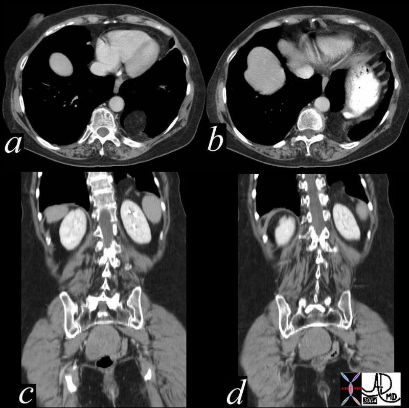
The left retrocardiac mass of fat density seen in the axial CTscans a, and b and the coronal scans in c and d, is reminiscent of fat herniating through a diaphragmatic defect. The finding is characteristic of a Bochdalek hernia Courtesy Ashley Davidoff MD 39235c01
Pneumopericardium
31-M-bilat-PTX-and-ppneumopericardium-03.jpg
TRAUMATIC PNEUMOPERICARDIUM and BILATERAL PNEUMOTHORACES
Ashley Davidoff MD
31-M-bilat-PTX-and-ppneumopericardium-01.jpg
TRAUMATIC PNEUMOPERICARDIUM and BILATERAL PNEUMOTHORACES
Ashley Davidoff MD
31-M-bilat-PTX-and-ppneumopericardium-02.jpg
TRAUMATIC PNEUMOPERICARDIUM and BILATERAL PNEUMOTHORACES
Ashley Davidoff MD
DOMElement Object
(
    [schemaTypeInfo] =>
    [tagName] => table
    [firstElementChild] => (object value omitted)
    [lastElementChild] => (object value omitted)
    [childElementCount] => 1
    [previousElementSibling] => (object value omitted)
    [nextElementSibling] => (object value omitted)
    [nodeName] => table
    [nodeValue] =>


Pulmonary Hypertension – Scleroderma


This unfortunate 32 year old female with scleroderma has many causes for chest pain but the cause under discussion is a relatively uncommon cause for chest pain – pulmonary hypertension.  This is best diagnosed with echocardiogram or cardiac catherization but in this CT the large pulmonary artery (blue – which should be the same size of the red aorta normally) is indicative of pulmonary hypertension.  Her heart is enlarged (a) and she has a pericardial effusion, and bilateral pleural effusions (d) both of which can cause pain.  In addition she has an enlarged patulous esophagus (seen as a black slit behind the heart (d) characteristic of scleroderma, and associated with reflux which is another cause for chest pain.
30464c12 32 female lungs pleura heart cardiac RA RV right ventricle right atrium pericardium esophagus ILD basal interstitial lung disease pericardial effusion pleural effusion cardiomegaly enlarged esophagus patulous esophagus gallbladder wall edema congestive cardiac failure RHF right heart failure rifht ventricular enlargement RVE RAE right atrial enlargement pulmonary hypertension cor pulmonale dx scleroderma Courtesy Ashley Davidoff MD


    [nodeType] => 1
    [parentNode] => (object value omitted)
    [childNodes] => (object value omitted)
    [firstChild] => (object value omitted)
    [lastChild] => (object value omitted)
    [previousSibling] => (object value omitted)
    [nextSibling] => (object value omitted)
    [attributes] => (object value omitted)
    [ownerDocument] => (object value omitted)
    [namespaceURI] =>
    [prefix] =>
    [localName] => table
    [baseURI] =>
    [textContent] =>


Pulmonary Hypertension – Scleroderma


This unfortunate 32 year old female with scleroderma has many causes for chest pain but the cause under discussion is a relatively uncommon cause for chest pain – pulmonary hypertension.  This is best diagnosed with echocardiogram or cardiac catherization but in this CT the large pulmonary artery (blue – which should be the same size of the red aorta normally) is indicative of pulmonary hypertension.  Her heart is enlarged (a) and she has a pericardial effusion, and bilateral pleural effusions (d) both of which can cause pain.  In addition she has an enlarged patulous esophagus (seen as a black slit behind the heart (d) characteristic of scleroderma, and associated with reflux which is another cause for chest pain.
30464c12 32 female lungs pleura heart cardiac RA RV right ventricle right atrium pericardium esophagus ILD basal interstitial lung disease pericardial effusion pleural effusion cardiomegaly enlarged esophagus patulous esophagus gallbladder wall edema congestive cardiac failure RHF right heart failure rifht ventricular enlargement RVE RAE right atrial enlargement pulmonary hypertension cor pulmonale dx scleroderma Courtesy Ashley Davidoff MD


)
DOMElement Object
(
    [schemaTypeInfo] =>
    [tagName] => td
    [firstElementChild] => (object value omitted)
    [lastElementChild] => (object value omitted)
    [childElementCount] => 2
    [previousElementSibling] =>
    [nextElementSibling] =>
    [nodeName] => td
    [nodeValue] =>
This unfortunate 32 year old female with scleroderma has many causes for chest pain but the cause under discussion is a relatively uncommon cause for chest pain – pulmonary hypertension.  This is best diagnosed with echocardiogram or cardiac catherization but in this CT the large pulmonary artery (blue – which should be the same size of the red aorta normally) is indicative of pulmonary hypertension.  Her heart is enlarged (a) and she has a pericardial effusion, and bilateral pleural effusions (d) both of which can cause pain.  In addition she has an enlarged patulous esophagus (seen as a black slit behind the heart (d) characteristic of scleroderma, and associated with reflux which is another cause for chest pain.
30464c12 32 female lungs pleura heart cardiac RA RV right ventricle right atrium pericardium esophagus ILD basal interstitial lung disease pericardial effusion pleural effusion cardiomegaly enlarged esophagus patulous esophagus gallbladder wall edema congestive cardiac failure RHF right heart failure rifht ventricular enlargement RVE RAE right atrial enlargement pulmonary hypertension cor pulmonale dx scleroderma Courtesy Ashley Davidoff MD
    [nodeType] => 1
    [parentNode] => (object value omitted)
    [childNodes] => (object value omitted)
    [firstChild] => (object value omitted)
    [lastChild] => (object value omitted)
    [previousSibling] => (object value omitted)
    [nextSibling] => (object value omitted)
    [attributes] => (object value omitted)
    [ownerDocument] => (object value omitted)
    [namespaceURI] =>
    [prefix] =>
    [localName] => td
    [baseURI] =>
    [textContent] =>
This unfortunate 32 year old female with scleroderma has many causes for chest pain but the cause under discussion is a relatively uncommon cause for chest pain – pulmonary hypertension.  This is best diagnosed with echocardiogram or cardiac catherization but in this CT the large pulmonary artery (blue – which should be the same size of the red aorta normally) is indicative of pulmonary hypertension.  Her heart is enlarged (a) and she has a pericardial effusion, and bilateral pleural effusions (d) both of which can cause pain.  In addition she has an enlarged patulous esophagus (seen as a black slit behind the heart (d) characteristic of scleroderma, and associated with reflux which is another cause for chest pain.
30464c12 32 female lungs pleura heart cardiac RA RV right ventricle right atrium pericardium esophagus ILD basal interstitial lung disease pericardial effusion pleural effusion cardiomegaly enlarged esophagus patulous esophagus gallbladder wall edema congestive cardiac failure RHF right heart failure rifht ventricular enlargement RVE RAE right atrial enlargement pulmonary hypertension cor pulmonale dx scleroderma Courtesy Ashley Davidoff MD
)
DOMElement Object
(
    [schemaTypeInfo] =>
    [tagName] => td
    [firstElementChild] => (object value omitted)
    [lastElementChild] => (object value omitted)
    [childElementCount] => 2
    [previousElementSibling] =>
    [nextElementSibling] =>
    [nodeName] => td
    [nodeValue] =>
Pulmonary Hypertension – Scleroderma

    [nodeType] => 1
    [parentNode] => (object value omitted)
    [childNodes] => (object value omitted)
    [firstChild] => (object value omitted)
    [lastChild] => (object value omitted)
    [previousSibling] => (object value omitted)
    [nextSibling] => (object value omitted)
    [attributes] => (object value omitted)
    [ownerDocument] => (object value omitted)
    [namespaceURI] =>
    [prefix] =>
    [localName] => td
    [baseURI] =>
    [textContent] =>
Pulmonary Hypertension – Scleroderma

)


