
In a Nutshell
- Overall volume
- Shape
- LV Ovoid with icecream cone shaped cavity
- Linear Size (4,5,4,5 rule)
- LV +/- 5cms (transverse) not >6
- Wall thickness
- Measure in diastole
- LV 1.2 to 1.4cms
- Measure in diastole
Linear Measurements
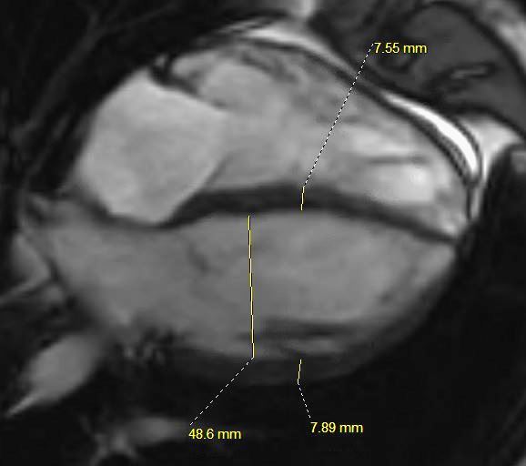
This is the MRI of a 19-year-old male who presented with syncope and the study was performed to identify a possible arrhythmogenic focus
White blood imaging using 4 chamber view shows a normal sized heart in diastole
The septum of the LV in diastole is 8mm, and the free wall is 8mms (upper limits normal is 1.2 -1.4cms. The transverse measurement of the LV cavity is 4.9cms with upper limits normal being about 5 cms.
Ashley Davidoff MD
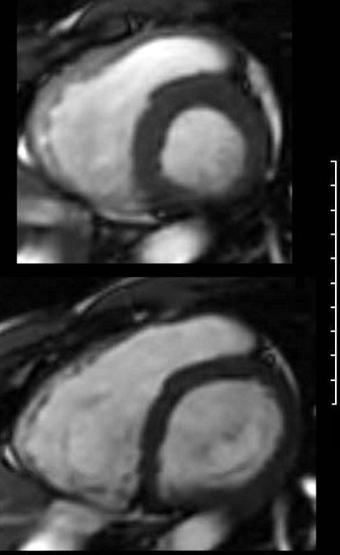
This is the MRI of a 19-year-old male who presented with syncope and the study was performed to identify a possible arrhythmogenic focus
White blood imaging using short axis view shows a normal sized heart in systole (above) and diastole (below). The left and right ventricles show normal wall thicknesses and the volume of the chambers in systole are about 2/3 the volume in diastole (normal). There is no obvious dyskinetic segment of the RVOT.
Ashley Davidoff MD
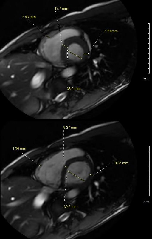
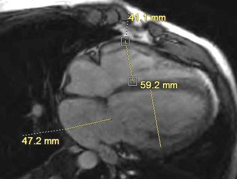
This is the MRI of a 19-year-old male who presented with syncope and the study was performed to identify a possible arrhythmogenic focus.
White blood imaging using short axis view shows a normal sized heart in systole (above) and diastole (below). The transverse dimension of the LV is 4cms in diastole which is normal. The septum of the LV in diastole (lower image) is less than 9.2mms, and the free wall is 8.7 mms (upper limits normal is 1.2cms. The wall of the RV is barely seen in diastole and measures about. In systole the residual volume of the RV is about 1/3 the diastolic volume indicating an approximate ejection of 2/3 = 66% ejection fraction (EF). Similarly, at peak LV systole the residual volume of the LV is about 1/3 the diastolic volume indicating an approximate ejection of 2/3 = 66% ejection fraction (EF).
Ashley Davidoff MD
3 Chamber LVOT
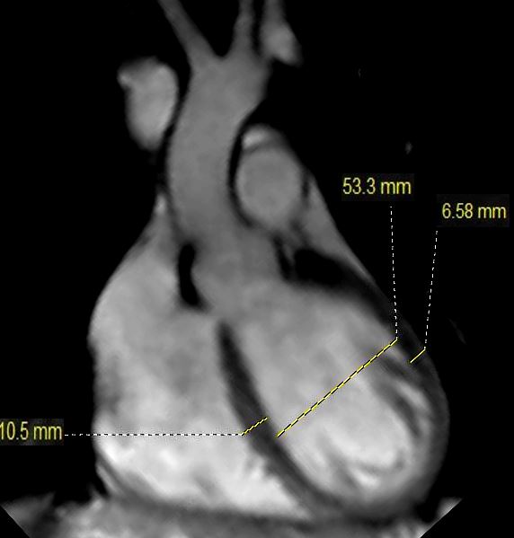
This is the MRI of a 19-year-old male who presented with syncope and the study was performed to identify a possible arrhythmogenic focus
White blood imaging of the LVOT view shows a normal sized ovoid LV in diastole. The septal thickness in diastole is 10.5mms, and bulges toward the RV while the free wall dimension is 6.6 mms. The LV cavity measures 5.3cms which is upper limits normal.
Ashley Davidoff MD
MRI confirmed the presence of a dilated cardiomyopathy, small pericardial effusion, without evidence of LGE, global hypokinesis and EF of about 20%
Ashley Davidoff MD