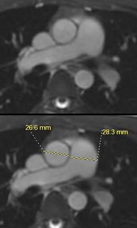
White blood imaging of the MPA and tubular portion of the ascending aorta at the level of the MPA bifurcation in the axial projection shows a normal sized MPA measuring 2.8cms (normal up to 3cms). The tubular portion of the ascending aorta measures 2.7cms (normal up to 3.5cms)
Ashley Davidoff MD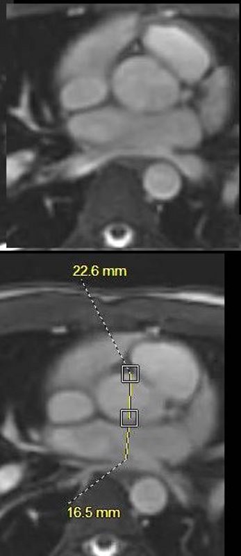
This is the MRI of a 19-year-old male who presented with syncope and the study was performed to identify a possible arrhythmogenic focus
White blood imaging of the LA in the axial projection shows a normal sized LA measuring 1.7cms (normal up to 4cms). In this plane the LA is relatively small, but normal. It is usually slightly larger than the proximal ascending aorta at this level. The aorta measures 2.3cms. Note the rectangular shape of the LA.
Ashley Davidoff MD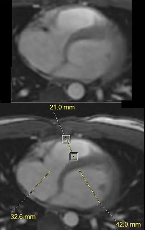
This is the MRI of a 19-year-old male who presented with syncope and the study was performed to identify a possible arrhythmogenic focus
White blood imaging of the RA RV and LV at a slightly inferior cut in the axial projection shows a normal sized RA measuring 3.3cms (normal up to 5cms). The internal diameter of the LV measures 4.2cms (+/- 5cms is normal) and the RV measures 2.1cms. (normal RV +/- 4cms).
Ashley Davidoff MD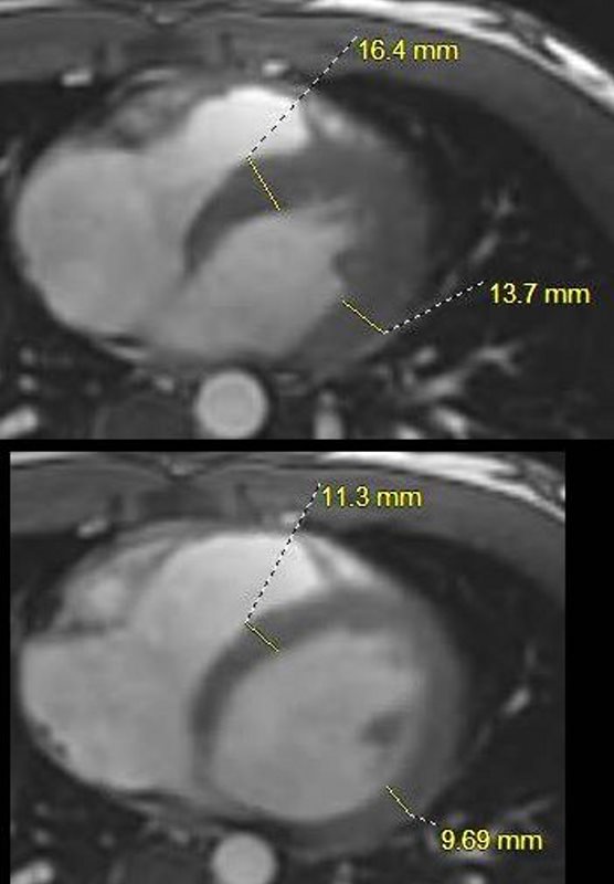
This is the MRI of a 19-year-old male who presented with syncope and the study was performed to identify a possible arrhythmogenic focus
White blood imaging of the LV in the axial projection in systole above and diastole below. In diastole the septum measures 1.1cms and the free wall measures 9.7mms (normal +/- 1.2- 1.4 cms).
Ashley Davidoff MD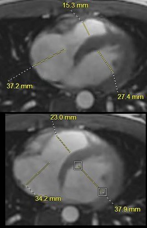
This is the MRI of a 19-year-old male who presented with syncope and the study was performed to identify a possible arrhythmogenic focus
White blood imaging of the RA, LV and RV in the axial projection in systole above and diastole below. In diastole the RA measures 3.4cms (n= up to about 5cms) the RV measures 2.3cms (n= up to about 4 or 4.5cms) and the LV wall measures 3.8cms (normal up to 5 or 5.5cms). The volume of the RV is about 2/3 the size of the LV and the RA volume appears about 1/3 the size of the RV.
Ashley Davidoff MD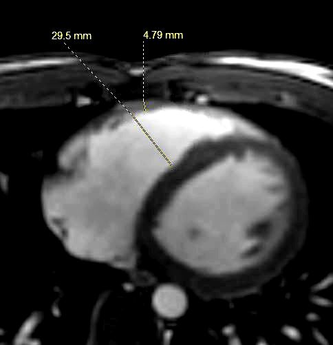
This is the MRI of a 19-year-old male who presented with syncope and the study was performed to identify a possible arrhythmogenic focus
White blood imaging of the RV in the axial projection in diastole shows a transverse diameter of the RV of 2.95cms., (up to 4-5cms) which is normal and an RV wall thickness 4.8mm (normal up to about 5mms. ) The volume of the RV is about 2/3 the size of the LV.
Ashley Davidoff MD
