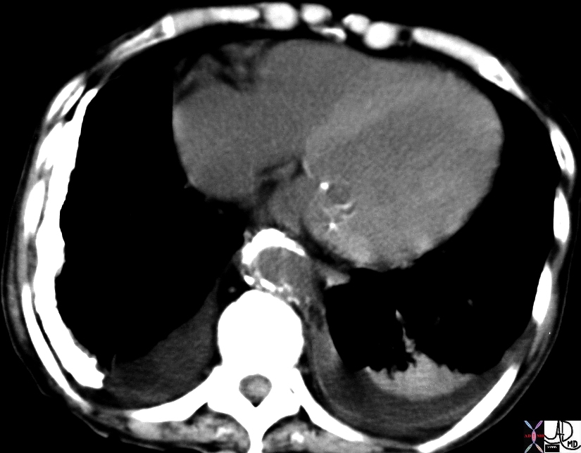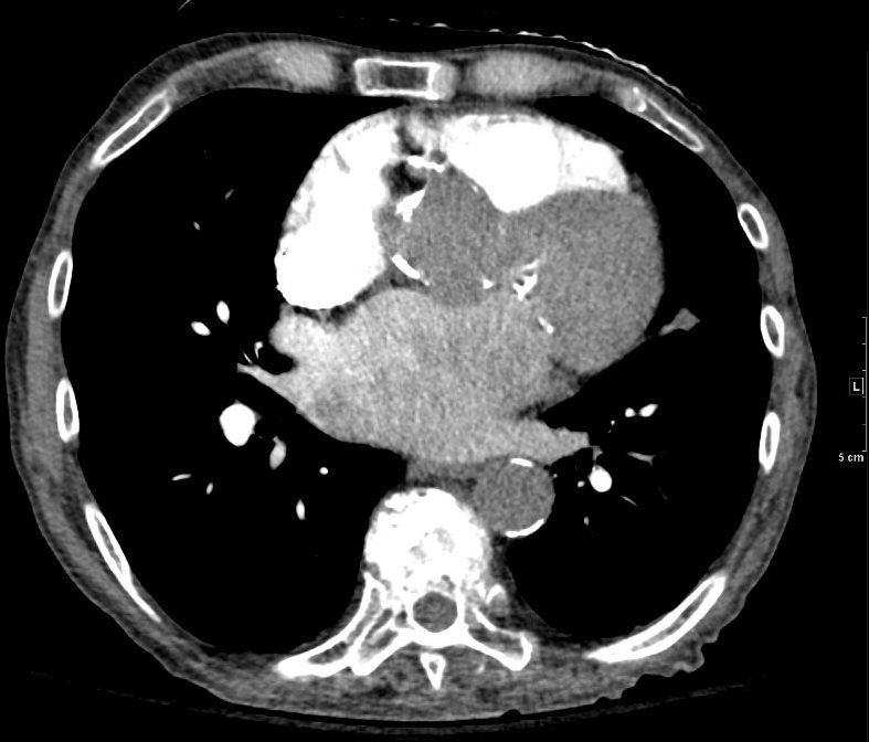
87 year old male with findings consistent with mitral stenosis , characterised by calcified mitral valve, enlarged left atrium, normal sized left ventricle,with enlarged right atrium, right ventricle and main pulmonary artery.
Pure mitral stenosis is most commonly caused by rheumatic heart disease
Ashley Davidoff MD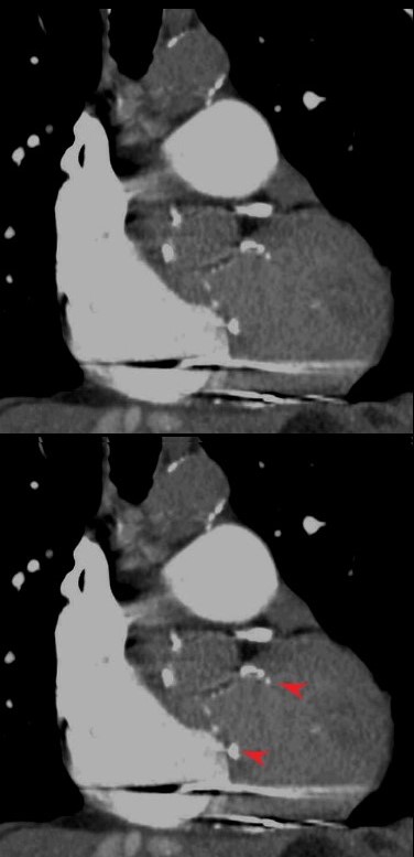
MITRAL VALVE CALCIFICATION
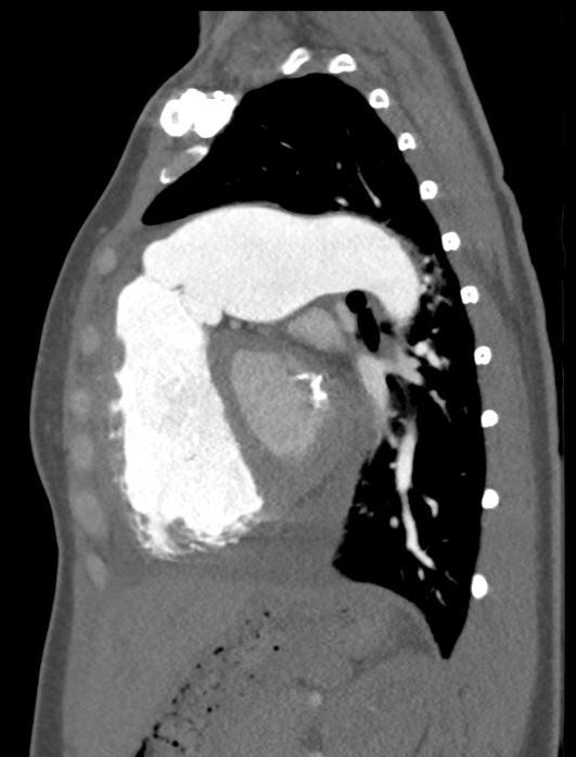
This chest CT through the middle of the left ventricle (LV) shows some calcification at the base of the mitral valve (MV) focal calcification of the anterior leaflet and and increase density of both leaflets. This patient had no known mitral valve disease and the findings most likely represent dystrophic calcification due to mucinous degeneration.
Courtesy Ashley Davidoff MD. 19729 code heart mitral valve MV calcification cardiac imaging radiology CTscan
MITRAL VALVE CALCIFICATION POSSIBLY RHEUMATIC IN ORIGIN
This chest CT through the middle of the left ventricle (LV) shows some calcification at the base of the mitral valve (MV) focal calcification of the anterior leaflet and and increase density of both leaflets. This patient had no known mitral valve disease and the findings most likely represent dystrophic calcification due to mucinous degeneration.
KEY WORDS heart pleural calcification cardiac interventricular septum IVS LV mitral valve calcification rheumatic heart disease probable chronic hemothorax or empyema anemia fx relatively dense interventricular septum due to anemia CT scan 19730 Courtesy Ashley Davidoff MD
SLE and PULMONARY HYPERTENSION without ILD
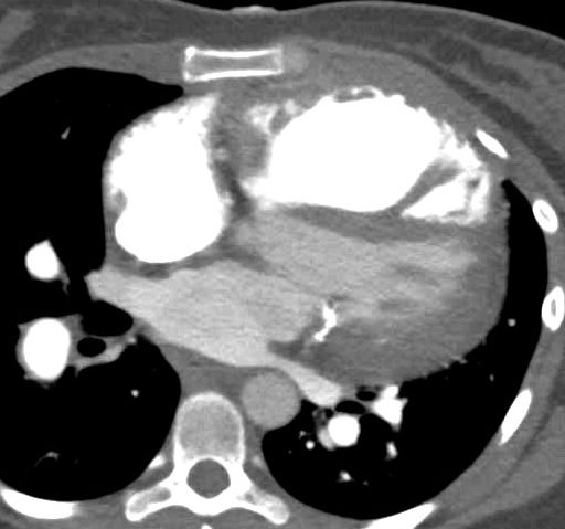
27-year-old female presents with dyspnea and a past history of SLE, Raynaud?s disease, and Lupus nephritis.
The CT scan confirms an enlarged MPA, RPA, RA and RV, and shows calcification on the posterior leaflet of the mitral valve consistent with Libman Sacks vegetation.
Ashley Davidoff MD


