
Mitral Valve Anatomy
The Common Vein Copyright 2008
Definition
- The mitral valve is a complex,
- bileaflet finely coordinated structure
- usually located
- cephalad in comparison with the tricuspid valve.
- Structurally the mitral valvular complex comprises of the
- fibro elastic annulus,
- two leaflets, the tendinous cords,
- two papillary muscles with their left ventricular attachments
Functionally it serves to prevents backflow to the left atrium during ventricular systole.
Diseases of the mitral valve may be either due to obstruction as in mitral stenosis or incompetence as in mitral regurgitation, both of which may be either congenital or acquired.
- Diagnosis of a mitral valvular disease a
- mitral stenosis
- low pitched mid diastolic rumbling murmur in the apical area
- mitral regurgitation
- high pitched pan systolic murmur
- On chest radiography, the characteristic findings of mitral stenosis are
- pulmonary congestion,
- enlargement of the main pulmonary arteries, and
- enlargement of the left atrium without cardiomegaly while
- mitral regurgitation is
- cardiomegaly.
- mitral stenosis
- electrocardiogram (ECG)
- left atrial enlargement or
- atrial fibrillation in both conditions.
- Two dimensional echocardiogram is the
- noninvasive diagnostic test of choice for both lesions.
Medical therapy for congestive heart failure or infective endocarditis, minimally invasive techniques and surgical options are available.
Structural Considerations
The complex mitral valve includes a tensor apparatus ( Fibrous annulus, LV myocardium between the annulus and attachment of base of papillary muscles, papillary muscles and the chordae) and a valvular apparatus ( two mitral leaflets )
“Mitral leaflets: The term “mitral” was introduced by Andreas Vesalius3 to describe the orientation of the two leaflets of the left atrioventricular valve owing to their resemblance to a plan view of the bishop’s mitre.
The two leaflets anterior (aortic) and posterior (mural)are notably different in shape and circumferential length with neither attached to the septum , unlike the tricuspid valve which has a septal leaflet1 . The anterior (aortic) leaflet has a rounded free edge, occupies a third of the annular circumference and a base to free edge length 2 or more times the posterior. Consequently, the anterior leaflet is more mobile, while the posterior leaflet fulfills a secondary or supporting role. The posterior mural leaflet is long and narrow lining the remainder of the circumference. However Both leaflets are thick at the bases and tips, with central thinning and with nearly identical surface ares.
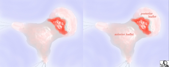
The mitral valve consists of two leaflets; an anterior shield shaped leaflet that extends deep into the LV, and a posterior leaflet that occupies a greater percentage of the circumference of the annulus, but is more shallow in its extension into LV.
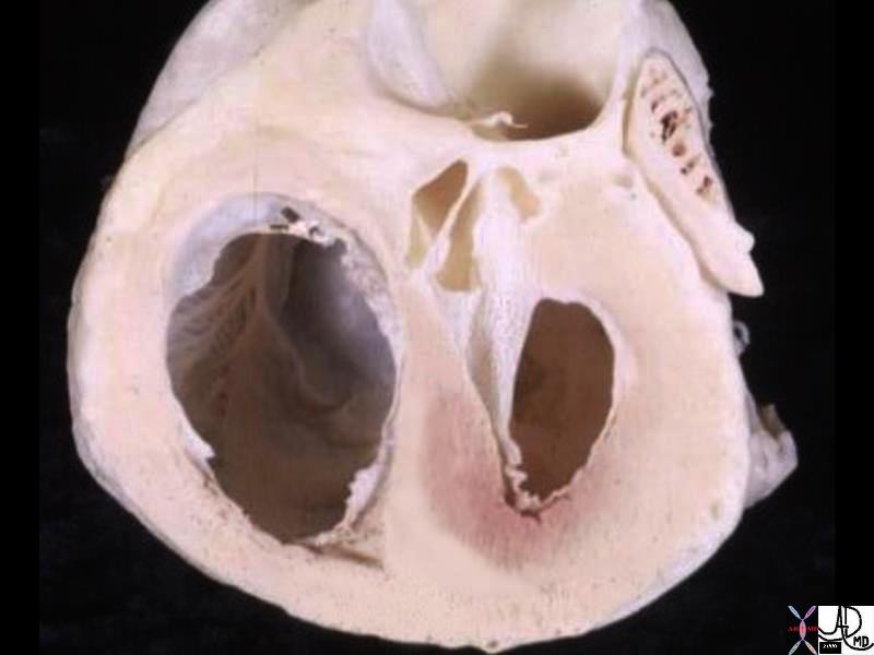
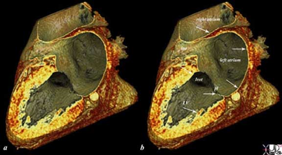
code cardiac heart normal MV mitral valve anatomy Courtesy of Ashley Davidoff M.D.32116
Ashley Davidoff MD TheCommonVein.net 15513
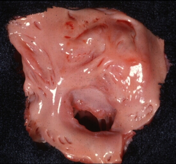
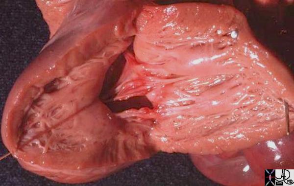
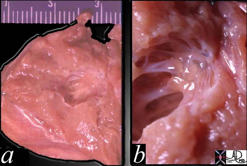
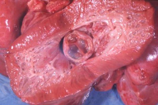
The anatomic specimen taken from the left atrium through the mitral annulus shows the broad anterior leaflet of the mitral valve ..
Courtesy of Ashley Davidoff M.D. code cardiac heart normal MV mitral valve anatomy 32106
Ashley Davidoff MD TheCommonVein.net 06546
Ashley Davidoff MD
01592
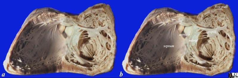
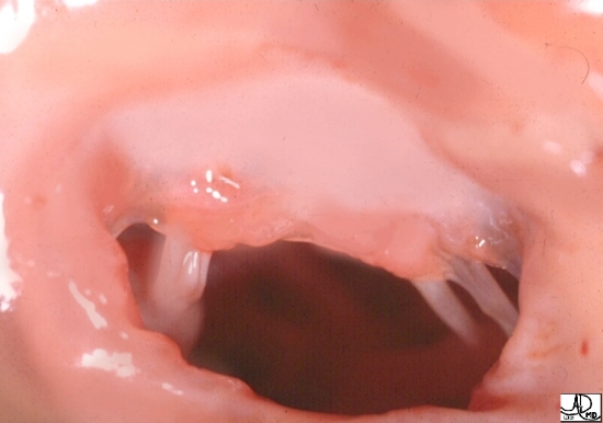
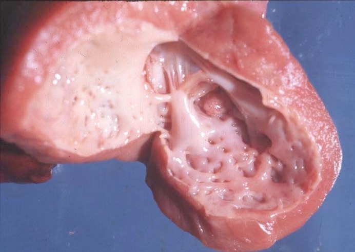
This normal anatomical specimen has been sectioned in long axis along the ventricular septum; then opened like a book with the septal wall seen on the left side of the image, and the free wall with the pair of papillary muscles seen on the right of the image. Note the fibrous continuity of the anterior leaflet of the mitral valve with the cusps of the aortic valve..
code 15389c05.8s Courtesy Ashley Davidoff MD copyright 2009
Ashley Davidoff MD
- Papillary muscles and LV myocardium:
- two papillary muscles –
- anterolateral and
- dual blood supply-
- first obtuse marginal branch from the left circumflex and the
- first diagonal branch from the left anterior descending artery.
- dual blood supply-
- posteromedial
- supplied by a single artery arising from either the right coronary artery or from the third obtuse marginal of the left circumflex artery
- anterolateral and
- two papillary muscles –
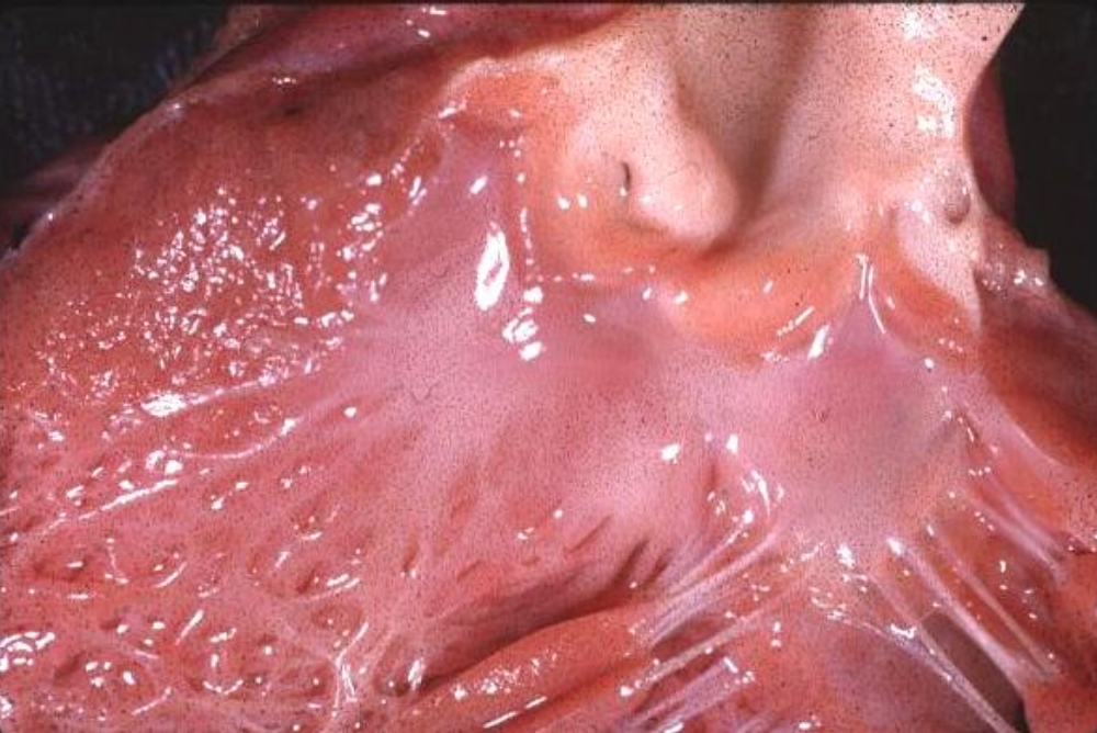
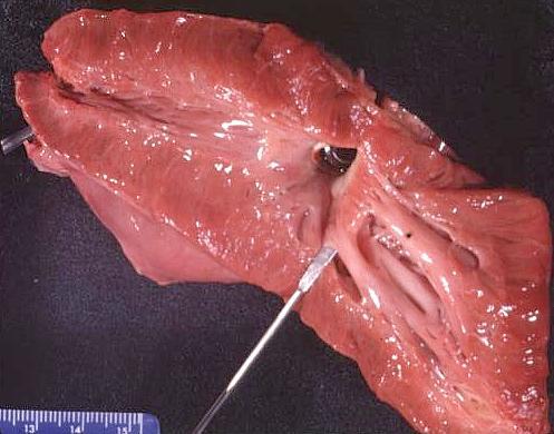
This anatomic specimen shows the LV with septal side to the patients right and free wall side to the patients left. Note the relatively smooth walled septal surface and the papillary muscle apparatus attached to the free wall side. There are two groups of papillary muscles neither of which are attached to the septum. The anterior leaflet of the MV is in full view and note that it is in fibrous continuity with the left and non coronary cusps of the aortic valve.
Courtesy Ashley Davidoff MD 01817
- Alterations in the size and shape of the left ventricle can
- distort papillary muscles,
- resulting in MR.
- Similarly rupture of the papillary muscle subsequent to infarction
- leads to mitral regurgitation.
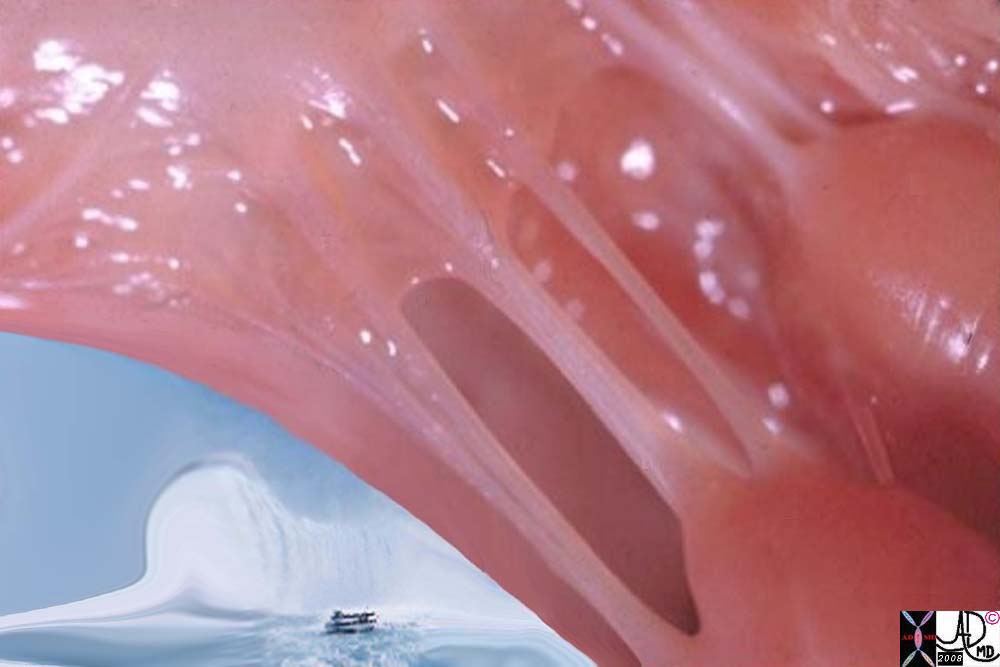
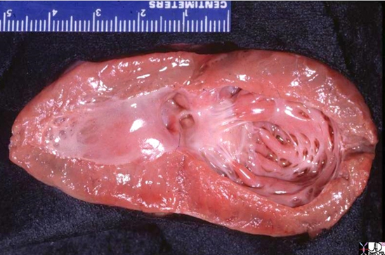
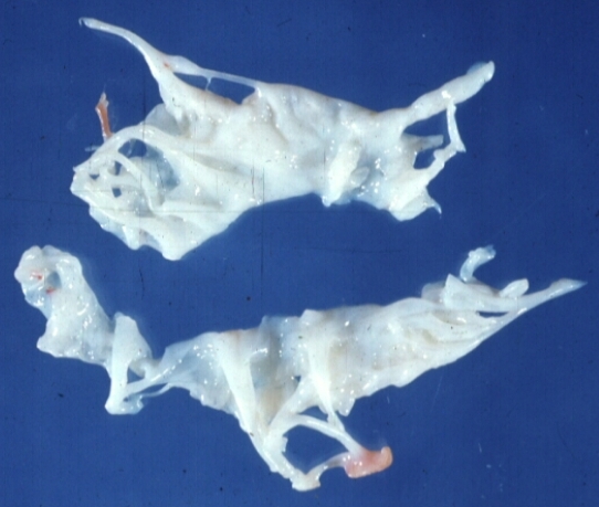
The anatomic specimen taken from the left ventricle alongside the pillars of the papillary muscles and chordae and the waterfall of blood as it comes from the left atrium into the left ventricle is conceptualized.
32118b03 heart papillary muscles chordae tendinae left ventricle maid of the mist mitral valve sounds of the blood flow Davidoff art Davidoff photography copyright 2019
- Mitral Annulus :
-
- fibrous ellipsoidal ,
- D shaped tissue
- fulcrum for the leaflets
- sphincteric contraction
- During systole
- annulus is elliptical
- diastole
- round in, with maximal dimensions in end-diastole
- (area 7.1 cm2, diameter 3.0 cm, circumference 9.3 cm)
-
The outflow tract
The anterior leaflet of the mitral valve.
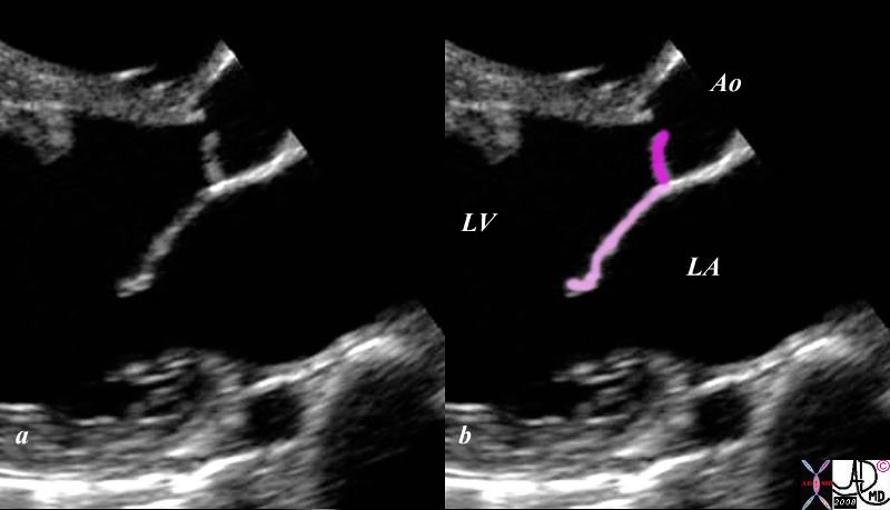
The Normal Mitral Aortic Complex
This gray scale echo of the heart shows the left ventricle, anterior and posterior leaflets of the mitral valve, the aortic valve and the base of the aorta. The echo demonstrates the intimate anatomic and physiological relationship of the anterior leaflet of the mitral valve (light pink) and the left aortic cusp(dark pink)..
( Courtesy Philips Medical Systems 33132 code cardiac heart echo LV MV aorta mitral aortic complex normal anatomy 33132c02.8s
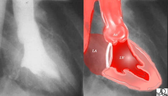
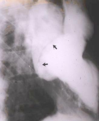
Normal LV Angiogram
This normal left ventriculogram is taken in the classical right anterior oblique (RAO) projection, enabling an excellent profile for the LV and the mitral valve plane. The annulus of the valve is inferred as a white ring in the color overlay.
Courtesy of Ashley Davidoff M.D.32120
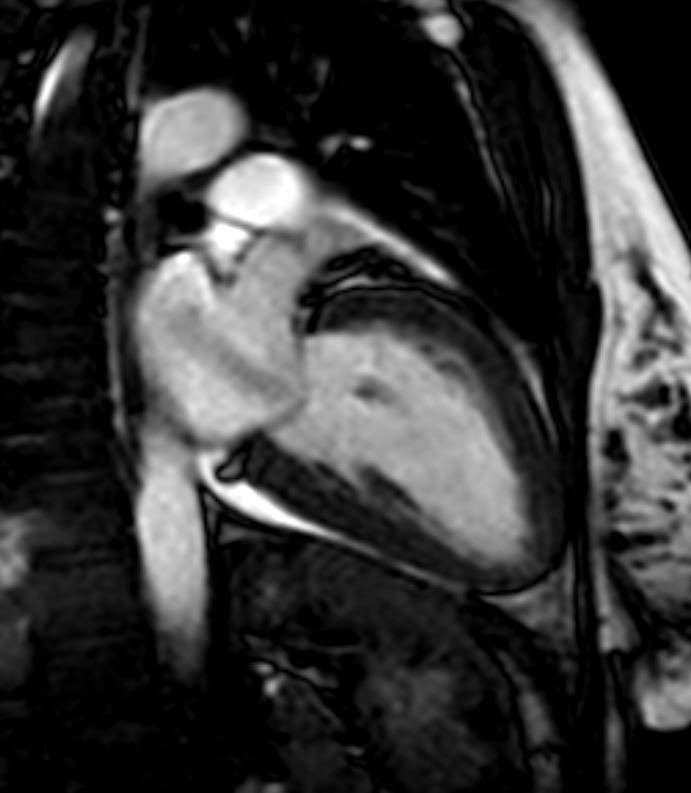
CT scan
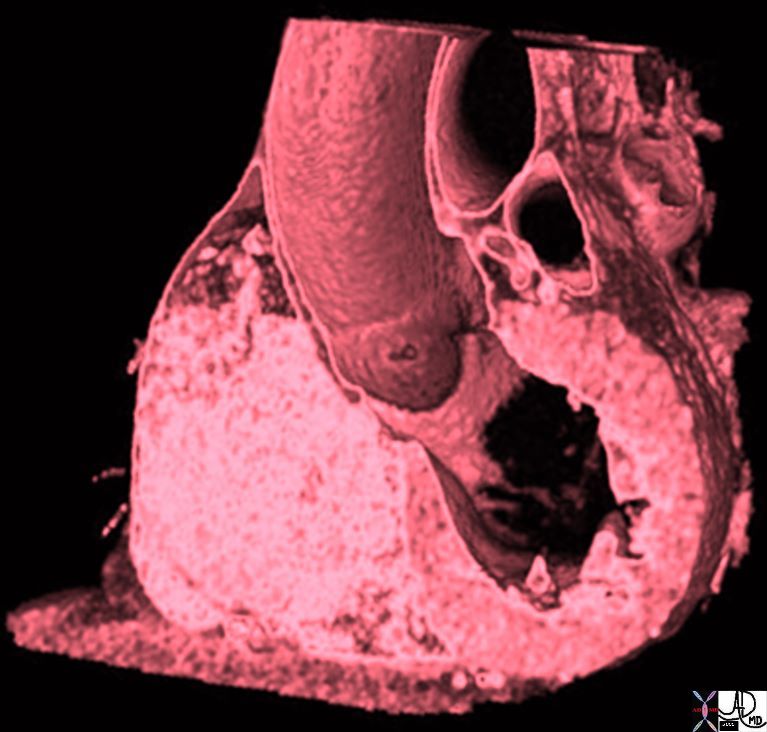
Thickness
The sagittally reconstructed CT study of the heart demonstrates the normal LV thickness, chamber size, mitral valve with continuity to the aortic valve. The smaller of the two hollow tubes to the left of the aorta represents the left atrial appendage and the larger more superior structure represents the main pulmonary artery
.
Davidoff MD 47823 copyright 2009 all rights reserved
The reconstructed CT scan is the left ventricle (LV), left atrium (LA) and right atrium (RA). The thin endocardium (white arrows) throughout the heart is one continuous sheet of tissue connected across the whole circulatory system and it is more fibrous in nature. The thin white layer is seen in the left atrium, left atrial appendage (LAA) over the posterior leaflet of the mitral valve (pl) in the left ventricle (LV) and in the right atrium. The surface of the left ventricle, left ventricular outflow tract (LVOT) and right atrium are also lined by the endothelium.Davidoff, M.D.
47824c02.8s
MRI

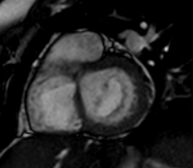
Ashley Davidoff MD TheCommonVein.net diastole-open-MRI-62f
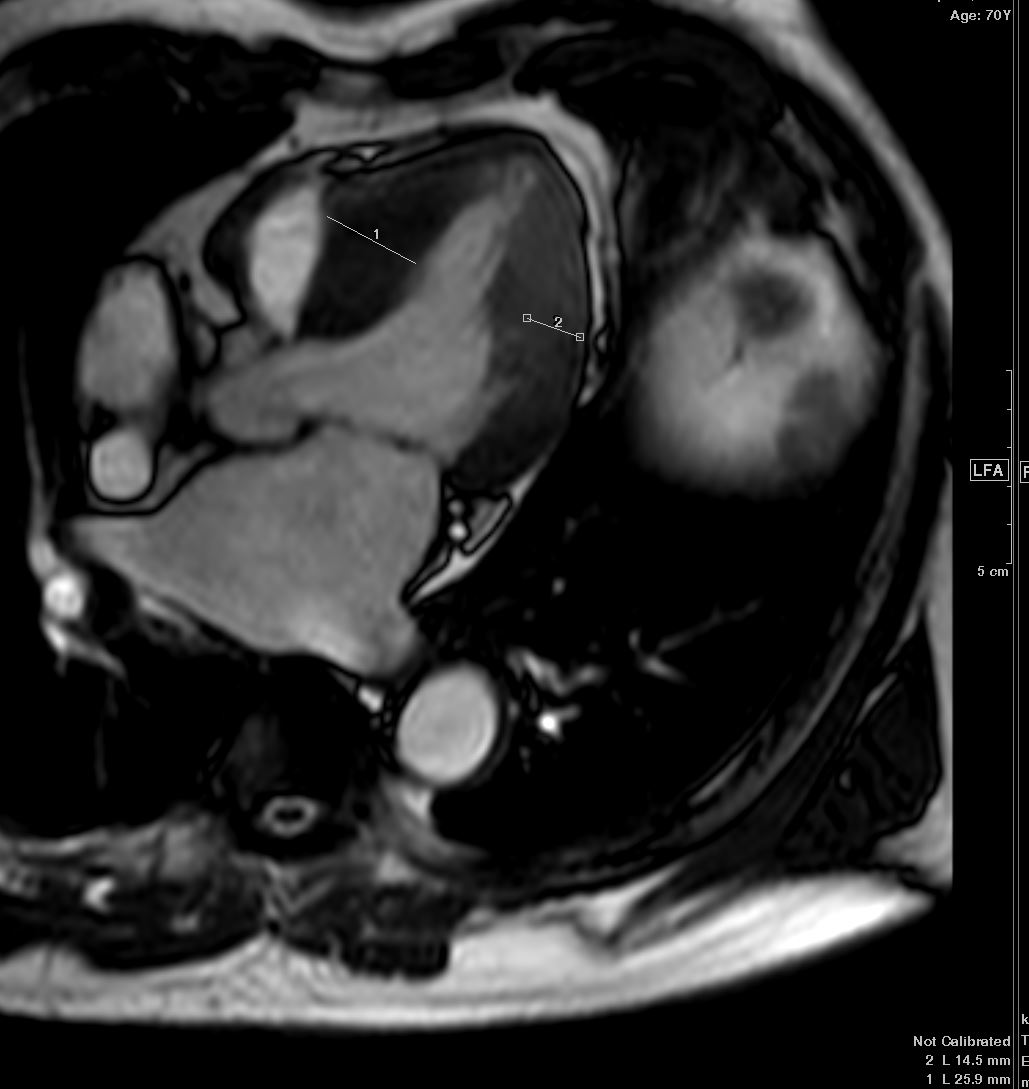
Mitral Valve in Systole and Diastole Mild LVH
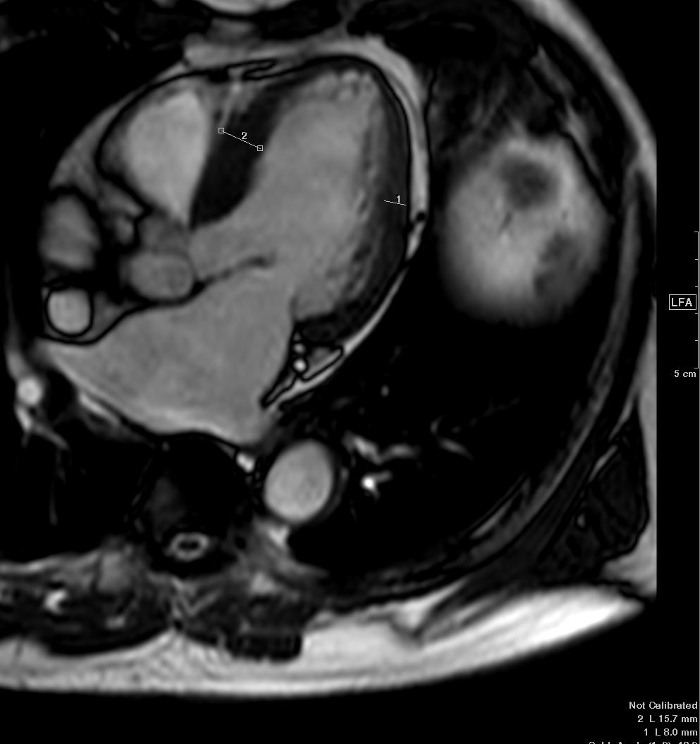
The Normal LV in diastole should be less than 12mm and in systole less than about 18mm. In this case the septum appears slighter thick (15.7mm) but the free wall is 8mm
Note the Mitral Valve is open
Ashley Davidoff MD TheCommonVein.net wall-thickness-001b
The Normal LV in diastole should be less than 12mm and in systole less than about 18mm.
Note the Mitral Valve is open
Ashley Davidoff MD TheCommonVein.net wall-thickness-001
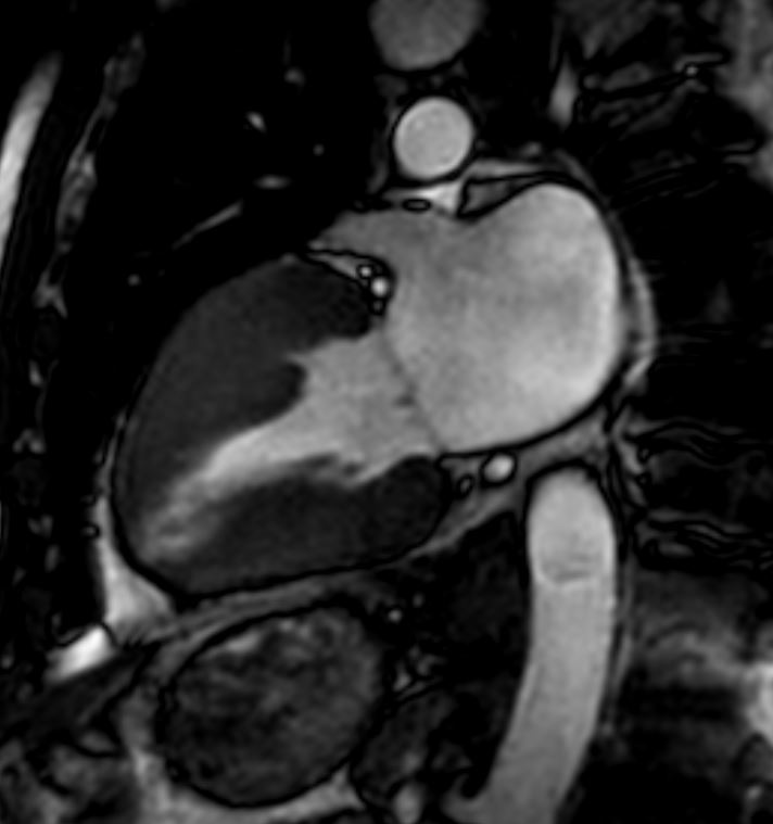
Thickening in Systole
The LV in systole shows concentric thickening and this case demonstrates LVH Septum measures 25mm and the free wall measures 14.5 mm
Note the mitral valve is closed
Ashley Davidoff MD TheCommonVein.net wall-thickness-002b
The Normal LV in systole shows thickening
Note the mitral valve is closed
Ashley Davidoff MD TheCommonVein.net wall-thickness-002
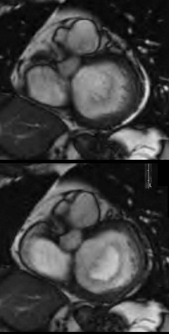
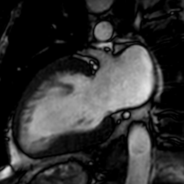
CONGESTIVE CARDIOMYOPATHY
61 male, with alcoholic congestive cardiomyopathy.
MRI of the heart in short axis, shows the LV in systole (above) with the mitral valve (MV) closed and in diastole (below) with the open mitral valve.
Ashley Davidoff MD
Diseases
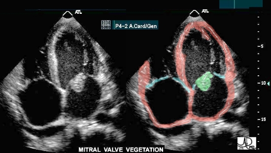
The four chambered echocardiogram of the heart shows an echogenic mass representing a mitral valve vegetation characteristic of endocarditis.
key words
heart cardiac MV mass SBE bacterial endocarditis vegetation infection imaging cardiac echo overlay
Courtesy of Philips Medical Systems, Ultrasound, and modified by Ashley Davidoff M.D 32121
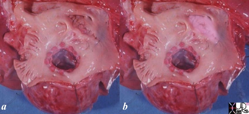
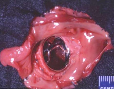
Complete Endocardial Cushion Defect
The pathology specimen shows a complete endocardial cushion defect with a cleft mitral valve and a VSD. The atrial chambers can be seen through the cleft of the mitral valve and a ASD primum was present as well
Key words
heart cardiac LV left ventricle IVS interventricular septum mitral valve endocardial cushions complete AV canal defect cleft mitral valve VSD endocardial cushion defect congenital heart disease gross pathology Courtesy Ashley Davidoff MD 01863b01
40 year old female with SLE and congestive cardiomyopathy in this 2 chambered view shows a jet of mitral regurgitation caused by to ventricular remodeling leading to reduced leaflet coaptation and valvular insufficiency.
Ashley Davidoff MD TheCommonVein.net mitral-regurgitation-001-MRI
Ashley Davidoff MD
TheCommonVein.net 01870
Mitral Atresia
Hypoplastic Left Heart Syndrome and Mitral Atresia Small LV
Ashley Davidoff MD TheCommonVein.net 08061
Mitral Stenosis
Congenital
Mild form of Hypoplastic Left Heart Syndrome
Ashley Davidoff MD TheCommonVein.net
Ashley Davidoff MD TheCommonVein.net 15515
Rheumatic Heart Disease
Historical Perspective Evaluation of the Left Atrium Circa 1975
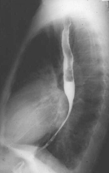
Ashley Davidoff MD TheCommonVein.net 01841
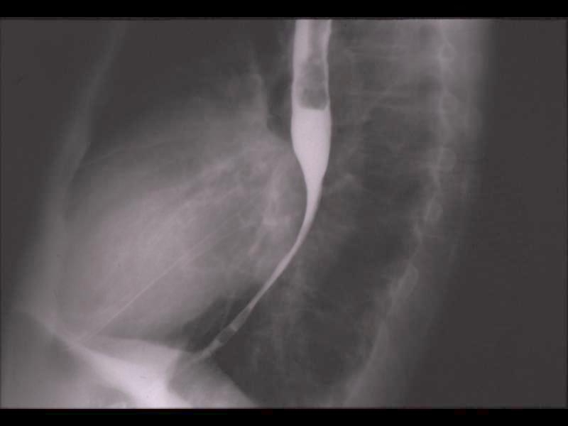
Ashley Davidoff MD TheCommonVein.net
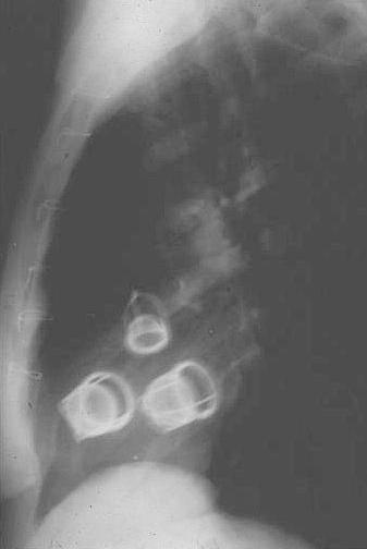
Thickened Papillary Muscles Shortened Chordae
Ashley Davidoff MD TheCommonVein.net 15518
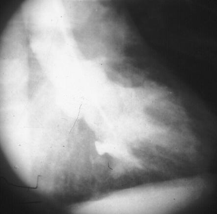
Thickened Papillary Muscles Shortened Chordae
Ashley Davidoff MD TheCommonVein.net 01875
Axial CT through the heart shows a papillary muscle close to the insertion of the valve indicating shortened chordae. This is a finding associated with rheumatic heart disease. Echo showed evidence of both mitral stenosis and mitral regurgitation.
Ashley Davidoff MD TheCommonVein.net 135548
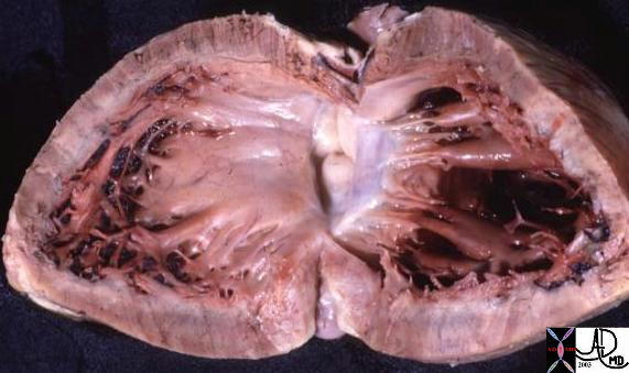
Axial CT through the heart shows a normal papillary muscle and normal chordae.
Ashley Davidoff MD TheCommonVein.net 135505
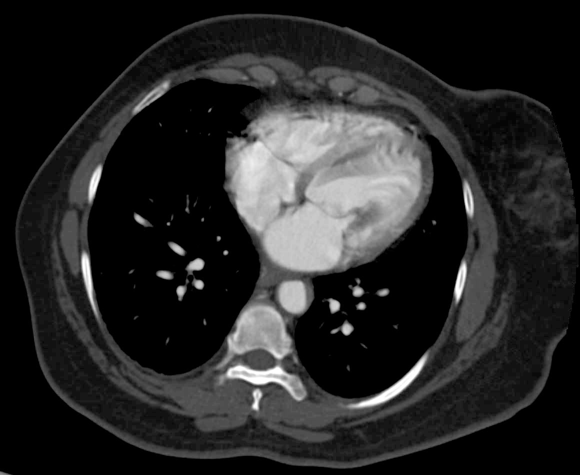
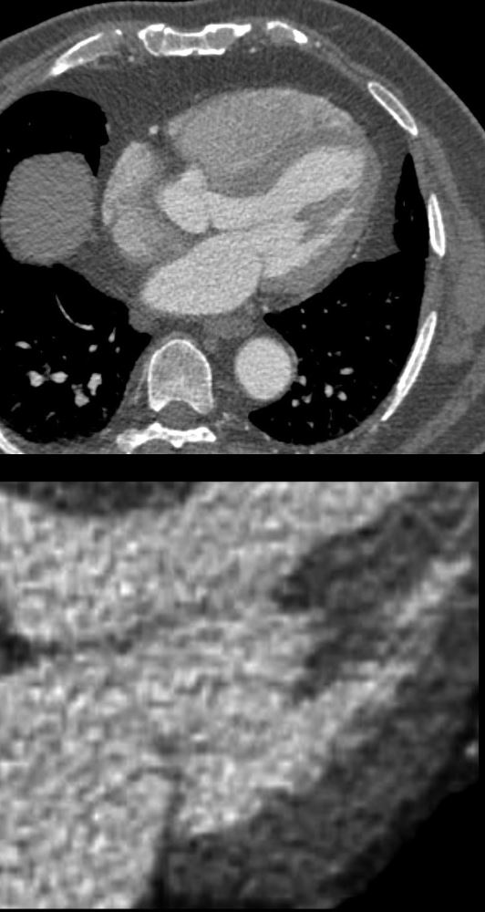
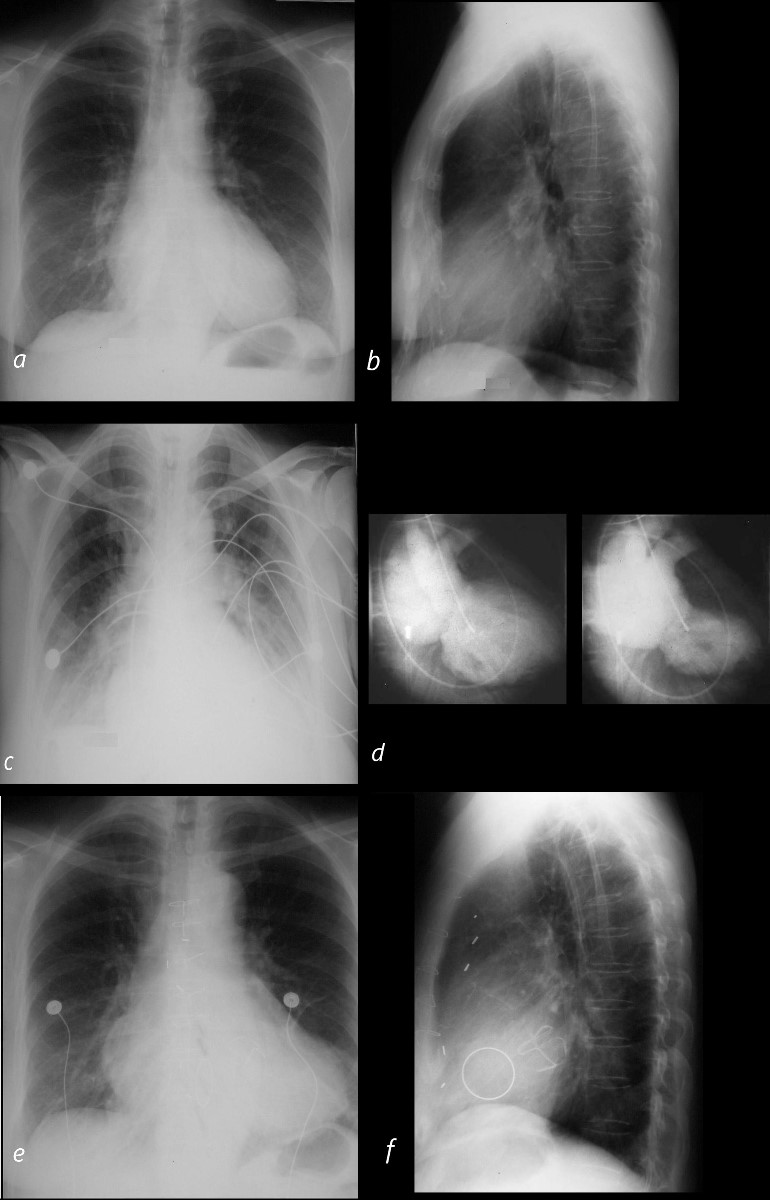
87 year old male with findings consistent with mitral stenosis , characterised by calcified mitral valve, enlarged left atrium, normal sized left ventricle, with enlarged right atrium, right ventricle and main pulmonary artery.
Pure mitral stenosis is most commonly caused by rheumatic heart disease
Ashley Davidoff MD
87 year old male with findings consistent with mitral stenosis , characterised by calcified mitral valve, enlarged left atrium, normal sized left ventricle,with enlarged right atrium, right ventricle and main pulmonary artery.
Pure mitral stenosis is most commonly caused by rheumatic heart disease
Ashley Davidoff MD
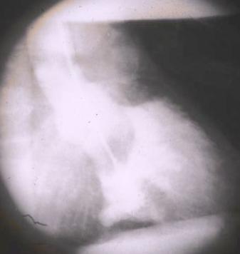
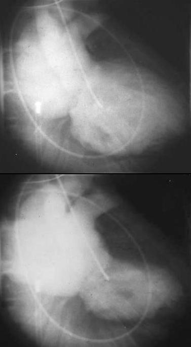
50 year old female with a history of rheumatic mitral stenosis
CT with contrast in the axial plane shows a mildly thickened mitral valve in systole
Courtesy Ashley Davidoff MD TheCommonVein.net 136519
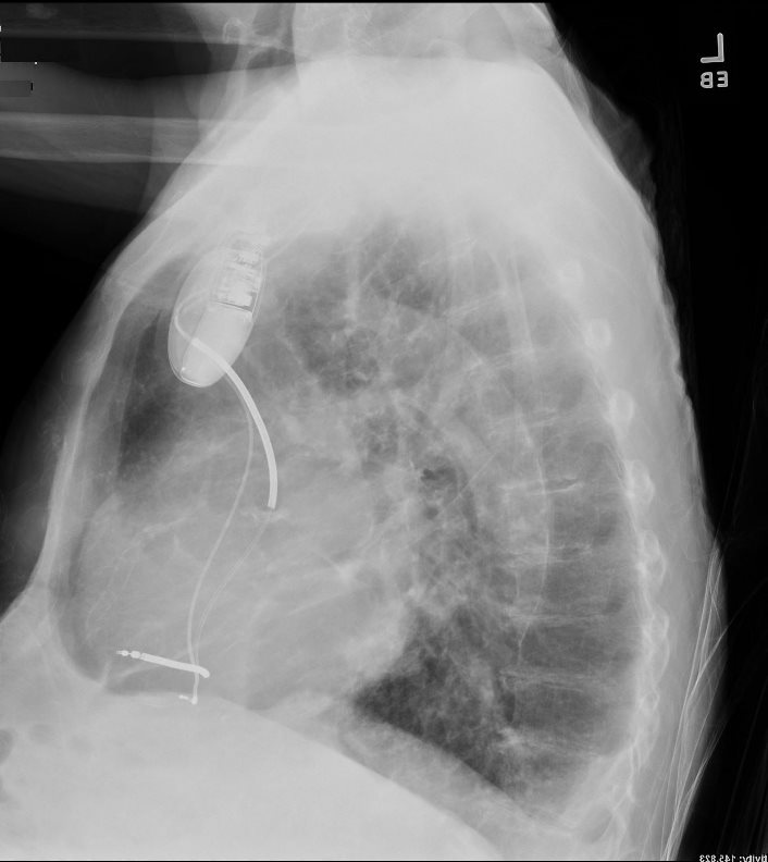
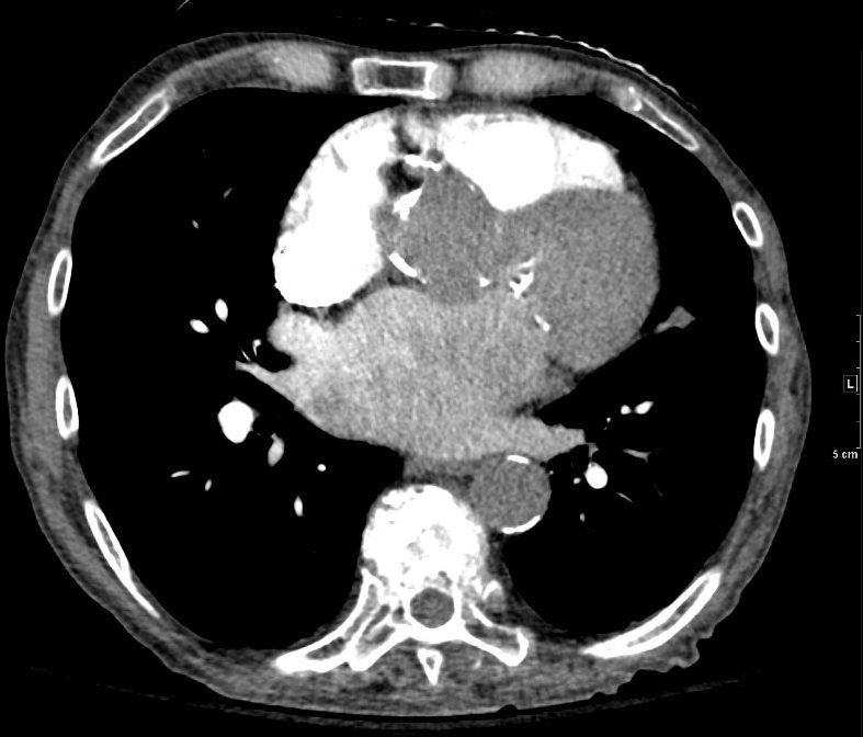
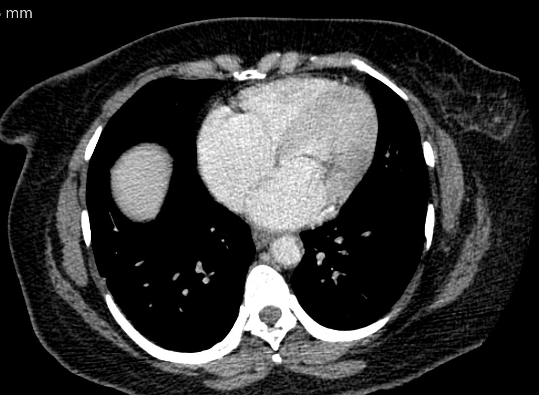
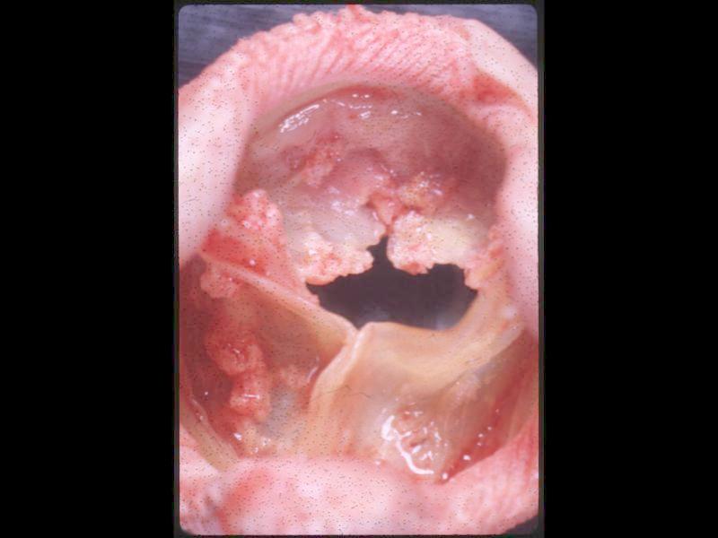
Endocarditis
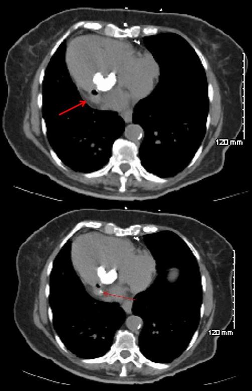
78 year old female with dextrocardia, presents 2 weeks after laparoscopic surgery with a fever. Prior CT showed dextrocardia with MAC. Repeat CT at the time of the fever, shows an air fluid on the lateral aspect of the MAC with disrupted calcification. Blood cultures were positive and a diagnosis of anaerobic gas forming endocarditis was made. She subsequently required MVR
Ashley Davidoff MD thecommonvein.net
Libman Sacks Vegetation
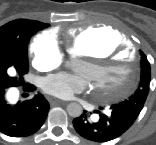
SLE and PULMONARY HYPERTENSION without ILD
27-year-old female presents with dyspnea and a past history of SLE, Raynaud?s disease, and Lupus nephritis.
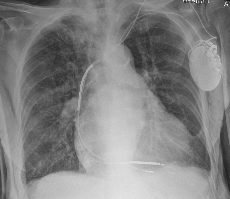
Chest X-ray shows cardiomegaly with right ventricular configuration on the PA and an enlarged main and probably left pulmonary and an enlarged descending RPA. The lateral confirms the enlarged RV and raises the possibility of LV enlargement.
The CT scan confirms an enlarged MPA, RPA, RA and RV, and shows calcification on the posterior leaflet of the mitral valve consistent with Libman Sacks vegetation. There is mild ground glass opacity at the lung bases but no sign of ILD.
A current non contrast abdominal CT shows a pericardial effusion and normal sized kidneys
Ashley Davidoff MD
Ruptured Papillary Muscle Post MI
Ashley Davidoff MD TheCommonVein.net
Ashley Davidoff MD TheCommonVein.net 01831cL.1k
Treatment
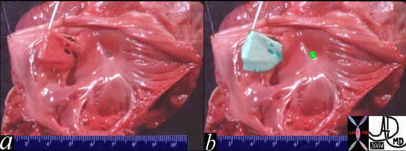
This anatomical specimen is from an unfortunate pediatric patient who had a mitral valve replaced and an ASD secundum closed. The pink overlay in b shows a suture line opposing the borders of the ASD secundum. A prosthetic mitral valve is noted.
key words
heart mitral valve atrial septal defect secundum repair treatment surgery Courtesy Ashley Davidoff MD copyright 2019 06704c01.8s
Porcine valve
Ashley Davidoff MD TheCommonVein.net 06711

Porcine valve
Ashley Davidoff MD TheCommonVein.net 06712b01.400
Bjork Shiley valve
Ashley Davidoff MD TheCommonVein.net 06713
Ashley Davidoff MD
TheCommonVein.net 06721
References:
1.Ho SY. Anatomy of mitral valve. Heart 2002;88(Suppl IV):iv5?iv10.
2.Condado JA and Gimon MV. Catheter-Based Approach to Mitral Regurgitation. (J Interven Cardiol 2003;16:523?534.
- Walmsley T. The heart. In: Sharpey-Schafer E, Symington J,Bryce TH, eds. Quain?s Elements of Anatomy, 11th ed. Vol 4,part 3. London: Longmans, Greens & Co, 1929, p. 42.