heart0067-low res
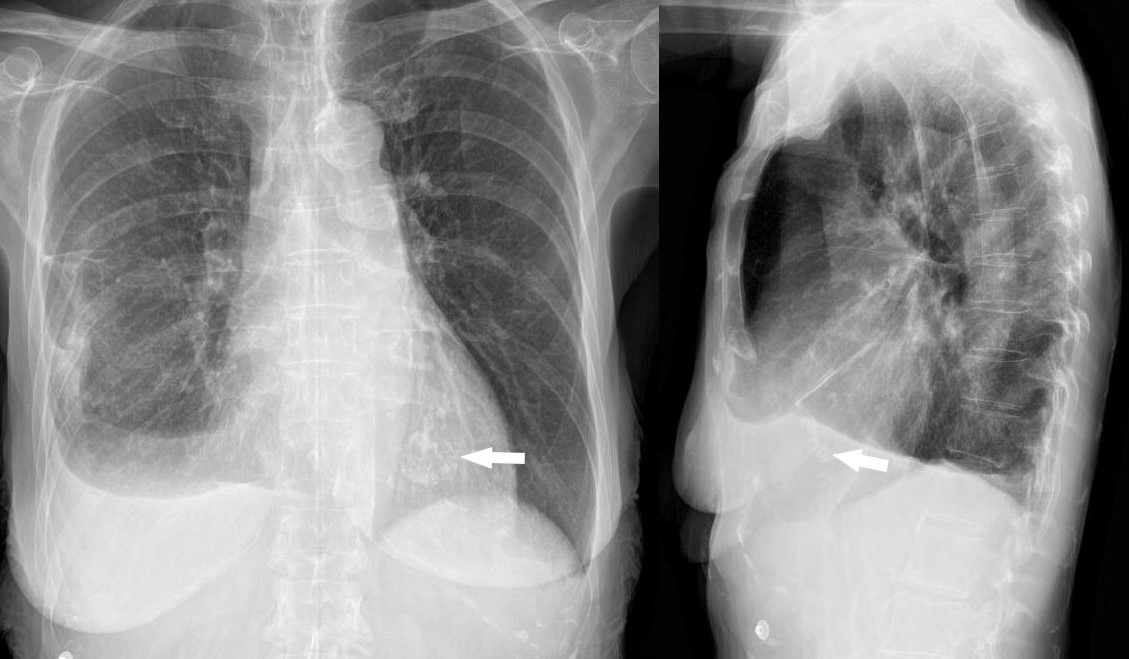
The patient is a 73 year old female with apical hypertrophic disease. She has a history of diastolic heart failure, pulmonary hypertension, CREST, esophageal stricture, COPD and chronic renal failure
heart0068b-low res
Ashley Davidoff MD
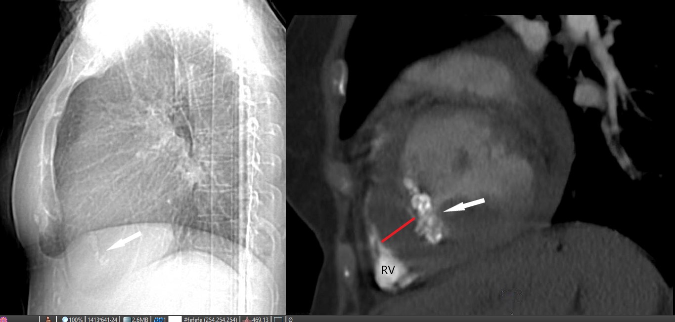
The patient is a 73-year-old female with apical hypertrophic disease. She has a history of diastolic heart failure, pulmonary hypertension, CREST, esophageal stricture, COPD and chronic renal failure
heart0069b02 Ashley Davidoff MD
heart0070L-low-res-1024x615.jpg
The CT axial images shows an enlarged pulmonary artery (PA) indicating pulmonary hypertension, and I an enlarged left atrium (LAE, b) , a small left ventricular (LV) cavity (red circle , c) calcified foci in the LV (c and d white arrows, and a small pericardial effusion (red arrow d)
The patient is a 73-year-old female with apical hypertrophic disease. She has a history of diastolic heart failure, pulmonary hypertension, CREST, esophageal stricture, COPD and chronic renal failure
Ashley Davidoff MD
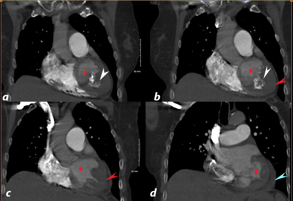
The CT coronally reconstructed images show a small left ventricular (LV) cavity (red circle ,a,b,c,d) intracavitary calcification (white arrow a,b), apical LV hypertrophy (LVH, red arrows b, and c) and a small pericardial effusion (blue arrow, d)
The patient is a 73-year-old female with apical hypertrophic disease. She has a history of diastolic heart failure, pulmonary hypertension, CREST, esophageal stricture, COPD and chronic renal failure
Ashley Davidoff MD
References and Links
- TCV

