History of the Heart
The Common Vein
Copyright 2009
Introduction
The heart has a profound history in the worlds of structure and function as well as in the ideas of its metaphysical implications to the different cultures of the world. In ancient times the science and the metaphysical were inextricably linked, eventually separating as the renaissance opened up the vistas of science. However remnants of the metaphysical remain in our conversation and actions to this day.
Egyptians (2500-1000BC)
For the ancient physicians the pulse was the most obvious external manifestation of the cardiovascular system, and represented the beginning of the intrigue with the heart and the study of the cardiovascular system.
Ancient Egyptians are the first people to have documented observations of the pulse in a papyrus dating from 1500BC. Its force, frequency and character were noted to reflect the patients state of health, and they connected the pulse with the patients blood vessels.
Thte ieb was the Egyptian term for the heart which was believed to be the center of life and morality. The Egyptians believed that the heart was weighed after death against the feather of Maat who was the Goddess of justice. If the heart was lighter than the feather, the person’s soul would join Osiris in the afterlife. In the event that heart weighed more than the feather, the heart would be eaten by the demon Ammut, and the soul of the person would would be lost.
Hippocrates (460 to 377 BC)
Hippocrates of Cos, a Greek physician who lived about 400 BC, was also aware of the pulse, its relationship to the blood vessels, and he made the connection of the vessels with the heart. The obvious relationship of the pulse with activity inside the body was observed and diagnoses were often based on these observations.
Hippocratess first ascribed a person’s pulse to the blood vessels and found that these blood vessels traced to the heart. Hippocrates, the Father of Medicine, wrote, “The vessels which spread themselves over the whole body, filling it with spirit, juice, and motion, are all of them but branches of an original vessel. I protest, I do not know where it begins or where it ends, for in a circle there is neither a beginning nor an end.” Although Hippocrates did not have the opportunity to view the body with today’s imaging modalities, the assumptions that he made in 470 B.C. were fairly accurate.
Aristotle was an astute observer of nature, but many of his observations regarding the cardiovascular system were erroneous, and had significant historical, cultural, and scientific consequences. He attached enormous relevance to the heart, observing correctly that it was the centre of the cardiovascular system, but erroneously ascribed the heart as the seat of intelligence, and the source of body heat. In addition his belief that the heart was the first organ to develop and the last to show activity in death was held as fact for many years.
4th Century BC – Aristotle
During the 4th century BC Aristotle identified the heart as the most important organ of the body based on his observations of the heart of the chick embryo. He felt it was the seat of intelligence, motion and sensation – though he felt it was a hot and dry organ.
From a structural point of view he viewed it as a three chambered organ which was central to all the organs, and that the other organs were designed to cool the heart.
Aristotle also saw the heart as the first in life and last in death organ.
In about 300 BC, a medical school was created in Alexandria in Egypt, which had been conquered and named after Alexandra the Great. Alexandria was a favorite residence for Greek aristocracy, and the medical school became funded and supported by the Greek dynasty, and stood out worldwide as an institution of remarkable academic quality.
Galen 131-201 AD –
In the second century AD Galen wrote a treatise called “On the Usefulness of the Parts of the Body” were he supported the idea that the heart was the source of body heat, and as the organ that was closest to the soul.
“The heart is, as it were, the hearthstone and source of the innate heat by which the animal is goverened.”
From a structural perspective he noted that “The heart is a hard flesh, not easily injured. In hardness, tension, general strength, and resistance to injury, the fibers of the heart far surpass all others, for no other instrument performs such contnuous, hard work as the heart.” He also noted that ” The complexity of fibers…was prepared by Nature to perform a variety of functions…. enlarging when it desires to attract what is useful, clasping its contents when it is time to enjoy what has been attracted, and contracting when it desires to expel residues.”Galen observes, “…blood is drawn from the right ventricle into the left, owing to there being perforations in the septum between them.” This structural concept was perpetuated for 1500 years without challenge, until direct examination in the Renaissance period revealed more accurate facts.
Galen did however describe the valves of the heartand noted the difference between the arteries and veins.
Galen, also Greek in origin, was a student in Alexandria in the second century AD. His influence on the medical world had profound influence not only in the world of medicine and science but also in Western literature where his ideas affected the writings of those such as Chaucer and Shakespeare.
In essence he taught that there were three major organs; the heart, brain and liver
Ingested food was rightly understood to be absorbed by the intestinal tract, and transported to the liver where it was incorporated into the blood and infused with an essence he called ?the natural spirits?. From the liver, believed by Galen to be the center of the venous system, the natural spirits were distributed to the body by the veins. This idea of course reveals ignorance on two fronts;
lack of understanding of the connection of the veins with the heart.
ignorance of the direction of systemic venous flow .
His idea of venous flow was a pattern of ebb and flow, perhaps based on observation veins of the neck.of the
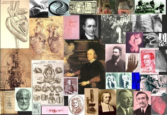
Collage of Cardiac History
|
| The collage shows the history of the heart from Hippocrates Aristotle and Galen, in the bottom left, through Vesalius and da Vinci (top left) via Harvey – (centre), Laenecc (stethoscope (middle top right) Roentgen Einthoven,(EKG) Sones (catheterization), Forsstman, (physiology and pressure measurement of the heart) Barnard (transplant), and Hounsfield CT scanning bottom right).
The circulaton goes round and around, fetching and taking, in circles and cycles, always moving in pulsatile fashion, mostly forward, but sometimes a little backward. This is the circulation. Although Hippocrates had a hint of a continuum and a cycle, his perceptions were not fully realized until Harvey’s work in the early 1600’s. Harvey’s work stands central to all that happened before, and all that came after. (Image composite by Ashley Davidoff M.D.) 3804 |
Galen struggled with the idea of how the blood went from the right side to the left side and arrived at the concept that
??..blood is drawn from the right ventricle into the left, owing to there being perforations in the septum between them?
The hormonal theory was advanced by Galen ascribing faculties logic to the brain, nutrition to the liver, and the vitality to the heart, which was central to function of the body at large.
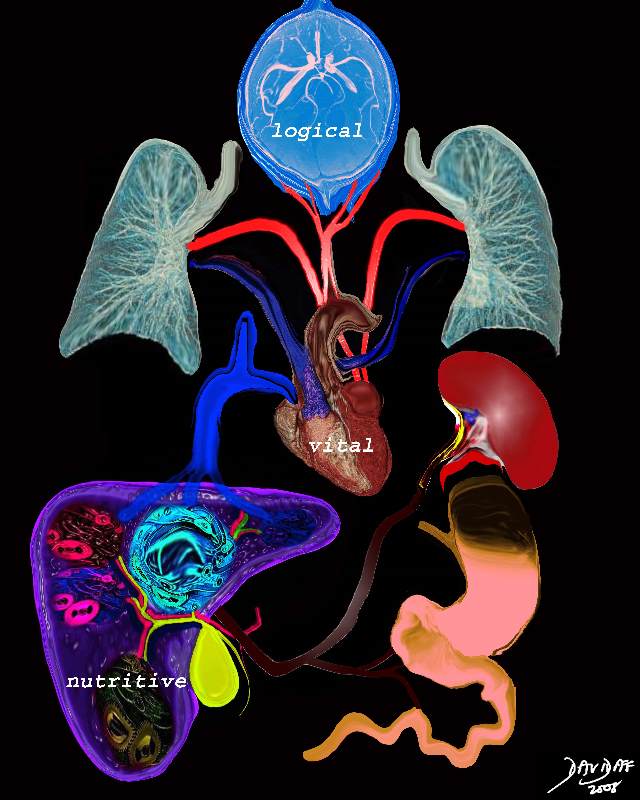
Galen
|
|
Heat plays a central role part in the theory of Galen. The three ?faculties? of the body are the nutritive, vital and logical faculties. The nutritive faculty is related to the stomach which “cooks” the food and converts it into chyle. The chyle is transported to the liver by the portal vein. In the liver further heat converts the food into blood and adds natural spirit. Some of the blood is transported via the veins to the heart where more heat is added to create vital spirit. The blood becomes thinner is distributed to the body by the arteries giving warmth and enables growth. The vital spirit is measured through the pulse. The brain adds psychic pneuma, which provides the rational and logical faculty in the form of thought will and choice. These are distributed to the body via the nerves. The logical faculty reigns supreme and is followed in orderof importance by the vital and nutrtive faculties. The transport systems of the body include the nerves which transmit the logical faculty, the arteries which transport the vital spirit, and the veins which transport the blood with nutritive faculty from the liver.
Galen faculties of the body nutrition portal vein stomach liver vein heart vital faculty pneuma lungs brain logical faculty animal spirits Davidoff art Copyright 2008 13169c18b01.8s
|
Teotihuacan Culture AD 100-900
The Teotihuacan culture was an ancient Mexican culture who believed in the teyolia, which was the spiritual force associated with the heart, and was considered the essence of life. This culture believed that some of the forces in the body were able to leave the body without detriment, but that the teyolia was essential for life. Its absence inferred death.
Christian Theology 1000 1400 AD
Avicenna – Beginning of the 11 the Century
Avicenna studued the books of ancient medicine and wrote his Canon of Medicine integrating Aristotle and Galens ideas. Central to his ideas on the heart was the noton that the heart :….the root of all faculties and gives the faculties of nutrition, life, apprehesion, and movement to several other members.” He also believed that the heart was the ” vital power or innate heat”, that it was central to inteligence and that it was the organ that controlled the other organs.
The Renaissance introduced a renewed and fresh look at the antomy and resulted in the beinning of understanding based on direct observation. During this time 4 chambers were identified.
Da Vinci- 1452 – 1519
Da Vinci’s knowledge obviously advanced from his early diagram where the atria and ventricles were disconnected to his later diagram where excquisite detail of the atrial appendages, coronary arteries and pulmonary valve are depicted in exquisiste detail.
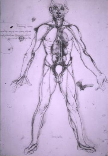
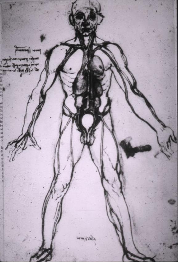
Atria and Ventricles Separated
|
| In this diagram of the heart by da Vinci, its position in the chest is exemplified. The superior vena cava and inferior vena cava enter into an atrial chamber which is discrete and remote from the ventricular chambers which lie upward and leftward, and which give . rise to the arteries. This drawing is likely from earlier times since subsequent drawings of the heart suggest that da Vinci understood the connection of the atria and ventricles.
44411 or 13161
|
The idea of the heart composed of two ventricles separated by a septum an ancient Galenic view, and retained by da Vinci is exempified in the following drawing.
|
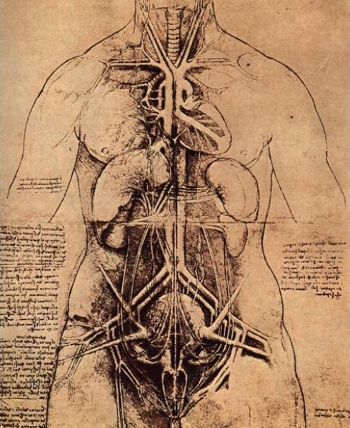
Appearance of the Heart Dominated by Two Ventricles and an Interventricular Septum
|
| This image shows heart, aorta, and caval system running side by side. The venous system is not shown to enter into a separate atrium
Courtesy Leonardo da Vinci 51899
|
|
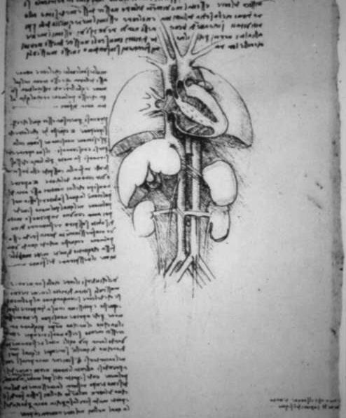
Veins Shown to Enter the Right Atrium and Connect to the Right Ventricle
|
| Drawing of the lungs and the heart by Leonardo da Vinci. In this drawing da Vinci has the veins, right atrium, and right ventricle connected correctly.
13154 code normal heart lung liver spleen kidneys aorta IVC anatomy historical drawing |
|
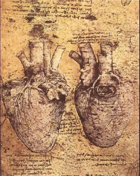
The Atrial Appendages Ventricles Pulmonary Valve and Vessels of the Heart
|
| This drawing by Leonardo da Vinci, is the most detailed early image of the heart, and particularly of the pulmonary valve and coronary arteries. The left atrial appendage made quite an impression on the old master. The image on the left is a detailed view of the left coronary artery showing the LAD, diagonals, circumflex, obtuse marginal, and left atrial and or S-A nodal branch. The RVOT tract is beautifully seen in this view as well.
Courtesy Leonardo daVinci 51896b
|
Da Vinci’s idea on the structure of the heart is best described in the following quote “The heart of itself is not the beginning of life but is a vessel made of dense muscle vivified and nourished by an artery and a vein as are the other muscles. The heart is of such density that fire can scarcely damage it.”
He started to ponder on the function of the heart as witnessed by the following observations of the beating heart. “At one and the same time, in one and the same subject, two opposite motions cannot take place, that is repentance and desire. Therefore, if the right upper(atrium) and lower ventricles are one and the same, it is necessary that the whole should cause at the same time one and the same effect and not two effects arising from diametrically opposite purposes as one sees in the case of the right ventricle with the lower, for whenever the lower contracts, the upper dilates to accommodate the blood which has been driven out of the lower ventricle.”
Vesalius 1514?1564
Vesalius also dissected the body and came to deeper understanding based on his observations. He challenged Galen?s idea of the septum suggested that the ventricular septum was impermeable. He also puzzled on the pulmonary circulaltion.
1628 William Harvey
William Harvey was an English Physician, first described blood circulation. He wrote in 1653: “The heart is situated at the 4th and 5th ribs. Therefore [it is] the principal part because [it is in] the principal place, as in the center of a circle, the middle of the necessary body.” In 1628, he wrote: “The heart’s one role is the transmission of the blood and its propulsion,, by means of the arteries, to the extremities everywhere.”
1706 Raymond de Vieussens
Raymond de Vieussens was a French anatomy professor, first described the structure of the heart’s chambers and vessels.
1733 Stephen Hales
Stephen Hales was an English clergyman and scientist, who first measured blood pressure.
1816 Rene T. H. Laennec
Rene T. H. Laennec a French physician, invents the stethoscope.
1903 Willem Einthoven
Willem Einthoven was a Dutch physiologist, who developd the electrocardiograph.
1912 James B. Herrick
James B. Herrick was an American physician, who first described heart disease resulting from hardening of the arteries.
1938 Robert E. Gross
Robert E. Gross was an American surgeon, who was the first to perform heart surgery.
1951 Charles Hufnagel
Charles Hufnagel was an American surgeon, who developed a plastic valve to repair an aortic valve.
1952 F. John Lewis
F. John Lewis was an American surgeon, who first performed successful open heart surgery.
1953 John H. Gibbon
John H. Gibbon was an American surgeon, who first used a mechanical heart and blood purifier.
1961 J. R. Jude
J. R. Jude was an American cardiologist, who led a team to perform the first external cardiac massage to restart a heart.
1965 Michael DeBakey and Adrian Kantrowitz
Michael DeBAkey and Adrian Kantowitz American surgeons, implanted a mechanical devices to help augment the function of a diseased heart.
1967 Christiaan Barnard
Christiaan Barnard was a South African surgeon, who performed the first whole heart transplant from one person to another.
1982 Willem DeVries
Willem DeVries an American surgeon, implants a permanent artificial heart, designed by Robert Jarvik, an American physician, into a patient.
Stanford Project
DOMElement Object
(
    [schemaTypeInfo] =>
    [tagName] => table
    [firstElementChild] => (object value omitted)
    [lastElementChild] => (object value omitted)
    [childElementCount] => 1
    [previousElementSibling] => (object value omitted)
    [nextElementSibling] => (object value omitted)
    [nodeName] => table
    [nodeValue] =>


The Atrial Appendages Ventricles Pulmonary Valve and Vessels of the Heart


This drawing by Leonardo da Vinci, is the most detailed early image of the heart, and particularly of the pulmonary valve and coronary arteries. The left atrial appendage made quite an impression on the old master. The image on the left is a detailed view of the left coronary artery showing the LAD, diagonals, circumflex, obtuse marginal, and left atrial and or S-A nodal branch. The RVOT tract is beautifully seen in this view as well.
Courtesy Leonardo daVinci 51896b


    [nodeType] => 1
    [parentNode] => (object value omitted)
    [childNodes] => (object value omitted)
    [firstChild] => (object value omitted)
    [lastChild] => (object value omitted)
    [previousSibling] => (object value omitted)
    [nextSibling] => (object value omitted)
    [attributes] => (object value omitted)
    [ownerDocument] => (object value omitted)
    [namespaceURI] =>
    [prefix] =>
    [localName] => table
    [baseURI] =>
    [textContent] =>


The Atrial Appendages Ventricles Pulmonary Valve and Vessels of the Heart


This drawing by Leonardo da Vinci, is the most detailed early image of the heart, and particularly of the pulmonary valve and coronary arteries. The left atrial appendage made quite an impression on the old master. The image on the left is a detailed view of the left coronary artery showing the LAD, diagonals, circumflex, obtuse marginal, and left atrial and or S-A nodal branch. The RVOT tract is beautifully seen in this view as well.
Courtesy Leonardo daVinci 51896b


)
DOMElement Object
(
    [schemaTypeInfo] =>
    [tagName] => td
    [firstElementChild] => (object value omitted)
    [lastElementChild] => (object value omitted)
    [childElementCount] => 1
    [previousElementSibling] =>
    [nextElementSibling] =>
    [nodeName] => td
    [nodeValue] => This drawing by Leonardo da Vinci, is the most detailed early image of the heart, and particularly of the pulmonary valve and coronary arteries. The left atrial appendage made quite an impression on the old master. The image on the left is a detailed view of the left coronary artery showing the LAD, diagonals, circumflex, obtuse marginal, and left atrial and or S-A nodal branch. The RVOT tract is beautifully seen in this view as well.
Courtesy Leonardo daVinci 51896b

    [nodeType] => 1
    [parentNode] => (object value omitted)
    [childNodes] => (object value omitted)
    [firstChild] => (object value omitted)
    [lastChild] => (object value omitted)
    [previousSibling] => (object value omitted)
    [nextSibling] => (object value omitted)
    [attributes] => (object value omitted)
    [ownerDocument] => (object value omitted)
    [namespaceURI] =>
    [prefix] =>
    [localName] => td
    [baseURI] =>
    [textContent] => This drawing by Leonardo da Vinci, is the most detailed early image of the heart, and particularly of the pulmonary valve and coronary arteries. The left atrial appendage made quite an impression on the old master. The image on the left is a detailed view of the left coronary artery showing the LAD, diagonals, circumflex, obtuse marginal, and left atrial and or S-A nodal branch. The RVOT tract is beautifully seen in this view as well.
Courtesy Leonardo daVinci 51896b

)
DOMElement Object
(
    [schemaTypeInfo] =>
    [tagName] => td
    [firstElementChild] => (object value omitted)
    [lastElementChild] => (object value omitted)
    [childElementCount] => 2
    [previousElementSibling] =>
    [nextElementSibling] =>
    [nodeName] => td
    [nodeValue] =>

The Atrial Appendages Ventricles Pulmonary Valve and Vessels of the Heart

    [nodeType] => 1
    [parentNode] => (object value omitted)
    [childNodes] => (object value omitted)
    [firstChild] => (object value omitted)
    [lastChild] => (object value omitted)
    [previousSibling] => (object value omitted)
    [nextSibling] => (object value omitted)
    [attributes] => (object value omitted)
    [ownerDocument] => (object value omitted)
    [namespaceURI] =>
    [prefix] =>
    [localName] => td
    [baseURI] =>
    [textContent] =>

The Atrial Appendages Ventricles Pulmonary Valve and Vessels of the Heart

)
DOMElement Object
(
    [schemaTypeInfo] =>
    [tagName] => table
    [firstElementChild] => (object value omitted)
    [lastElementChild] => (object value omitted)
    [childElementCount] => 1
    [previousElementSibling] => (object value omitted)
    [nextElementSibling] => (object value omitted)
    [nodeName] => table
    [nodeValue] =>


Veins Shown to Enter the Right Atrium and Connect to the Right Ventricle


Drawing of the lungs and the heart by Leonardo da Vinci.  In this drawing da Vinci has the veins, right atrium, and right ventricle connected correctly.
13154 code normal heart lung liver spleen kidneys aorta IVC anatomy historical drawing


    [nodeType] => 1
    [parentNode] => (object value omitted)
    [childNodes] => (object value omitted)
    [firstChild] => (object value omitted)
    [lastChild] => (object value omitted)
    [previousSibling] => (object value omitted)
    [nextSibling] => (object value omitted)
    [attributes] => (object value omitted)
    [ownerDocument] => (object value omitted)
    [namespaceURI] =>
    [prefix] =>
    [localName] => table
    [baseURI] =>
    [textContent] =>


Veins Shown to Enter the Right Atrium and Connect to the Right Ventricle


Drawing of the lungs and the heart by Leonardo da Vinci.  In this drawing da Vinci has the veins, right atrium, and right ventricle connected correctly.
13154 code normal heart lung liver spleen kidneys aorta IVC anatomy historical drawing


)
DOMElement Object
(
    [schemaTypeInfo] =>
    [tagName] => td
    [firstElementChild] => (object value omitted)
    [lastElementChild] => (object value omitted)
    [childElementCount] => 1
    [previousElementSibling] =>
    [nextElementSibling] =>
    [nodeName] => td
    [nodeValue] => Drawing of the lungs and the heart by Leonardo da Vinci.  In this drawing da Vinci has the veins, right atrium, and right ventricle connected correctly.
13154 code normal heart lung liver spleen kidneys aorta IVC anatomy historical drawing
    [nodeType] => 1
    [parentNode] => (object value omitted)
    [childNodes] => (object value omitted)
    [firstChild] => (object value omitted)
    [lastChild] => (object value omitted)
    [previousSibling] => (object value omitted)
    [nextSibling] => (object value omitted)
    [attributes] => (object value omitted)
    [ownerDocument] => (object value omitted)
    [namespaceURI] =>
    [prefix] =>
    [localName] => td
    [baseURI] =>
    [textContent] => Drawing of the lungs and the heart by Leonardo da Vinci.  In this drawing da Vinci has the veins, right atrium, and right ventricle connected correctly.
13154 code normal heart lung liver spleen kidneys aorta IVC anatomy historical drawing
)
DOMElement Object
(
    [schemaTypeInfo] =>
    [tagName] => td
    [firstElementChild] => (object value omitted)
    [lastElementChild] => (object value omitted)
    [childElementCount] => 2
    [previousElementSibling] =>
    [nextElementSibling] =>
    [nodeName] => td
    [nodeValue] =>

Veins Shown to Enter the Right Atrium and Connect to the Right Ventricle


    [nodeType] => 1
    [parentNode] => (object value omitted)
    [childNodes] => (object value omitted)
    [firstChild] => (object value omitted)
    [lastChild] => (object value omitted)
    [previousSibling] => (object value omitted)
    [nextSibling] => (object value omitted)
    [attributes] => (object value omitted)
    [ownerDocument] => (object value omitted)
    [namespaceURI] =>
    [prefix] =>
    [localName] => td
    [baseURI] =>
    [textContent] =>

Veins Shown to Enter the Right Atrium and Connect to the Right Ventricle


)
DOMElement Object
(
    [schemaTypeInfo] =>
    [tagName] => table
    [firstElementChild] => (object value omitted)
    [lastElementChild] => (object value omitted)
    [childElementCount] => 1
    [previousElementSibling] => (object value omitted)
    [nextElementSibling] => (object value omitted)
    [nodeName] => table
    [nodeValue] =>


Appearance of the Heart Dominated by Two Ventricles and an Interventricular Septum


This image shows  heart, aorta, and caval system running side by side.   The venous system is not shown to enter into a separate atrium
Courtesy Leonardo da Vinci 51899


    [nodeType] => 1
    [parentNode] => (object value omitted)
    [childNodes] => (object value omitted)
    [firstChild] => (object value omitted)
    [lastChild] => (object value omitted)
    [previousSibling] => (object value omitted)
    [nextSibling] => (object value omitted)
    [attributes] => (object value omitted)
    [ownerDocument] => (object value omitted)
    [namespaceURI] =>
    [prefix] =>
    [localName] => table
    [baseURI] =>
    [textContent] =>


Appearance of the Heart Dominated by Two Ventricles and an Interventricular Septum


This image shows  heart, aorta, and caval system running side by side.   The venous system is not shown to enter into a separate atrium
Courtesy Leonardo da Vinci 51899


)
DOMElement Object
(
    [schemaTypeInfo] =>
    [tagName] => td
    [firstElementChild] => (object value omitted)
    [lastElementChild] => (object value omitted)
    [childElementCount] => 1
    [previousElementSibling] =>
    [nextElementSibling] =>
    [nodeName] => td
    [nodeValue] => This image shows  heart, aorta, and caval system running side by side.   The venous system is not shown to enter into a separate atrium
Courtesy Leonardo da Vinci 51899

    [nodeType] => 1
    [parentNode] => (object value omitted)
    [childNodes] => (object value omitted)
    [firstChild] => (object value omitted)
    [lastChild] => (object value omitted)
    [previousSibling] => (object value omitted)
    [nextSibling] => (object value omitted)
    [attributes] => (object value omitted)
    [ownerDocument] => (object value omitted)
    [namespaceURI] =>
    [prefix] =>
    [localName] => td
    [baseURI] =>
    [textContent] => This image shows  heart, aorta, and caval system running side by side.   The venous system is not shown to enter into a separate atrium
Courtesy Leonardo da Vinci 51899

)
DOMElement Object
(
    [schemaTypeInfo] =>
    [tagName] => td
    [firstElementChild] => (object value omitted)
    [lastElementChild] => (object value omitted)
    [childElementCount] => 2
    [previousElementSibling] =>
    [nextElementSibling] =>
    [nodeName] => td
    [nodeValue] =>

Appearance of the Heart Dominated by Two Ventricles and an Interventricular Septum

    [nodeType] => 1
    [parentNode] => (object value omitted)
    [childNodes] => (object value omitted)
    [firstChild] => (object value omitted)
    [lastChild] => (object value omitted)
    [previousSibling] => (object value omitted)
    [nextSibling] => (object value omitted)
    [attributes] => (object value omitted)
    [ownerDocument] => (object value omitted)
    [namespaceURI] =>
    [prefix] =>
    [localName] => td
    [baseURI] =>
    [textContent] =>

Appearance of the Heart Dominated by Two Ventricles and an Interventricular Septum

)
DOMElement Object
(
    [schemaTypeInfo] =>
    [tagName] => table
    [firstElementChild] => (object value omitted)
    [lastElementChild] => (object value omitted)
    [childElementCount] => 1
    [previousElementSibling] => (object value omitted)
    [nextElementSibling] => (object value omitted)
    [nodeName] => table
    [nodeValue] =>


Atria and Ventricles Separated


In this diagram of the heart by da Vinci, its position in the chest is exemplified.  The superior vena cava and inferior vena cava enter  into an atrial  chamber which is discrete and remote from the ventricular chambers which lie upward and leftward, and which give .  rise to the arteries.  This drawing is likely from earlier times since subsequent drawings of the heart suggest that da Vinci understood the connection of the atria and ventricles.
44411 or 13161


    [nodeType] => 1
    [parentNode] => (object value omitted)
    [childNodes] => (object value omitted)
    [firstChild] => (object value omitted)
    [lastChild] => (object value omitted)
    [previousSibling] => (object value omitted)
    [nextSibling] => (object value omitted)
    [attributes] => (object value omitted)
    [ownerDocument] => (object value omitted)
    [namespaceURI] =>
    [prefix] =>
    [localName] => table
    [baseURI] =>
    [textContent] =>


Atria and Ventricles Separated


In this diagram of the heart by da Vinci, its position in the chest is exemplified.  The superior vena cava and inferior vena cava enter  into an atrial  chamber which is discrete and remote from the ventricular chambers which lie upward and leftward, and which give .  rise to the arteries.  This drawing is likely from earlier times since subsequent drawings of the heart suggest that da Vinci understood the connection of the atria and ventricles.
44411 or 13161


)
DOMElement Object
(
    [schemaTypeInfo] =>
    [tagName] => td
    [firstElementChild] => (object value omitted)
    [lastElementChild] => (object value omitted)
    [childElementCount] => 1
    [previousElementSibling] =>
    [nextElementSibling] =>
    [nodeName] => td
    [nodeValue] => In this diagram of the heart by da Vinci, its position in the chest is exemplified.  The superior vena cava and inferior vena cava enter  into an atrial  chamber which is discrete and remote from the ventricular chambers which lie upward and leftward, and which give .  rise to the arteries.  This drawing is likely from earlier times since subsequent drawings of the heart suggest that da Vinci understood the connection of the atria and ventricles.
44411 or 13161

    [nodeType] => 1
    [parentNode] => (object value omitted)
    [childNodes] => (object value omitted)
    [firstChild] => (object value omitted)
    [lastChild] => (object value omitted)
    [previousSibling] => (object value omitted)
    [nextSibling] => (object value omitted)
    [attributes] => (object value omitted)
    [ownerDocument] => (object value omitted)
    [namespaceURI] =>
    [prefix] =>
    [localName] => td
    [baseURI] =>
    [textContent] => In this diagram of the heart by da Vinci, its position in the chest is exemplified.  The superior vena cava and inferior vena cava enter  into an atrial  chamber which is discrete and remote from the ventricular chambers which lie upward and leftward, and which give .  rise to the arteries.  This drawing is likely from earlier times since subsequent drawings of the heart suggest that da Vinci understood the connection of the atria and ventricles.
44411 or 13161

)
DOMElement Object
(
    [schemaTypeInfo] =>
    [tagName] => td
    [firstElementChild] => (object value omitted)
    [lastElementChild] => (object value omitted)
    [childElementCount] => 2
    [previousElementSibling] =>
    [nextElementSibling] =>
    [nodeName] => td
    [nodeValue] =>

Atria and Ventricles Separated

    [nodeType] => 1
    [parentNode] => (object value omitted)
    [childNodes] => (object value omitted)
    [firstChild] => (object value omitted)
    [lastChild] => (object value omitted)
    [previousSibling] => (object value omitted)
    [nextSibling] => (object value omitted)
    [attributes] => (object value omitted)
    [ownerDocument] => (object value omitted)
    [namespaceURI] =>
    [prefix] =>
    [localName] => td
    [baseURI] =>
    [textContent] =>

Atria and Ventricles Separated

)
https://beta.thecommonvein.net/wp-content/uploads/2023/06/44411.jpg https://beta.thecommonvein.net/wp-content/uploads/2023/05/13161.jpg
http://www.thecommonvein.net/media/44411.jpg
DOMElement Object
(
    [schemaTypeInfo] =>
    [tagName] => table
    [firstElementChild] => (object value omitted)
    [lastElementChild] => (object value omitted)
    [childElementCount] => 1
    [previousElementSibling] => (object value omitted)
    [nextElementSibling] => (object value omitted)
    [nodeName] => table
    [nodeValue] =>


Galen


Heat plays a central role part in the theory of Galen. The three ?faculties? of the body are the nutritive, vital and logical faculties. The nutritive faculty is related to the stomach which “cooks” the food and converts it into chyle. The chyle is transported to the liver by the portal vein. In the liver further heat converts the food into blood and adds natural spirit. Some of the blood is transported via the veins to the heart where more heat is added to create vital spirit. The blood becomes thinner is distributed to the body by the arteries giving warmth and enables growth. The vital spirit is measured through the pulse. The brain adds psychic pneuma, which provides the rational and logical faculty in the form of thought will and choice. These are distributed to the body via the nerves. The logical faculty reigns supreme and is followed in orderof importance by the vital and nutrtive faculties. The transport systems of the body include the nerves which transmit the logical faculty, the arteries which transport the vital spirit, and the veins which transport the blood with nutritive faculty from the liver.
Galen faculties of the body nutrition portal vein stomach liver vein heart vital faculty pneuma lungs brain logical faculty animal spirits Davidoff art Copyright 2008 13169c18b01.8s


    [nodeType] => 1
    [parentNode] => (object value omitted)
    [childNodes] => (object value omitted)
    [firstChild] => (object value omitted)
    [lastChild] => (object value omitted)
    [previousSibling] => (object value omitted)
    [nextSibling] => (object value omitted)
    [attributes] => (object value omitted)
    [ownerDocument] => (object value omitted)
    [namespaceURI] =>
    [prefix] =>
    [localName] => table
    [baseURI] =>
    [textContent] =>


Galen


Heat plays a central role part in the theory of Galen. The three ?faculties? of the body are the nutritive, vital and logical faculties. The nutritive faculty is related to the stomach which “cooks” the food and converts it into chyle. The chyle is transported to the liver by the portal vein. In the liver further heat converts the food into blood and adds natural spirit. Some of the blood is transported via the veins to the heart where more heat is added to create vital spirit. The blood becomes thinner is distributed to the body by the arteries giving warmth and enables growth. The vital spirit is measured through the pulse. The brain adds psychic pneuma, which provides the rational and logical faculty in the form of thought will and choice. These are distributed to the body via the nerves. The logical faculty reigns supreme and is followed in orderof importance by the vital and nutrtive faculties. The transport systems of the body include the nerves which transmit the logical faculty, the arteries which transport the vital spirit, and the veins which transport the blood with nutritive faculty from the liver.
Galen faculties of the body nutrition portal vein stomach liver vein heart vital faculty pneuma lungs brain logical faculty animal spirits Davidoff art Copyright 2008 13169c18b01.8s


)
DOMElement Object
(
    [schemaTypeInfo] =>
    [tagName] => td
    [firstElementChild] => (object value omitted)
    [lastElementChild] => (object value omitted)
    [childElementCount] => 2
    [previousElementSibling] =>
    [nextElementSibling] =>
    [nodeName] => td
    [nodeValue] =>
Heat plays a central role part in the theory of Galen. The three ?faculties? of the body are the nutritive, vital and logical faculties. The nutritive faculty is related to the stomach which “cooks” the food and converts it into chyle. The chyle is transported to the liver by the portal vein. In the liver further heat converts the food into blood and adds natural spirit. Some of the blood is transported via the veins to the heart where more heat is added to create vital spirit. The blood becomes thinner is distributed to the body by the arteries giving warmth and enables growth. The vital spirit is measured through the pulse. The brain adds psychic pneuma, which provides the rational and logical faculty in the form of thought will and choice. These are distributed to the body via the nerves. The logical faculty reigns supreme and is followed in orderof importance by the vital and nutrtive faculties. The transport systems of the body include the nerves which transmit the logical faculty, the arteries which transport the vital spirit, and the veins which transport the blood with nutritive faculty from the liver.
Galen faculties of the body nutrition portal vein stomach liver vein heart vital faculty pneuma lungs brain logical faculty animal spirits Davidoff art Copyright 2008 13169c18b01.8s

    [nodeType] => 1
    [parentNode] => (object value omitted)
    [childNodes] => (object value omitted)
    [firstChild] => (object value omitted)
    [lastChild] => (object value omitted)
    [previousSibling] => (object value omitted)
    [nextSibling] => (object value omitted)
    [attributes] => (object value omitted)
    [ownerDocument] => (object value omitted)
    [namespaceURI] =>
    [prefix] =>
    [localName] => td
    [baseURI] =>
    [textContent] =>
Heat plays a central role part in the theory of Galen. The three ?faculties? of the body are the nutritive, vital and logical faculties. The nutritive faculty is related to the stomach which “cooks” the food and converts it into chyle. The chyle is transported to the liver by the portal vein. In the liver further heat converts the food into blood and adds natural spirit. Some of the blood is transported via the veins to the heart where more heat is added to create vital spirit. The blood becomes thinner is distributed to the body by the arteries giving warmth and enables growth. The vital spirit is measured through the pulse. The brain adds psychic pneuma, which provides the rational and logical faculty in the form of thought will and choice. These are distributed to the body via the nerves. The logical faculty reigns supreme and is followed in orderof importance by the vital and nutrtive faculties. The transport systems of the body include the nerves which transmit the logical faculty, the arteries which transport the vital spirit, and the veins which transport the blood with nutritive faculty from the liver.
Galen faculties of the body nutrition portal vein stomach liver vein heart vital faculty pneuma lungs brain logical faculty animal spirits Davidoff art Copyright 2008 13169c18b01.8s

)
DOMElement Object
(
    [schemaTypeInfo] =>
    [tagName] => td
    [firstElementChild] => (object value omitted)
    [lastElementChild] => (object value omitted)
    [childElementCount] => 2
    [previousElementSibling] =>
    [nextElementSibling] =>
    [nodeName] => td
    [nodeValue] =>
Galen

    [nodeType] => 1
    [parentNode] => (object value omitted)
    [childNodes] => (object value omitted)
    [firstChild] => (object value omitted)
    [lastChild] => (object value omitted)
    [previousSibling] => (object value omitted)
    [nextSibling] => (object value omitted)
    [attributes] => (object value omitted)
    [ownerDocument] => (object value omitted)
    [namespaceURI] =>
    [prefix] =>
    [localName] => td
    [baseURI] =>
    [textContent] =>
Galen

)
DOMElement Object
(
    [schemaTypeInfo] =>
    [tagName] => table
    [firstElementChild] => (object value omitted)
    [lastElementChild] => (object value omitted)
    [childElementCount] => 1
    [previousElementSibling] => (object value omitted)
    [nextElementSibling] => (object value omitted)
    [nodeName] => table
    [nodeValue] =>


Collage of Cardiac History


The collage shows the history of the heart from Hippocrates Aristotle and Galen, in the bottom left, through Vesalius and da Vinci (top left) via Harvey – (centre), Laenecc (stethoscope (middle top right) Roentgen Einthoven,(EKG) Sones (catheterization), Forsstman, (physiology and pressure measurement of the heart) Barnard (transplant), and Hounsfield CT scanning bottom right).
The circulaton  goes round and around, fetching and taking, in circles and cycles, always moving in pulsatile fashion, mostly forward, but sometimes a little backward. This is the circulation. Although Hippocrates had a hint of a continuum and a cycle, his perceptions were not fully realized until Harvey’s work in the early 1600’s. Harvey’s work stands central to all that happened before, and all that came after. (Image composite by Ashley Davidoff M.D.) 3804


    [nodeType] => 1
    [parentNode] => (object value omitted)
    [childNodes] => (object value omitted)
    [firstChild] => (object value omitted)
    [lastChild] => (object value omitted)
    [previousSibling] => (object value omitted)
    [nextSibling] => (object value omitted)
    [attributes] => (object value omitted)
    [ownerDocument] => (object value omitted)
    [namespaceURI] =>
    [prefix] =>
    [localName] => table
    [baseURI] =>
    [textContent] =>


Collage of Cardiac History


The collage shows the history of the heart from Hippocrates Aristotle and Galen, in the bottom left, through Vesalius and da Vinci (top left) via Harvey – (centre), Laenecc (stethoscope (middle top right) Roentgen Einthoven,(EKG) Sones (catheterization), Forsstman, (physiology and pressure measurement of the heart) Barnard (transplant), and Hounsfield CT scanning bottom right).
The circulaton  goes round and around, fetching and taking, in circles and cycles, always moving in pulsatile fashion, mostly forward, but sometimes a little backward. This is the circulation. Although Hippocrates had a hint of a continuum and a cycle, his perceptions were not fully realized until Harvey’s work in the early 1600’s. Harvey’s work stands central to all that happened before, and all that came after. (Image composite by Ashley Davidoff M.D.) 3804


)
DOMElement Object
(
    [schemaTypeInfo] =>
    [tagName] => td
    [firstElementChild] => (object value omitted)
    [lastElementChild] => (object value omitted)
    [childElementCount] => 1
    [previousElementSibling] =>
    [nextElementSibling] =>
    [nodeName] => td
    [nodeValue] => The collage shows the history of the heart from Hippocrates Aristotle and Galen, in the bottom left, through Vesalius and da Vinci (top left) via Harvey – (centre), Laenecc (stethoscope (middle top right) Roentgen Einthoven,(EKG) Sones (catheterization), Forsstman, (physiology and pressure measurement of the heart) Barnard (transplant), and Hounsfield CT scanning bottom right).
The circulaton  goes round and around, fetching and taking, in circles and cycles, always moving in pulsatile fashion, mostly forward, but sometimes a little backward. This is the circulation. Although Hippocrates had a hint of a continuum and a cycle, his perceptions were not fully realized until Harvey’s work in the early 1600’s. Harvey’s work stands central to all that happened before, and all that came after. (Image composite by Ashley Davidoff M.D.) 3804
    [nodeType] => 1
    [parentNode] => (object value omitted)
    [childNodes] => (object value omitted)
    [firstChild] => (object value omitted)
    [lastChild] => (object value omitted)
    [previousSibling] => (object value omitted)
    [nextSibling] => (object value omitted)
    [attributes] => (object value omitted)
    [ownerDocument] => (object value omitted)
    [namespaceURI] =>
    [prefix] =>
    [localName] => td
    [baseURI] =>
    [textContent] => The collage shows the history of the heart from Hippocrates Aristotle and Galen, in the bottom left, through Vesalius and da Vinci (top left) via Harvey – (centre), Laenecc (stethoscope (middle top right) Roentgen Einthoven,(EKG) Sones (catheterization), Forsstman, (physiology and pressure measurement of the heart) Barnard (transplant), and Hounsfield CT scanning bottom right).
The circulaton  goes round and around, fetching and taking, in circles and cycles, always moving in pulsatile fashion, mostly forward, but sometimes a little backward. This is the circulation. Although Hippocrates had a hint of a continuum and a cycle, his perceptions were not fully realized until Harvey’s work in the early 1600’s. Harvey’s work stands central to all that happened before, and all that came after. (Image composite by Ashley Davidoff M.D.) 3804
)
DOMElement Object
(
    [schemaTypeInfo] =>
    [tagName] => td
    [firstElementChild] => (object value omitted)
    [lastElementChild] => (object value omitted)
    [childElementCount] => 2
    [previousElementSibling] =>
    [nextElementSibling] =>
    [nodeName] => td
    [nodeValue] =>
Collage of Cardiac History


    [nodeType] => 1
    [parentNode] => (object value omitted)
    [childNodes] => (object value omitted)
    [firstChild] => (object value omitted)
    [lastChild] => (object value omitted)
    [previousSibling] => (object value omitted)
    [nextSibling] => (object value omitted)
    [attributes] => (object value omitted)
    [ownerDocument] => (object value omitted)
    [namespaceURI] =>
    [prefix] =>
    [localName] => td
    [baseURI] =>
    [textContent] =>
Collage of Cardiac History


)


