
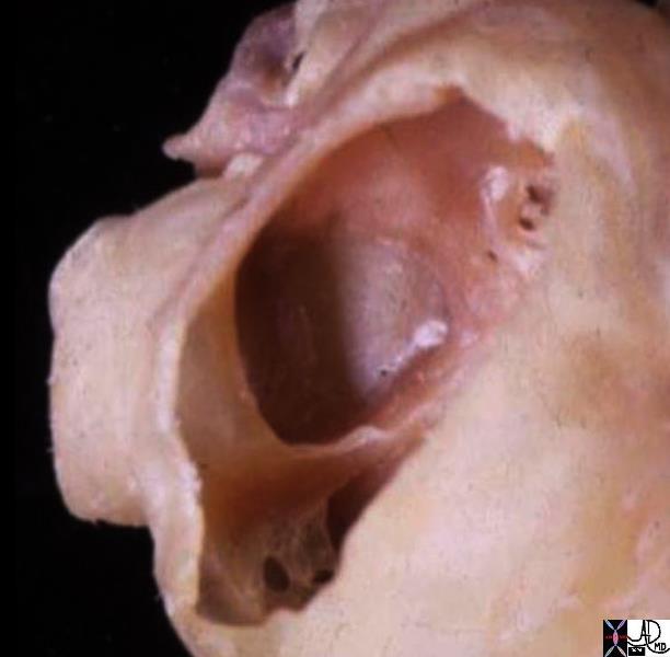
In this anatomic specimen the SVC and IVC are well seen and their lateral relationship to the right atrium is appreciated. They are embryological remnants of the sinus venosus and their similarity and conjoined structural origin and functionality can be well appreciated in this image. Davidoff MD
Key Words
heart cardiac RA right atrium thebesian valves coronary sinus Eustachian valve sulcus terminalis fossa ovalis foramen ovale septum secundum superior limbic band IVC inferior vena cava anatomy normal gross anatomy post mortem specimen
Courtesy Ashley Davidoff MD 2019 01707.6
key words
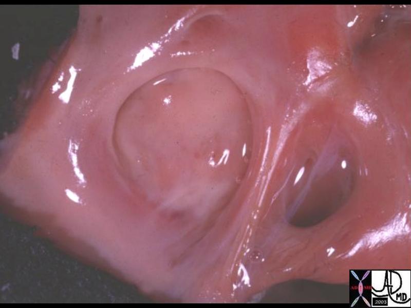
heart cardiac atrial septum septum primum septum secundum sinus venosus right atrium normal anatomy coronary sinus thebesian valve Eustachian valve coronary sinus triangle of Koch atrioventricular node A-V node gross pathology
Courtesy Ashley Davidoff MD 01671