The Common Vein Copyright 2008
Definition
Esophagitis is an inflammatory response of the esophagus to multiple causes, the most common of which is due to reflux of acidic gastric contents through an incompetent lower esophageal sphincter into the distal esophagus. It is thus considered in this instance as a chemical burn.
It is characterized by a reddened inflammed and swollen and often erosive mucosa, resulting in pain that may be epigastric or substernal. This presentation may simulate cardiac pain.
Long standing esophagitis may be complicated by a peptic stricture or Barrett’s esophagus which is a premalignant condition.
The diagnosis is suspected clinically by the classical presentation of substernal burning in the presence of symptoms of reflux, waterbrash (hypersalivation), and halitosis, with relief from antacid therapy. Documentation of the disease is best achieved by endoscopy, with double contrast barium swallow being less sensitive to the disease.
The mainstay of therapy for GERD with esophagitis remains the proton pump inhibitors (PPI) and the H2 Blockers.
|
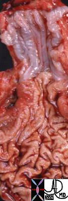
The Normal (left) and Abnormal (right) Red and Swollen GE junction – Reflux Esophagitis |
|
| The GE junction is a distinct border as seen normally in the left image. The distal esophagus is normally a pearl white hue, while the gastric mucosa is a pink hue. In esophagitis the hyperemic inflammatory changes make the border less distinct (right image)
01239b01.800 esophagus stomach GE junction gastroesophageaal junction inflammed inflammation GERD red swollen reflux esophagitis grosspathology Courtesy Ashley Davidoff MD |
Quinidine Ulcer |
| Courtesy Ashley Davidoff MD 01258 01258b01 esophagus + quinidine ulcer barium swallow inflammation upper GI UGI imaging radiology contrast X-Ray |
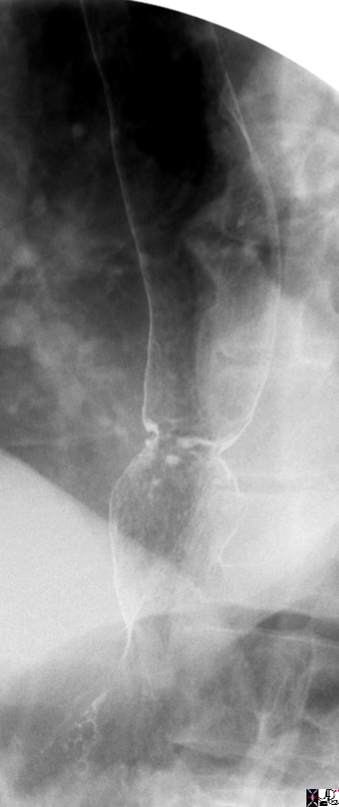
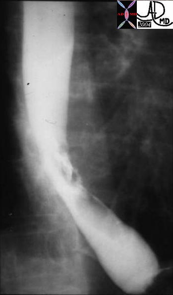
 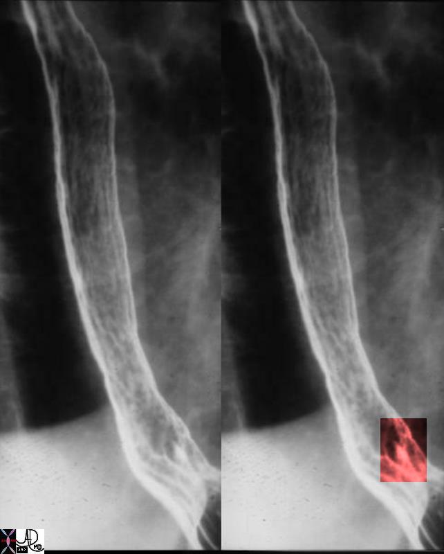 Linear Erosion at the GE Junction Linear Erosion at the GE Junction |
| 01252c Courtesy Ashley Davidoff MD 01252 code esophagus + fx linear erosion + dx reflux esophagitis + barium swallow upper GI UGI imaging radiology contrast X-Ray |
DOMElement Object
(
    [schemaTypeInfo] =>
    [tagName] => table
    [firstElementChild] => (object value omitted)
    [lastElementChild] => (object value omitted)
    [childElementCount] => 1
    [previousElementSibling] => (object value omitted)
    [nextElementSibling] => (object value omitted)
    [nodeName] => table
    [nodeValue] =>


Benign Serpigenous Obstructing Ulcer


76398c03 esophagus dysphagia odynophagia esophagus esophageal ulcer giant ulcer heaped edges 13mm barium pill stuck held up endoscopy showed a serpigenous ulcer pathology showed esophagitis interesting with the heaped edges impresio was that it was malignant barium swallow CourtesyAshley Davidoff MD 76398c01


    [nodeType] => 1
    [parentNode] => (object value omitted)
    [childNodes] => (object value omitted)
    [firstChild] => (object value omitted)
    [lastChild] => (object value omitted)
    [previousSibling] => (object value omitted)
    [nextSibling] => (object value omitted)
    [attributes] => (object value omitted)
    [ownerDocument] => (object value omitted)
    [namespaceURI] =>
    [prefix] =>
    [localName] => table
    [baseURI] =>
    [textContent] =>


Benign Serpigenous Obstructing Ulcer


76398c03 esophagus dysphagia odynophagia esophagus esophageal ulcer giant ulcer heaped edges 13mm barium pill stuck held up endoscopy showed a serpigenous ulcer pathology showed esophagitis interesting with the heaped edges impresio was that it was malignant barium swallow CourtesyAshley Davidoff MD 76398c01


)
DOMElement Object
(
    [schemaTypeInfo] =>
    [tagName] => td
    [firstElementChild] =>
    [lastElementChild] =>
    [childElementCount] => 0
    [previousElementSibling] =>
    [nextElementSibling] =>
    [nodeName] => td
    [nodeValue] => 76398c03 esophagus dysphagia odynophagia esophagus esophageal ulcer giant ulcer heaped edges 13mm barium pill stuck held up endoscopy showed a serpigenous ulcer pathology showed esophagitis interesting with the heaped edges impresio was that it was malignant barium swallow CourtesyAshley Davidoff MD 76398c01
    [nodeType] => 1
    [parentNode] => (object value omitted)
    [childNodes] => (object value omitted)
    [firstChild] => (object value omitted)
    [lastChild] => (object value omitted)
    [previousSibling] => (object value omitted)
    [nextSibling] => (object value omitted)
    [attributes] => (object value omitted)
    [ownerDocument] => (object value omitted)
    [namespaceURI] =>
    [prefix] =>
    [localName] => td
    [baseURI] =>
    [textContent] => 76398c03 esophagus dysphagia odynophagia esophagus esophageal ulcer giant ulcer heaped edges 13mm barium pill stuck held up endoscopy showed a serpigenous ulcer pathology showed esophagitis interesting with the heaped edges impresio was that it was malignant barium swallow CourtesyAshley Davidoff MD 76398c01
)
DOMElement Object
(
    [schemaTypeInfo] =>
    [tagName] => td
    [firstElementChild] => (object value omitted)
    [lastElementChild] => (object value omitted)
    [childElementCount] => 2
    [previousElementSibling] =>
    [nextElementSibling] =>
    [nodeName] => td
    [nodeValue] =>
Benign Serpigenous Obstructing Ulcer
    [nodeType] => 1
    [parentNode] => (object value omitted)
    [childNodes] => (object value omitted)
    [firstChild] => (object value omitted)
    [lastChild] => (object value omitted)
    [previousSibling] => (object value omitted)
    [nextSibling] => (object value omitted)
    [attributes] => (object value omitted)
    [ownerDocument] => (object value omitted)
    [namespaceURI] =>
    [prefix] =>
    [localName] => td
    [baseURI] =>
    [textContent] =>
Benign Serpigenous Obstructing Ulcer
)
DOMElement Object
(
    [schemaTypeInfo] =>
    [tagName] => table
    [firstElementChild] => (object value omitted)
    [lastElementChild] => (object value omitted)
    [childElementCount] => 1
    [previousElementSibling] => (object value omitted)
    [nextElementSibling] => (object value omitted)
    [nodeName] => table
    [nodeValue] =>


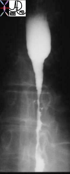
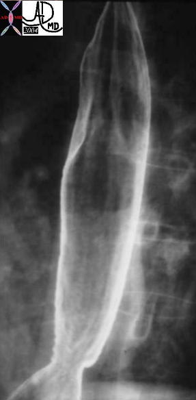
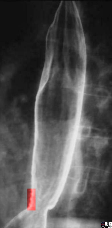
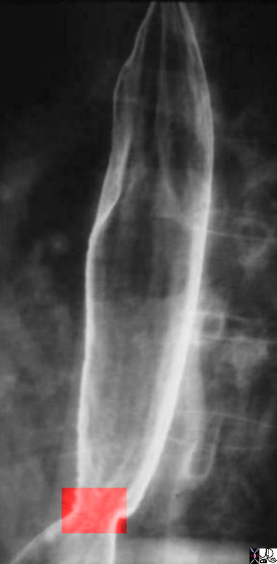
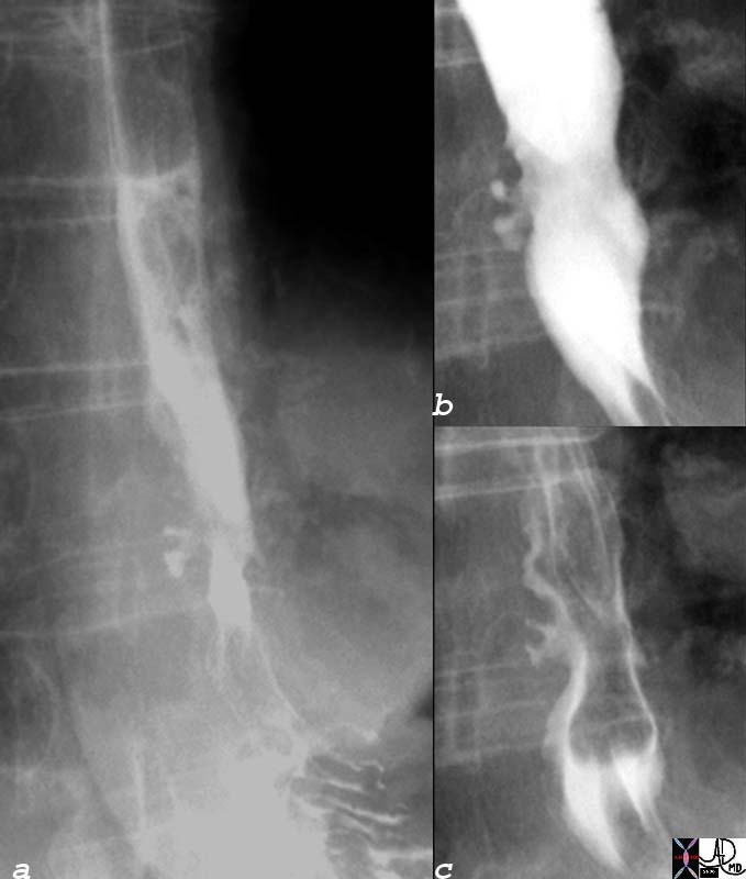
Esophagitis with Superficial Ulcerations (middle) and Stricture (right)


Courtesy Ashley Davidoff MD 01221 esophagus + reflux esophagitis stricture ulcer barium swallow upper GI UGI imaging radiology contrast X-Ray


    [nodeType] => 1
    [parentNode] => (object value omitted)
    [childNodes] => (object value omitted)
    [firstChild] => (object value omitted)
    [lastChild] => (object value omitted)
    [previousSibling] => (object value omitted)
    [nextSibling] => (object value omitted)
    [attributes] => (object value omitted)
    [ownerDocument] => (object value omitted)
    [namespaceURI] =>
    [prefix] =>
    [localName] => table
    [baseURI] =>
    [textContent] =>


Esophagitis with Superficial Ulcerations (middle) and Stricture (right)


Courtesy Ashley Davidoff MD 01221 esophagus + reflux esophagitis stricture ulcer barium swallow upper GI UGI imaging radiology contrast X-Ray


)
DOMElement Object
(
    [schemaTypeInfo] =>
    [tagName] => td
    [firstElementChild] =>
    [lastElementChild] =>
    [childElementCount] => 0
    [previousElementSibling] =>
    [nextElementSibling] =>
    [nodeName] => td
    [nodeValue] => Courtesy Ashley Davidoff MD 01221 esophagus + reflux esophagitis stricture ulcer barium swallow upper GI UGI imaging radiology contrast X-Ray
    [nodeType] => 1
    [parentNode] => (object value omitted)
    [childNodes] => (object value omitted)
    [firstChild] => (object value omitted)
    [lastChild] => (object value omitted)
    [previousSibling] => (object value omitted)
    [nextSibling] => (object value omitted)
    [attributes] => (object value omitted)
    [ownerDocument] => (object value omitted)
    [namespaceURI] =>
    [prefix] =>
    [localName] => td
    [baseURI] =>
    [textContent] => Courtesy Ashley Davidoff MD 01221 esophagus + reflux esophagitis stricture ulcer barium swallow upper GI UGI imaging radiology contrast X-Ray
)
DOMElement Object
(
    [schemaTypeInfo] =>
    [tagName] => td
    [firstElementChild] => (object value omitted)
    [lastElementChild] => (object value omitted)
    [childElementCount] => 4
    [previousElementSibling] =>
    [nextElementSibling] =>
    [nodeName] => td
    [nodeValue] =>


Esophagitis with Superficial Ulcerations (middle) and Stricture (right)
    [nodeType] => 1
    [parentNode] => (object value omitted)
    [childNodes] => (object value omitted)
    [firstChild] => (object value omitted)
    [lastChild] => (object value omitted)
    [previousSibling] => (object value omitted)
    [nextSibling] => (object value omitted)
    [attributes] => (object value omitted)
    [ownerDocument] => (object value omitted)
    [namespaceURI] =>
    [prefix] =>
    [localName] => td
    [baseURI] =>
    [textContent] =>


Esophagitis with Superficial Ulcerations (middle) and Stricture (right)
)
DOMElement Object
(
    [schemaTypeInfo] =>
    [tagName] => table
    [firstElementChild] => (object value omitted)
    [lastElementChild] => (object value omitted)
    [childElementCount] => 1
    [previousElementSibling] => (object value omitted)
    [nextElementSibling] => (object value omitted)
    [nodeName] => table
    [nodeValue] =>


Linear Erosion at the GE Junction


01252c Courtesy Ashley Davidoff MD 01252 code esophagus + fx linear erosion + dx reflux esophagitis + barium swallow upper GI UGI imaging radiology contrast X-Ray


    [nodeType] => 1
    [parentNode] => (object value omitted)
    [childNodes] => (object value omitted)
    [firstChild] => (object value omitted)
    [lastChild] => (object value omitted)
    [previousSibling] => (object value omitted)
    [nextSibling] => (object value omitted)
    [attributes] => (object value omitted)
    [ownerDocument] => (object value omitted)
    [namespaceURI] =>
    [prefix] =>
    [localName] => table
    [baseURI] =>
    [textContent] =>


Linear Erosion at the GE Junction


01252c Courtesy Ashley Davidoff MD 01252 code esophagus + fx linear erosion + dx reflux esophagitis + barium swallow upper GI UGI imaging radiology contrast X-Ray


)
DOMElement Object
(
    [schemaTypeInfo] =>
    [tagName] => td
    [firstElementChild] =>
    [lastElementChild] =>
    [childElementCount] => 0
    [previousElementSibling] =>
    [nextElementSibling] =>
    [nodeName] => td
    [nodeValue] => 01252c Courtesy Ashley Davidoff MD 01252 code esophagus + fx linear erosion + dx reflux esophagitis + barium swallow upper GI UGI imaging radiology contrast X-Ray
    [nodeType] => 1
    [parentNode] => (object value omitted)
    [childNodes] => (object value omitted)
    [firstChild] => (object value omitted)
    [lastChild] => (object value omitted)
    [previousSibling] => (object value omitted)
    [nextSibling] => (object value omitted)
    [attributes] => (object value omitted)
    [ownerDocument] => (object value omitted)
    [namespaceURI] =>
    [prefix] =>
    [localName] => td
    [baseURI] =>
    [textContent] => 01252c Courtesy Ashley Davidoff MD 01252 code esophagus + fx linear erosion + dx reflux esophagitis + barium swallow upper GI UGI imaging radiology contrast X-Ray
)
DOMElement Object
(
    [schemaTypeInfo] =>
    [tagName] => td
    [firstElementChild] => (object value omitted)
    [lastElementChild] => (object value omitted)
    [childElementCount] => 1
    [previousElementSibling] =>
    [nextElementSibling] =>
    [nodeName] => td
    [nodeValue] => Linear Erosion at the GE Junction
    [nodeType] => 1
    [parentNode] => (object value omitted)
    [childNodes] => (object value omitted)
    [firstChild] => (object value omitted)
    [lastChild] => (object value omitted)
    [previousSibling] => (object value omitted)
    [nextSibling] => (object value omitted)
    [attributes] => (object value omitted)
    [ownerDocument] => (object value omitted)
    [namespaceURI] =>
    [prefix] =>
    [localName] => td
    [baseURI] =>
    [textContent] => Linear Erosion at the GE Junction
)
DOMElement Object
(
    [schemaTypeInfo] =>
    [tagName] => table
    [firstElementChild] => (object value omitted)
    [lastElementChild] => (object value omitted)
    [childElementCount] => 1
    [previousElementSibling] => (object value omitted)
    [nextElementSibling] => (object value omitted)
    [nodeName] => table
    [nodeValue] =>


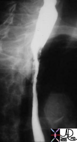
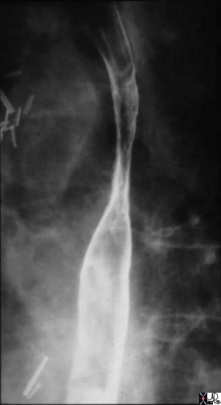
Mid Esophageal Stricture from Radiation Lung Cancer


Courtesy Ashley Davidoff MD 01251 01251 b01 code esophagus + fx stricture + dx radiation stricture + barium swallow upper GI UGI imaging radiology contrast X-Ray


    [nodeType] => 1
    [parentNode] => (object value omitted)
    [childNodes] => (object value omitted)
    [firstChild] => (object value omitted)
    [lastChild] => (object value omitted)
    [previousSibling] => (object value omitted)
    [nextSibling] => (object value omitted)
    [attributes] => (object value omitted)
    [ownerDocument] => (object value omitted)
    [namespaceURI] =>
    [prefix] =>
    [localName] => table
    [baseURI] =>
    [textContent] =>


Mid Esophageal Stricture from Radiation Lung Cancer


Courtesy Ashley Davidoff MD 01251 01251 b01 code esophagus + fx stricture + dx radiation stricture + barium swallow upper GI UGI imaging radiology contrast X-Ray


)
DOMElement Object
(
    [schemaTypeInfo] =>
    [tagName] => td
    [firstElementChild] =>
    [lastElementChild] =>
    [childElementCount] => 0
    [previousElementSibling] =>
    [nextElementSibling] =>
    [nodeName] => td
    [nodeValue] => Courtesy Ashley Davidoff MD 01251 01251 b01 code esophagus + fx stricture + dx radiation stricture + barium swallow upper GI UGI imaging radiology contrast X-Ray
    [nodeType] => 1
    [parentNode] => (object value omitted)
    [childNodes] => (object value omitted)
    [firstChild] => (object value omitted)
    [lastChild] => (object value omitted)
    [previousSibling] => (object value omitted)
    [nextSibling] => (object value omitted)
    [attributes] => (object value omitted)
    [ownerDocument] => (object value omitted)
    [namespaceURI] =>
    [prefix] =>
    [localName] => td
    [baseURI] =>
    [textContent] => Courtesy Ashley Davidoff MD 01251 01251 b01 code esophagus + fx stricture + dx radiation stricture + barium swallow upper GI UGI imaging radiology contrast X-Ray
)
DOMElement Object
(
    [schemaTypeInfo] =>
    [tagName] => td
    [firstElementChild] => (object value omitted)
    [lastElementChild] => (object value omitted)
    [childElementCount] => 3
    [previousElementSibling] =>
    [nextElementSibling] =>
    [nodeName] => td
    [nodeValue] =>

Mid Esophageal Stricture from Radiation Lung Cancer
    [nodeType] => 1
    [parentNode] => (object value omitted)
    [childNodes] => (object value omitted)
    [firstChild] => (object value omitted)
    [lastChild] => (object value omitted)
    [previousSibling] => (object value omitted)
    [nextSibling] => (object value omitted)
    [attributes] => (object value omitted)
    [ownerDocument] => (object value omitted)
    [namespaceURI] =>
    [prefix] =>
    [localName] => td
    [baseURI] =>
    [textContent] =>

Mid Esophageal Stricture from Radiation Lung Cancer
)
DOMElement Object
(
    [schemaTypeInfo] =>
    [tagName] => table
    [firstElementChild] => (object value omitted)
    [lastElementChild] => (object value omitted)
    [childElementCount] => 1
    [previousElementSibling] => (object value omitted)
    [nextElementSibling] => (object value omitted)
    [nodeName] => table
    [nodeValue] =>


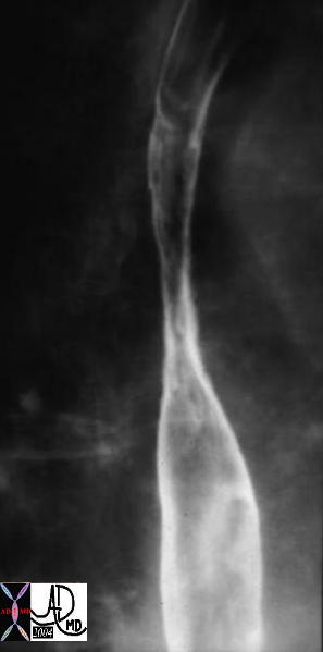
Lye Ingestion with Deep Focal Ulcers Grade 2b


Courtesy Ashley Davidoff MD 01250 code esophagus + fx irregular + fx long distal fx narrowed + fx ulceration + dx lye stricture upper GI UGI imaging radiology contrast X-Ray


    [nodeType] => 1
    [parentNode] => (object value omitted)
    [childNodes] => (object value omitted)
    [firstChild] => (object value omitted)
    [lastChild] => (object value omitted)
    [previousSibling] => (object value omitted)
    [nextSibling] => (object value omitted)
    [attributes] => (object value omitted)
    [ownerDocument] => (object value omitted)
    [namespaceURI] =>
    [prefix] =>
    [localName] => table
    [baseURI] =>
    [textContent] =>


Lye Ingestion with Deep Focal Ulcers Grade 2b


Courtesy Ashley Davidoff MD 01250 code esophagus + fx irregular + fx long distal fx narrowed + fx ulceration + dx lye stricture upper GI UGI imaging radiology contrast X-Ray


)
DOMElement Object
(
    [schemaTypeInfo] =>
    [tagName] => td
    [firstElementChild] =>
    [lastElementChild] =>
    [childElementCount] => 0
    [previousElementSibling] =>
    [nextElementSibling] =>
    [nodeName] => td
    [nodeValue] => Courtesy Ashley Davidoff MD 01250 code esophagus + fx irregular + fx long distal fx narrowed + fx ulceration + dx lye stricture upper GI UGI imaging radiology contrast X-Ray
    [nodeType] => 1
    [parentNode] => (object value omitted)
    [childNodes] => (object value omitted)
    [firstChild] => (object value omitted)
    [lastChild] => (object value omitted)
    [previousSibling] => (object value omitted)
    [nextSibling] => (object value omitted)
    [attributes] => (object value omitted)
    [ownerDocument] => (object value omitted)
    [namespaceURI] =>
    [prefix] =>
    [localName] => td
    [baseURI] =>
    [textContent] => Courtesy Ashley Davidoff MD 01250 code esophagus + fx irregular + fx long distal fx narrowed + fx ulceration + dx lye stricture upper GI UGI imaging radiology contrast X-Ray
)
DOMElement Object
(
    [schemaTypeInfo] =>
    [tagName] => td
    [firstElementChild] => (object value omitted)
    [lastElementChild] => (object value omitted)
    [childElementCount] => 2
    [previousElementSibling] =>
    [nextElementSibling] =>
    [nodeName] => td
    [nodeValue] =>
Lye Ingestion with Deep Focal Ulcers Grade 2b
    [nodeType] => 1
    [parentNode] => (object value omitted)
    [childNodes] => (object value omitted)
    [firstChild] => (object value omitted)
    [lastChild] => (object value omitted)
    [previousSibling] => (object value omitted)
    [nextSibling] => (object value omitted)
    [attributes] => (object value omitted)
    [ownerDocument] => (object value omitted)
    [namespaceURI] =>
    [prefix] =>
    [localName] => td
    [baseURI] =>
    [textContent] =>
Lye Ingestion with Deep Focal Ulcers Grade 2b
)
DOMElement Object
(
    [schemaTypeInfo] =>
    [tagName] => table
    [firstElementChild] => (object value omitted)
    [lastElementChild] => (object value omitted)
    [childElementCount] => 1
    [previousElementSibling] => (object value omitted)
    [nextElementSibling] => (object value omitted)
    [nodeName] => table
    [nodeValue] =>


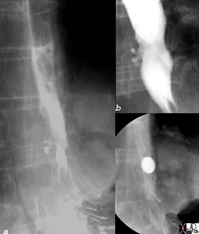
Quinidine Ulcer


Courtesy Ashley Davidoff MD 01258 01258b01 esophagus + quinidine ulcer barium swallow inflammation upper GI UGI imaging radiology contrast X-Ray


    [nodeType] => 1
    [parentNode] => (object value omitted)
    [childNodes] => (object value omitted)
    [firstChild] => (object value omitted)
    [lastChild] => (object value omitted)
    [previousSibling] => (object value omitted)
    [nextSibling] => (object value omitted)
    [attributes] => (object value omitted)
    [ownerDocument] => (object value omitted)
    [namespaceURI] =>
    [prefix] =>
    [localName] => table
    [baseURI] =>
    [textContent] =>


Quinidine Ulcer


Courtesy Ashley Davidoff MD 01258 01258b01 esophagus + quinidine ulcer barium swallow inflammation upper GI UGI imaging radiology contrast X-Ray


)
DOMElement Object
(
    [schemaTypeInfo] =>
    [tagName] => td
    [firstElementChild] =>
    [lastElementChild] =>
    [childElementCount] => 0
    [previousElementSibling] =>
    [nextElementSibling] =>
    [nodeName] => td
    [nodeValue] => Courtesy Ashley Davidoff MD 01258 01258b01 esophagus + quinidine ulcer barium swallow inflammation upper GI UGI imaging radiology contrast X-Ray
    [nodeType] => 1
    [parentNode] => (object value omitted)
    [childNodes] => (object value omitted)
    [firstChild] => (object value omitted)
    [lastChild] => (object value omitted)
    [previousSibling] => (object value omitted)
    [nextSibling] => (object value omitted)
    [attributes] => (object value omitted)
    [ownerDocument] => (object value omitted)
    [namespaceURI] =>
    [prefix] =>
    [localName] => td
    [baseURI] =>
    [textContent] => Courtesy Ashley Davidoff MD 01258 01258b01 esophagus + quinidine ulcer barium swallow inflammation upper GI UGI imaging radiology contrast X-Ray
)
DOMElement Object
(
    [schemaTypeInfo] =>
    [tagName] => td
    [firstElementChild] => (object value omitted)
    [lastElementChild] => (object value omitted)
    [childElementCount] => 3
    [previousElementSibling] =>
    [nextElementSibling] =>
    [nodeName] => td
    [nodeValue] =>

Quinidine Ulcer
    [nodeType] => 1
    [parentNode] => (object value omitted)
    [childNodes] => (object value omitted)
    [firstChild] => (object value omitted)
    [lastChild] => (object value omitted)
    [previousSibling] => (object value omitted)
    [nextSibling] => (object value omitted)
    [attributes] => (object value omitted)
    [ownerDocument] => (object value omitted)
    [namespaceURI] =>
    [prefix] =>
    [localName] => td
    [baseURI] =>
    [textContent] =>

Quinidine Ulcer
)
DOMElement Object
(
    [schemaTypeInfo] =>
    [tagName] => table
    [firstElementChild] => (object value omitted)
    [lastElementChild] => (object value omitted)
    [childElementCount] => 1
    [previousElementSibling] => (object value omitted)
    [nextElementSibling] => (object value omitted)
    [nodeName] => table
    [nodeValue] =>


Mid Esophageal necrotizingh Esophagitis
Stricture and Ulceration


Courtesy Ashley Davidoff MD 01233 code esophagus + fx narrowing + fx ulceration + dx necrotizing esophagitis + barium swallow upper GI UGI imaging radiology contrast X-Ray


    [nodeType] => 1
    [parentNode] => (object value omitted)
    [childNodes] => (object value omitted)
    [firstChild] => (object value omitted)
    [lastChild] => (object value omitted)
    [previousSibling] => (object value omitted)
    [nextSibling] => (object value omitted)
    [attributes] => (object value omitted)
    [ownerDocument] => (object value omitted)
    [namespaceURI] =>
    [prefix] =>
    [localName] => table
    [baseURI] =>
    [textContent] =>


Mid Esophageal necrotizingh Esophagitis
Stricture and Ulceration


Courtesy Ashley Davidoff MD 01233 code esophagus + fx narrowing + fx ulceration + dx necrotizing esophagitis + barium swallow upper GI UGI imaging radiology contrast X-Ray


)
DOMElement Object
(
    [schemaTypeInfo] =>
    [tagName] => td
    [firstElementChild] =>
    [lastElementChild] =>
    [childElementCount] => 0
    [previousElementSibling] =>
    [nextElementSibling] =>
    [nodeName] => td
    [nodeValue] => Courtesy Ashley Davidoff MD 01233 code esophagus + fx narrowing + fx ulceration + dx necrotizing esophagitis + barium swallow upper GI UGI imaging radiology contrast X-Ray
    [nodeType] => 1
    [parentNode] => (object value omitted)
    [childNodes] => (object value omitted)
    [firstChild] => (object value omitted)
    [lastChild] => (object value omitted)
    [previousSibling] => (object value omitted)
    [nextSibling] => (object value omitted)
    [attributes] => (object value omitted)
    [ownerDocument] => (object value omitted)
    [namespaceURI] =>
    [prefix] =>
    [localName] => td
    [baseURI] =>
    [textContent] => Courtesy Ashley Davidoff MD 01233 code esophagus + fx narrowing + fx ulceration + dx necrotizing esophagitis + barium swallow upper GI UGI imaging radiology contrast X-Ray
)
DOMElement Object
(
    [schemaTypeInfo] =>
    [tagName] => td
    [firstElementChild] => (object value omitted)
    [lastElementChild] => (object value omitted)
    [childElementCount] => 2
    [previousElementSibling] =>
    [nextElementSibling] =>
    [nodeName] => td
    [nodeValue] => Mid Esophageal necrotizingh Esophagitis
Stricture and Ulceration
    [nodeType] => 1
    [parentNode] => (object value omitted)
    [childNodes] => (object value omitted)
    [firstChild] => (object value omitted)
    [lastChild] => (object value omitted)
    [previousSibling] => (object value omitted)
    [nextSibling] => (object value omitted)
    [attributes] => (object value omitted)
    [ownerDocument] => (object value omitted)
    [namespaceURI] =>
    [prefix] =>
    [localName] => td
    [baseURI] =>
    [textContent] => Mid Esophageal necrotizingh Esophagitis
Stricture and Ulceration
)
DOMElement Object
(
    [schemaTypeInfo] =>
    [tagName] => table
    [firstElementChild] => (object value omitted)
    [lastElementChild] => (object value omitted)
    [childElementCount] => 1
    [previousElementSibling] => (object value omitted)
    [nextElementSibling] => (object value omitted)
    [nodeName] => table
    [nodeValue] =>


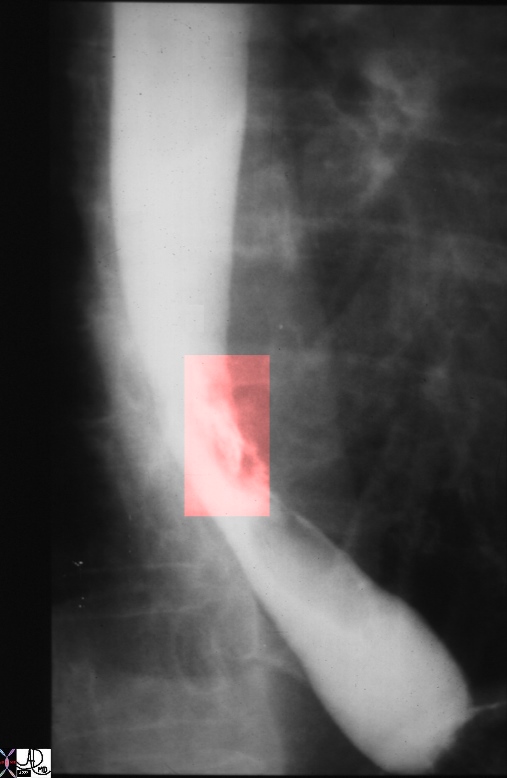
Reflux Esophagitis with Ulcers


70 year old male presents with burning substernal chest pain Barium swallow shows superficial ulceration at the GE junction and also small areas of ulceration along the distal esophagus
esophagus GE junction ulcer esophagitis barium swallow 83326.8s Courtesy Ashley Davidoff MD copyright 2009


    [nodeType] => 1
    [parentNode] => (object value omitted)
    [childNodes] => (object value omitted)
    [firstChild] => (object value omitted)
    [lastChild] => (object value omitted)
    [previousSibling] => (object value omitted)
    [nextSibling] => (object value omitted)
    [attributes] => (object value omitted)
    [ownerDocument] => (object value omitted)
    [namespaceURI] =>
    [prefix] =>
    [localName] => table
    [baseURI] =>
    [textContent] =>


Reflux Esophagitis with Ulcers


70 year old male presents with burning substernal chest pain Barium swallow shows superficial ulceration at the GE junction and also small areas of ulceration along the distal esophagus
esophagus GE junction ulcer esophagitis barium swallow 83326.8s Courtesy Ashley Davidoff MD copyright 2009


)
DOMElement Object
(
    [schemaTypeInfo] =>
    [tagName] => td
    [firstElementChild] => (object value omitted)
    [lastElementChild] => (object value omitted)
    [childElementCount] => 1
    [previousElementSibling] =>
    [nextElementSibling] =>
    [nodeName] => td
    [nodeValue] => 70 year old male presents with burning substernal chest pain Barium swallow shows superficial ulceration at the GE junction and also small areas of ulceration along the distal esophagus
esophagus GE junction ulcer esophagitis barium swallow 83326.8s Courtesy Ashley Davidoff MD copyright 2009
    [nodeType] => 1
    [parentNode] => (object value omitted)
    [childNodes] => (object value omitted)
    [firstChild] => (object value omitted)
    [lastChild] => (object value omitted)
    [previousSibling] => (object value omitted)
    [nextSibling] => (object value omitted)
    [attributes] => (object value omitted)
    [ownerDocument] => (object value omitted)
    [namespaceURI] =>
    [prefix] =>
    [localName] => td
    [baseURI] =>
    [textContent] => 70 year old male presents with burning substernal chest pain Barium swallow shows superficial ulceration at the GE junction and also small areas of ulceration along the distal esophagus
esophagus GE junction ulcer esophagitis barium swallow 83326.8s Courtesy Ashley Davidoff MD copyright 2009
)
DOMElement Object
(
    [schemaTypeInfo] =>
    [tagName] => td
    [firstElementChild] => (object value omitted)
    [lastElementChild] => (object value omitted)
    [childElementCount] => 2
    [previousElementSibling] =>
    [nextElementSibling] =>
    [nodeName] => td
    [nodeValue] =>
Reflux Esophagitis with Ulcers
    [nodeType] => 1
    [parentNode] => (object value omitted)
    [childNodes] => (object value omitted)
    [firstChild] => (object value omitted)
    [lastChild] => (object value omitted)
    [previousSibling] => (object value omitted)
    [nextSibling] => (object value omitted)
    [attributes] => (object value omitted)
    [ownerDocument] => (object value omitted)
    [namespaceURI] =>
    [prefix] =>
    [localName] => td
    [baseURI] =>
    [textContent] =>
Reflux Esophagitis with Ulcers
)
DOMElement Object
(
    [schemaTypeInfo] =>
    [tagName] => table
    [firstElementChild] => (object value omitted)
    [lastElementChild] => (object value omitted)
    [childElementCount] => 1
    [previousElementSibling] => (object value omitted)
    [nextElementSibling] => (object value omitted)
    [nodeName] => table
    [nodeValue] =>


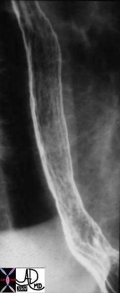
Normal (a) and Reflux Esophagitis (b)


76217c esophagus normal smooth mucosa featurless heterogeneous mucosa nodular mucosa diffuse reflux esophagitis double contrast barium swallow Courtesy Ashley Davidoff MD


    [nodeType] => 1
    [parentNode] => (object value omitted)
    [childNodes] => (object value omitted)
    [firstChild] => (object value omitted)
    [lastChild] => (object value omitted)
    [previousSibling] => (object value omitted)
    [nextSibling] => (object value omitted)
    [attributes] => (object value omitted)
    [ownerDocument] => (object value omitted)
    [namespaceURI] =>
    [prefix] =>
    [localName] => table
    [baseURI] =>
    [textContent] =>


Normal (a) and Reflux Esophagitis (b)


76217c esophagus normal smooth mucosa featurless heterogeneous mucosa nodular mucosa diffuse reflux esophagitis double contrast barium swallow Courtesy Ashley Davidoff MD


)
DOMElement Object
(
    [schemaTypeInfo] =>
    [tagName] => td
    [firstElementChild] =>
    [lastElementChild] =>
    [childElementCount] => 0
    [previousElementSibling] =>
    [nextElementSibling] =>
    [nodeName] => td
    [nodeValue] => 76217c esophagus normal smooth mucosa featurless heterogeneous mucosa nodular mucosa diffuse reflux esophagitis double contrast barium swallow Courtesy Ashley Davidoff MD
    [nodeType] => 1
    [parentNode] => (object value omitted)
    [childNodes] => (object value omitted)
    [firstChild] => (object value omitted)
    [lastChild] => (object value omitted)
    [previousSibling] => (object value omitted)
    [nextSibling] => (object value omitted)
    [attributes] => (object value omitted)
    [ownerDocument] => (object value omitted)
    [namespaceURI] =>
    [prefix] =>
    [localName] => td
    [baseURI] =>
    [textContent] => 76217c esophagus normal smooth mucosa featurless heterogeneous mucosa nodular mucosa diffuse reflux esophagitis double contrast barium swallow Courtesy Ashley Davidoff MD
)
DOMElement Object
(
    [schemaTypeInfo] =>
    [tagName] => td
    [firstElementChild] => (object value omitted)
    [lastElementChild] => (object value omitted)
    [childElementCount] => 2
    [previousElementSibling] =>
    [nextElementSibling] =>
    [nodeName] => td
    [nodeValue] =>
Normal (a) and Reflux Esophagitis (b)
    [nodeType] => 1
    [parentNode] => (object value omitted)
    [childNodes] => (object value omitted)
    [firstChild] => (object value omitted)
    [lastChild] => (object value omitted)
    [previousSibling] => (object value omitted)
    [nextSibling] => (object value omitted)
    [attributes] => (object value omitted)
    [ownerDocument] => (object value omitted)
    [namespaceURI] =>
    [prefix] =>
    [localName] => td
    [baseURI] =>
    [textContent] =>
Normal (a) and Reflux Esophagitis (b)
)
DOMElement Object
(
    [schemaTypeInfo] =>
    [tagName] => table
    [firstElementChild] => (object value omitted)
    [lastElementChild] => (object value omitted)
    [childElementCount] => 1
    [previousElementSibling] => (object value omitted)
    [nextElementSibling] => (object value omitted)
    [nodeName] => table
    [nodeValue] =>


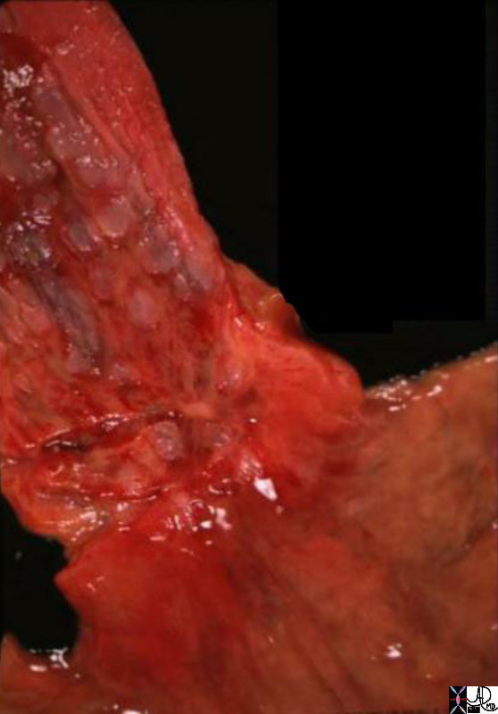
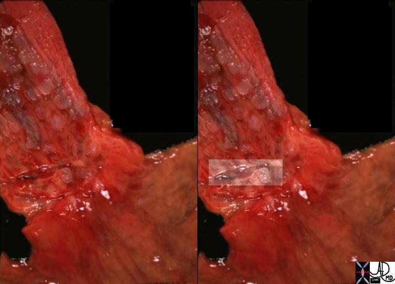
Linear Ulcer in a Hyperemic Background of Diffuse Esophagitis


On closer inspection of the GE junction, a linear ulcer is seen (overlaid in right image).  It is even painful to look at.
01239c esophagus stomach GE junction gastroesophageaal junction inflammed inflammation GERD red swollen reflux esophagitis linear ulcer grosspathology Courtesy Ashley Davidoff MD


    [nodeType] => 1
    [parentNode] => (object value omitted)
    [childNodes] => (object value omitted)
    [firstChild] => (object value omitted)
    [lastChild] => (object value omitted)
    [previousSibling] => (object value omitted)
    [nextSibling] => (object value omitted)
    [attributes] => (object value omitted)
    [ownerDocument] => (object value omitted)
    [namespaceURI] =>
    [prefix] =>
    [localName] => table
    [baseURI] =>
    [textContent] =>


Linear Ulcer in a Hyperemic Background of Diffuse Esophagitis


On closer inspection of the GE junction, a linear ulcer is seen (overlaid in right image).  It is even painful to look at.
01239c esophagus stomach GE junction gastroesophageaal junction inflammed inflammation GERD red swollen reflux esophagitis linear ulcer grosspathology Courtesy Ashley Davidoff MD


)
DOMElement Object
(
    [schemaTypeInfo] =>
    [tagName] => td
    [firstElementChild] => (object value omitted)
    [lastElementChild] => (object value omitted)
    [childElementCount] => 1
    [previousElementSibling] =>
    [nextElementSibling] =>
    [nodeName] => td
    [nodeValue] => On closer inspection of the GE junction, a linear ulcer is seen (overlaid in right image).  It is even painful to look at.
01239c esophagus stomach GE junction gastroesophageaal junction inflammed inflammation GERD red swollen reflux esophagitis linear ulcer grosspathology Courtesy Ashley Davidoff MD
    [nodeType] => 1
    [parentNode] => (object value omitted)
    [childNodes] => (object value omitted)
    [firstChild] => (object value omitted)
    [lastChild] => (object value omitted)
    [previousSibling] => (object value omitted)
    [nextSibling] => (object value omitted)
    [attributes] => (object value omitted)
    [ownerDocument] => (object value omitted)
    [namespaceURI] =>
    [prefix] =>
    [localName] => td
    [baseURI] =>
    [textContent] => On closer inspection of the GE junction, a linear ulcer is seen (overlaid in right image).  It is even painful to look at.
01239c esophagus stomach GE junction gastroesophageaal junction inflammed inflammation GERD red swollen reflux esophagitis linear ulcer grosspathology Courtesy Ashley Davidoff MD
)
DOMElement Object
(
    [schemaTypeInfo] =>
    [tagName] => td
    [firstElementChild] => (object value omitted)
    [lastElementChild] => (object value omitted)
    [childElementCount] => 1
    [previousElementSibling] =>
    [nextElementSibling] =>
    [nodeName] => td
    [nodeValue] => Linear Ulcer in a Hyperemic Background of Diffuse Esophagitis
    [nodeType] => 1
    [parentNode] => (object value omitted)
    [childNodes] => (object value omitted)
    [firstChild] => (object value omitted)
    [lastChild] => (object value omitted)
    [previousSibling] => (object value omitted)
    [nextSibling] => (object value omitted)
    [attributes] => (object value omitted)
    [ownerDocument] => (object value omitted)
    [namespaceURI] =>
    [prefix] =>
    [localName] => td
    [baseURI] =>
    [textContent] => Linear Ulcer in a Hyperemic Background of Diffuse Esophagitis
)
DOMElement Object
(
    [schemaTypeInfo] =>
    [tagName] => table
    [firstElementChild] => (object value omitted)
    [lastElementChild] => (object value omitted)
    [childElementCount] => 1
    [previousElementSibling] =>
    [nextElementSibling] => (object value omitted)
    [nodeName] => table
    [nodeValue] =>


    [nodeType] => 1
    [parentNode] => (object value omitted)
    [childNodes] => (object value omitted)
    [firstChild] => (object value omitted)
    [lastChild] => (object value omitted)
    [previousSibling] => (object value omitted)
    [nextSibling] => (object value omitted)
    [attributes] => (object value omitted)
    [ownerDocument] => (object value omitted)
    [namespaceURI] =>
    [prefix] =>
    [localName] => table
    [baseURI] =>
    [textContent] =>


)
DOMElement Object
(
    [schemaTypeInfo] =>
    [tagName] => td
    [firstElementChild] => (object value omitted)
    [lastElementChild] => (object value omitted)
    [childElementCount] => 1
    [previousElementSibling] =>
    [nextElementSibling] =>
    [nodeName] => td
    [nodeValue] =>


    [nodeType] => 1
    [parentNode] => (object value omitted)
    [childNodes] => (object value omitted)
    [firstChild] => (object value omitted)
    [lastChild] => (object value omitted)
    [previousSibling] => (object value omitted)
    [nextSibling] => (object value omitted)
    [attributes] => (object value omitted)
    [ownerDocument] => (object value omitted)
    [namespaceURI] =>
    [prefix] =>
    [localName] => td
    [baseURI] =>
    [textContent] =>


)
DOMElement Object
(
    [schemaTypeInfo] =>
    [tagName] => table
    [firstElementChild] => (object value omitted)
    [lastElementChild] => (object value omitted)
    [childElementCount] => 1
    [previousElementSibling] => (object value omitted)
    [nextElementSibling] => (object value omitted)
    [nodeName] => table
    [nodeValue] =>


The Normal (left) and Abnormal (right) Red and Swollen GE junction – Reflux Esophagitis


The GE junction is a distinct border as seen normally in the left image.  The distal esophagus is normally a pearl white hue, while the gastric mucosa is a pink hue.  In esophagitis the hyperemic inflammatory changes make the border less distinct (right image)
01239b01.800 esophagus stomach GE junction gastroesophageaal junction inflammed inflammation GERD red swollen reflux esophagitis grosspathology Courtesy Ashley Davidoff MD


    [nodeType] => 1
    [parentNode] => (object value omitted)
    [childNodes] => (object value omitted)
    [firstChild] => (object value omitted)
    [lastChild] => (object value omitted)
    [previousSibling] => (object value omitted)
    [nextSibling] => (object value omitted)
    [attributes] => (object value omitted)
    [ownerDocument] => (object value omitted)
    [namespaceURI] =>
    [prefix] =>
    [localName] => table
    [baseURI] =>
    [textContent] =>


The Normal (left) and Abnormal (right) Red and Swollen GE junction – Reflux Esophagitis


The GE junction is a distinct border as seen normally in the left image.  The distal esophagus is normally a pearl white hue, while the gastric mucosa is a pink hue.  In esophagitis the hyperemic inflammatory changes make the border less distinct (right image)
01239b01.800 esophagus stomach GE junction gastroesophageaal junction inflammed inflammation GERD red swollen reflux esophagitis grosspathology Courtesy Ashley Davidoff MD


)
DOMElement Object
(
    [schemaTypeInfo] =>
    [tagName] => td
    [firstElementChild] => (object value omitted)
    [lastElementChild] => (object value omitted)
    [childElementCount] => 1
    [previousElementSibling] =>
    [nextElementSibling] =>
    [nodeName] => td
    [nodeValue] => The GE junction is a distinct border as seen normally in the left image.  The distal esophagus is normally a pearl white hue, while the gastric mucosa is a pink hue.  In esophagitis the hyperemic inflammatory changes make the border less distinct (right image)
01239b01.800 esophagus stomach GE junction gastroesophageaal junction inflammed inflammation GERD red swollen reflux esophagitis grosspathology Courtesy Ashley Davidoff MD
    [nodeType] => 1
    [parentNode] => (object value omitted)
    [childNodes] => (object value omitted)
    [firstChild] => (object value omitted)
    [lastChild] => (object value omitted)
    [previousSibling] => (object value omitted)
    [nextSibling] => (object value omitted)
    [attributes] => (object value omitted)
    [ownerDocument] => (object value omitted)
    [namespaceURI] =>
    [prefix] =>
    [localName] => td
    [baseURI] =>
    [textContent] => The GE junction is a distinct border as seen normally in the left image.  The distal esophagus is normally a pearl white hue, while the gastric mucosa is a pink hue.  In esophagitis the hyperemic inflammatory changes make the border less distinct (right image)
01239b01.800 esophagus stomach GE junction gastroesophageaal junction inflammed inflammation GERD red swollen reflux esophagitis grosspathology Courtesy Ashley Davidoff MD
)
DOMElement Object
(
    [schemaTypeInfo] =>
    [tagName] => td
    [firstElementChild] => (object value omitted)
    [lastElementChild] => (object value omitted)
    [childElementCount] => 1
    [previousElementSibling] =>
    [nextElementSibling] =>
    [nodeName] => td
    [nodeValue] =>


    [nodeType] => 1
    [parentNode] => (object value omitted)
    [childNodes] => (object value omitted)
    [firstChild] => (object value omitted)
    [lastChild] => (object value omitted)
    [previousSibling] => (object value omitted)
    [nextSibling] => (object value omitted)
    [attributes] => (object value omitted)
    [ownerDocument] => (object value omitted)
    [namespaceURI] =>
    [prefix] =>
    [localName] => td
    [baseURI] =>
    [textContent] =>


)
DOMElement Object
(
    [schemaTypeInfo] =>
    [tagName] => td
    [firstElementChild] => (object value omitted)
    [lastElementChild] => (object value omitted)
    [childElementCount] => 2
    [previousElementSibling] =>
    [nextElementSibling] =>
    [nodeName] => td
    [nodeValue] =>


The Normal (left) and Abnormal (right) Red and Swollen GE junction – Reflux Esophagitis
    [nodeType] => 1
    [parentNode] => (object value omitted)
    [childNodes] => (object value omitted)
    [firstChild] => (object value omitted)
    [lastChild] => (object value omitted)
    [previousSibling] => (object value omitted)
    [nextSibling] => (object value omitted)
    [attributes] => (object value omitted)
    [ownerDocument] => (object value omitted)
    [namespaceURI] =>
    [prefix] =>
    [localName] => td
    [baseURI] =>
    [textContent] =>


The Normal (left) and Abnormal (right) Red and Swollen GE junction – Reflux Esophagitis
)

