D – Transposition of the Great Vessels
Copyright 2009
Atlas
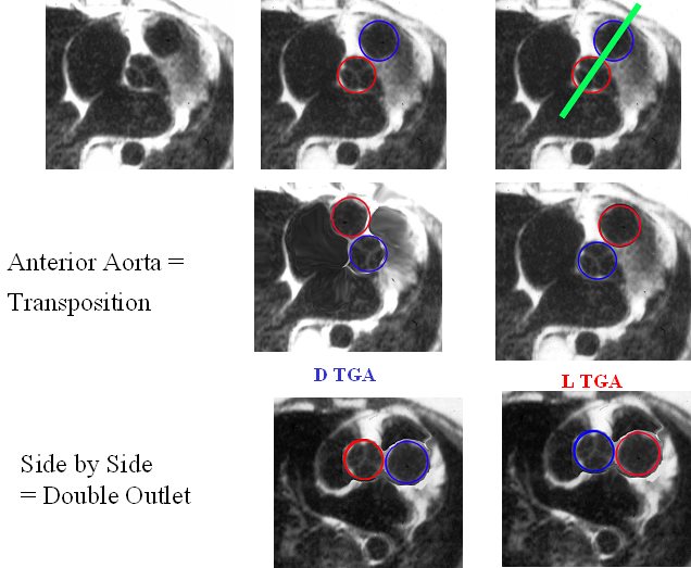
The image reflects the relationship of the aorta and pulmonary arteries in the normal patient, in DTGA, LTGA and DORV. In the normal patient with D loop the aorta (Ao) is posterior and to the right, and the pulmonary artery (PA) is anterior and to the right. In the patient with DTGA, the Ao is anterior and to the right and the PA is posterior and to the left. In an L loop the Ao is anterior and to the left and the PA is posterior and to the right. In double outlet right ventricle (DORV) the great vessels lie side by side and in DORV with a D loop the aorta is to the right and with an L loop the aorta is to the left
86778 01639 01639d02 01639d04 01639f03.jpg Anterior aorta = Transposition
Ashley Davidoff MD
TheCommonVein.net
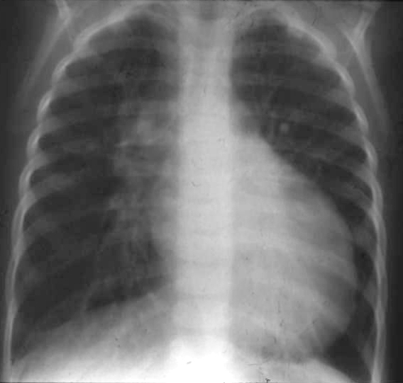
The CXR in frontal projection shows cardiomegaly, narrowed mediastinum and vascular congestion The cardiomegaly has a classic appearance of on egg on its side or egg on a string and together with the narrow mediastinum is characteristic of DTGA. The hypervascularity indicates a left to right shunt. The alteration of the positioning of the aorta and pulmonary artery in association with stress-induced thymic hypoplasia results in the narrowing of the mediastinum.
Ashley Davidoff MD TheCommonVein.net
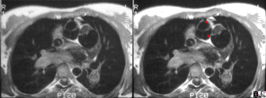
The axial CT shows findings characteristic of D transposition of the aorta with an anteriorly and rightward positioned aorta and posteriorly positioned pulmonary artery.
07249e01cs aorta pulmonary artery conus arteriosus displaced DTGA congenital heart disease heart cardiac MRI T1 weighted Courtesy Ashley Davidoff MD TheCommonVein.net
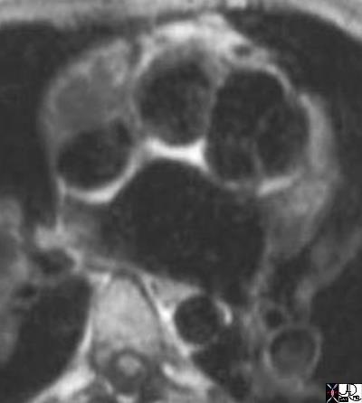
The axial CT slightly inferior below the aortic valve reveals a subaortic conus This finding is characteristic of D transposition of the aorta with a subaortic conus and an anteriorly and rightward positioned aorta and posteriorly positioned pulmonary artery.
07258b02 anterior aorta posterior pulmonary artery position connection subaortic conus D TGA TGV D transposition of the great vessels D transposition of the great arteries dextro rightward MRI Courtesy Ashley Davidoff MD TheCommonVein.net
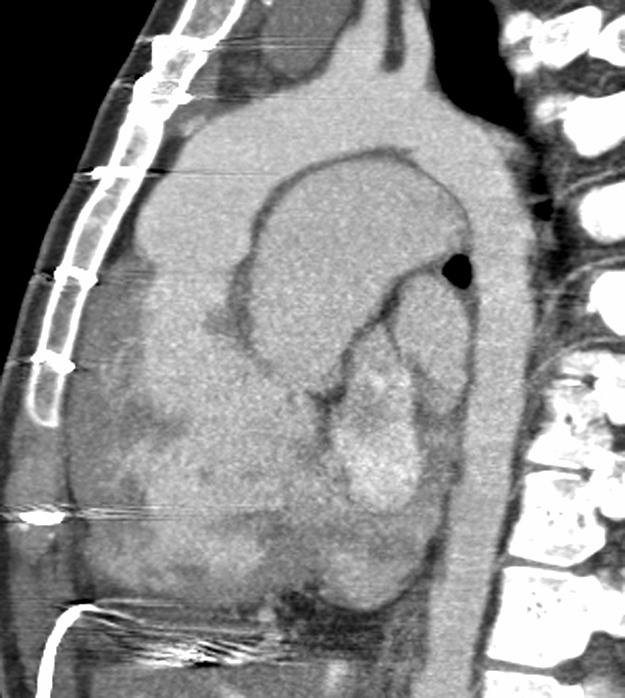
48321 CT of patient with an anterior aorta and a large posterior pulmonary artery TGA transposition heart aorta position 48320 48321 DTGA Courtesy Dr Hyun Woo Goo from Korea and Dr Laureen Sena
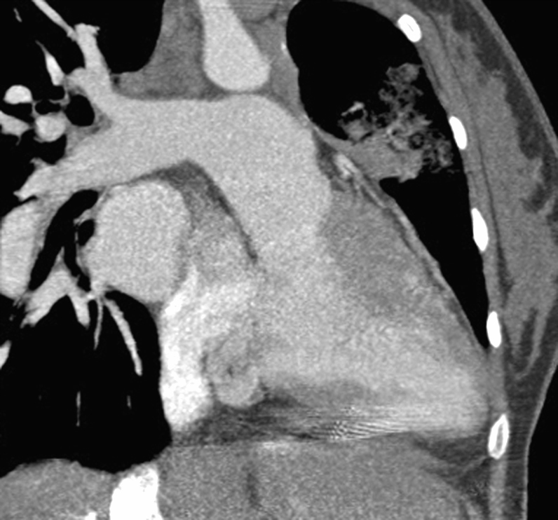
TGA transposition 48320 48321 DTGA
Courtesy Dr Hyun Woo Goo from Korea and Dr Laureen Sena
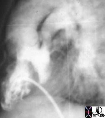
This is an angiogram of the RV, showing an anteriorly placed aorta, a VSD filling the LV, and a posteriorly positioned smaller MPA. The catheter courses via the IVC into the RV. The findings are consistent with TGA and in this case a D-TGA, though it is impossible to identify the position of the aortic valve in relation to the PA in this lateral projection. An associated VSD and subpulmonary stenosis and or PS is implied by the small sized PA. 01487 code CVS heart cardiac transpoistion of the great arteries DTGA VSD subpulmonary stenosis imaging radiology angiography
Keywords:
CVS heart cardiac transpoistion of the great arteries DTGA VSD subpulmonary stenosis imaging radiology angiography
Ashley Davidoff MD TheCommonVein.net
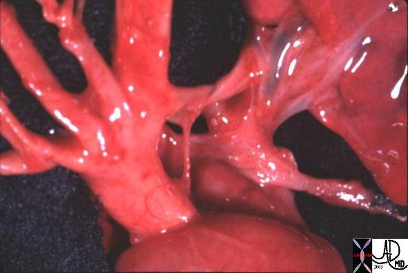
The aorta is anterior and to the right of the atretic main pulmonary artery and therefore represents DTGA. A patent ductus supplies the branch pulmonary arteries
Ashley Davidoff MD TheCommonVein.net
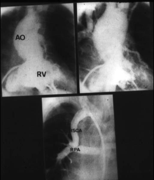
The aorta is anterior and to the right of the main pulmonary artery is not seen in this patient with known DTGA pulmonary atresia. The right subclavian artery has been connected to the small pulmonary arteries to enable blood flow to the lungs
Ashley Davidoff MD TheCommonVein.net
References
- TCV
- Conotruncal Abnormalities
- Embryology
- Anatomy of the Right Ventricle
- Double Outlet Right Ventricle
- Double Outlet Left Ventricle
- Tetralogy of Fallot
- Transposition of the Great Vessels
- Truncus Arteriosus
