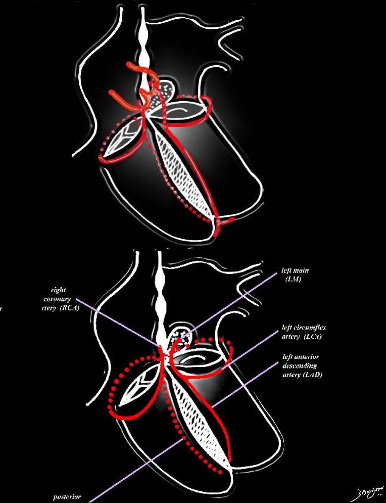
The heart has a left and right arterial system. The vessels are formed around the cross of the heart, “vertically” along the interventricular septum and interatrial septum, and “horizontally” in the atrioventricular (A-V) grooves. The left coronary artery arises from the left coronary ostium and supplies one branch, the left anterior descending artery, that travels anteriorly on the anterior aspect of the interventricular septum, and one branch that travels in the atrioventricular groove (left circumflex). The right coronary artery has a branch that courses along the right atrioventricular groove and usually continues as the posterior descending artery on the posterior aspect of the interventricular groove, and commonly gives rise to the A-V nodal branch, at the back of the crux of the heart, which travels in the vertical axis in the interatrial septum.
Courtesy of: Ashley Davidoff, M.D.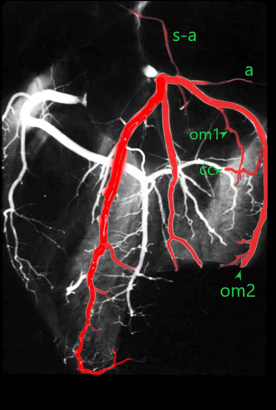
The anatomical specimen has been injected with barium and shows the circumflex (c) arising from the left main coronary artery. The obtuse marginal arteries are large vessels so the distal circumflex continuation (c c green arrow) is a small vessel that runs in the posterior A-V groove with the coronary sinus
The S-A nodal artery (s-a) sometimes arises from the circumflex as the first branch. Atrial branches (a) also arise from the proximal circumflex artery.
A number of obtuse marginal arteries (OM1 and OM2 ) supply the posterior and posterolateral parts of the LV and are almost always larger than the distal circumflex itself (c c).
Ashley Davidoff MD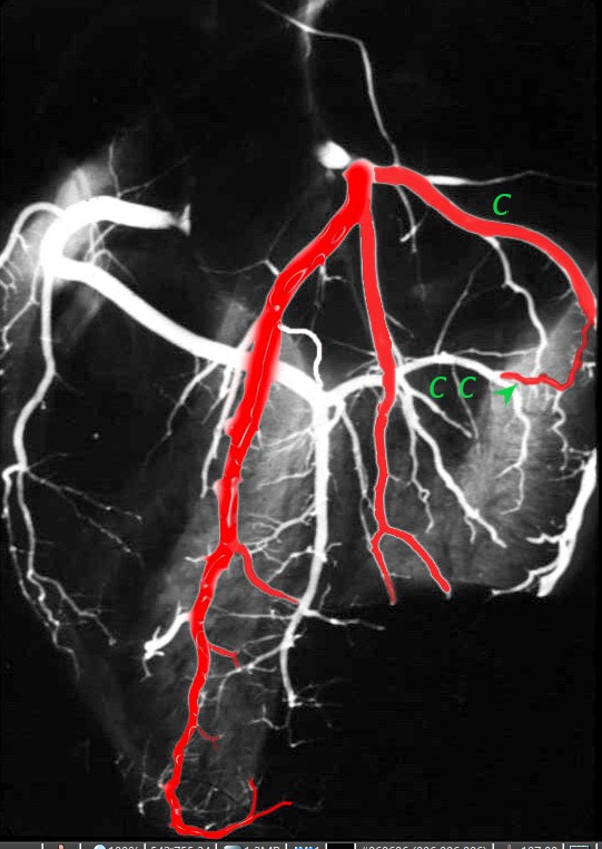
The anatomical specimen has been injected with barium and shows the circumflex (c) arising from the left main coronary artery. The obtuse marginal arteries are large vessels so the distal circumflex (c c green arrow) is a small vessel that runs in the posterior A-V groove with the coronary sinus
Ashley Davidoff MD
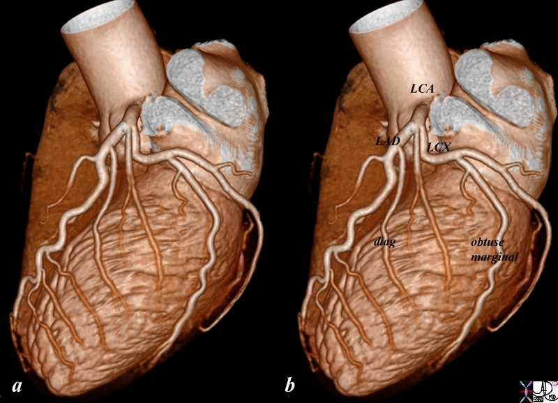
Reconstruction of the CTA of the left left coronary artery shows normal LM , LAD and diagonals and circumflex and obtuse marginals .
Ashley Davidoff MD thecommonvein.net
87260b01.8s
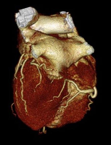
Reconstruction of the CTA of the distal circumflex and distal right coronary arteries in the posterior aspect of the heart showing the obtuse marginals supplying the posterior wall of the left ventricle (LV) and the distal RCA supplying the PDA and crossing over to give rise to the posterior left ventricular branch
Ashley Davidoff MD thecommonvein.net
45170
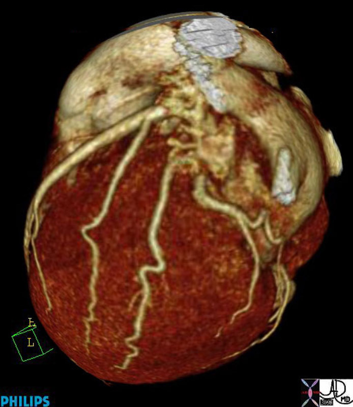
Reconstruction of the CTA of the circumflex coronary artery showing the obtuse marginals supplying the posterolateral wall of the left ventricle (LV)
Ashley Davidoff MD thecommonvein.net
45168b01.8s
He was anticoagulated and thrombus resolved on subsequent CT.
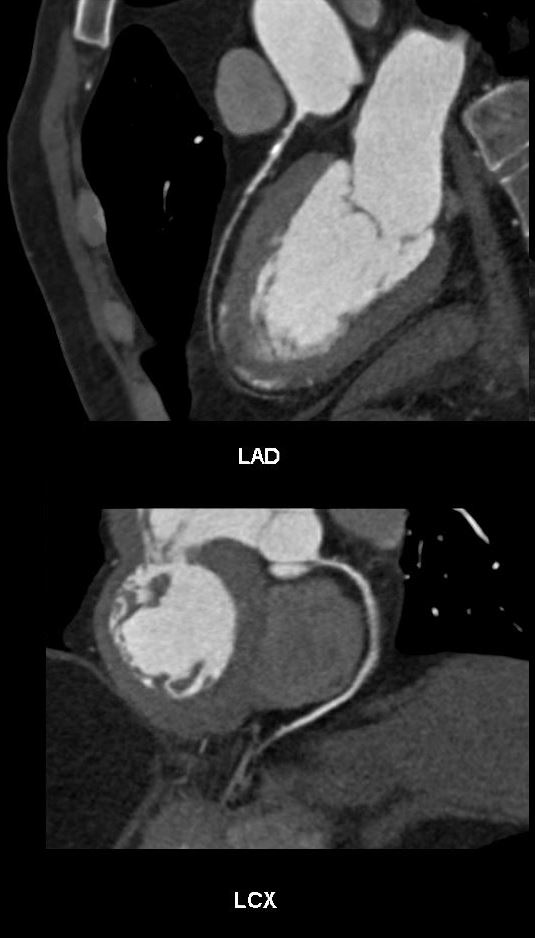
Reconstructions of the LAD above showed mild LAD disease. Both calcified plaque and non-calcified plaque were identified The calcified proximal lesion was felt to be mild on multiple views.
The circumflex artery (below) was normal
Ashley Davidoff thecommonvein.net
131083c
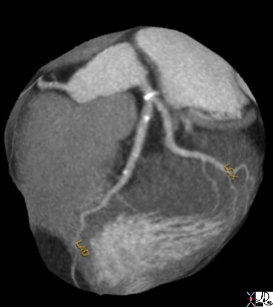
key words heart cardiac artery ramus medianus coronary artery circumflex coronary artery LAD left anterior descending coronary artery mild atherosclerosis calcific plaques CTscan volume rendering 3D Courtesy Ashley Davidoff MD thecommonvein.net
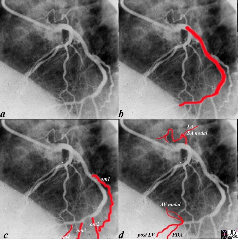
The angiogram of the LCA (a) shows the circumflex (b) arising from the left main coronary artery. In this left dominant system, there are 4 obtuse marginal arteries (c). Distally the circumflex continues and gives rise to the PDA, (d) AV nodal artery and posterior LV branches
Ashley Davidoff MD
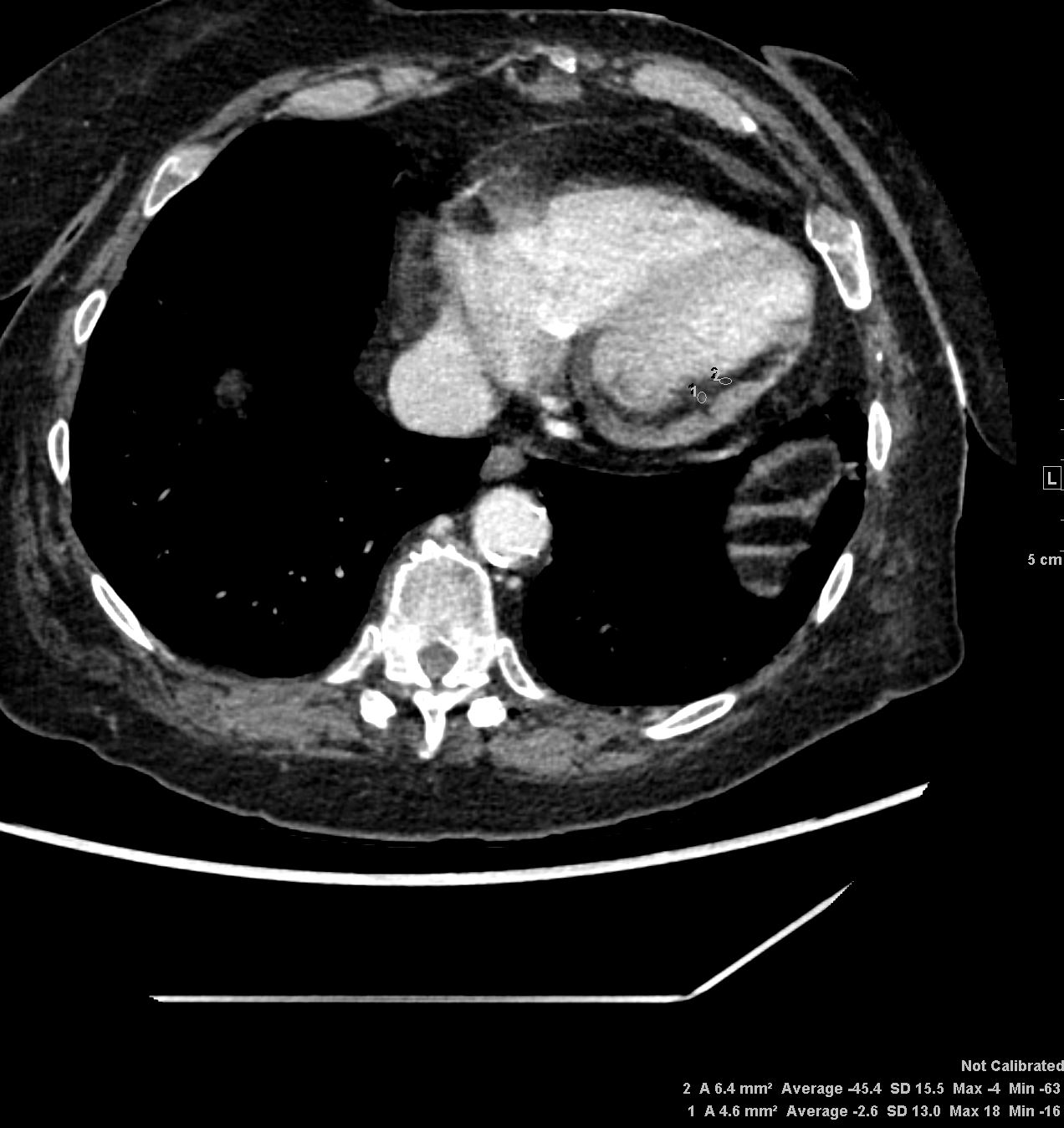
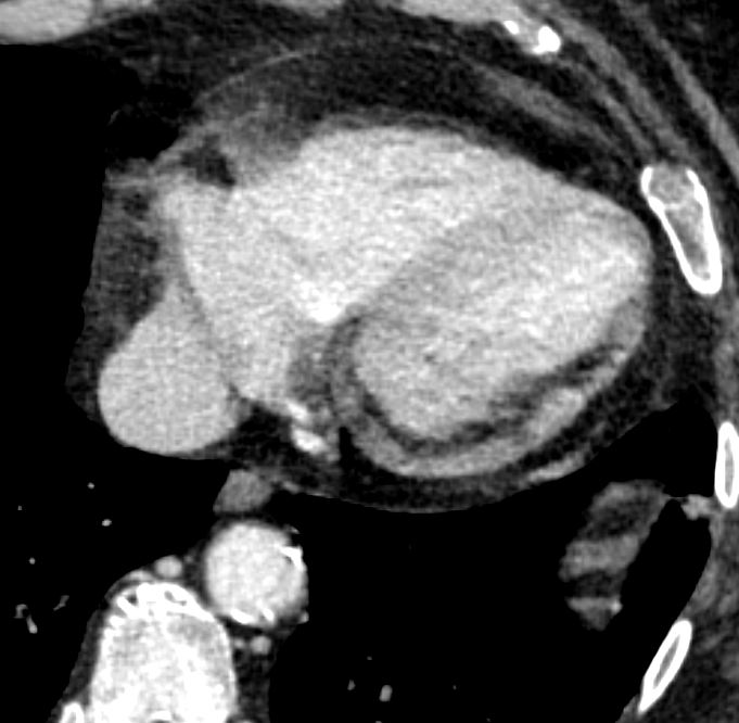
Axial CT through the LV shows subendocardial fat in the lateral wall of the left ventricle indicating prior myocardial infarction in the circumflex coronary artery territory
Ashley Davidoff MD TheCommonvein.net prior MI 001
Ashley Davidoff MD TheCommonvein.net prior MI 002
Aberrant Origin
Aberrant origin of the LCx from the RCA is generally felt to be of no clinical significance due to its posterior course to the left ventricle. This is the most common origin anomaly of the coronary arteries
