
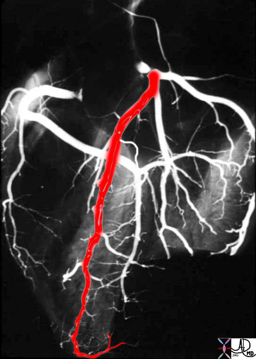
If we were to “crack open” the chest of the chest X-ray, the structures that would dominate this bloody, black and white scene, would be the right sided chambers. The right ventricle (RV) would be the dominant anterior chamber, and would form the dominant interface with the diaphragm. The right atrium (RA) would form the border with the right lung. The RA would of course be slightly posterior to the RV. The left border would be formed by the left ventricle. Most the left ventricle is hidden posteriorly in this view. The left anterior descending artery would be visible from this anterior view. It marks the position of the interventricular septum.
Ashley Davidoff MD
CORONARY CIRCULATION IN 3D ? ANATOMICAL SPECIMEN INJECTED WITH BARIUM LAD in LAO
33805aladP
44198d14 32134
CORONARY CIRCULATION IN 3D ? ANATOMICAL SPECIMEN INJECTED WITH BARIUM LAD in LAO SEPTALS and DIAGONALS
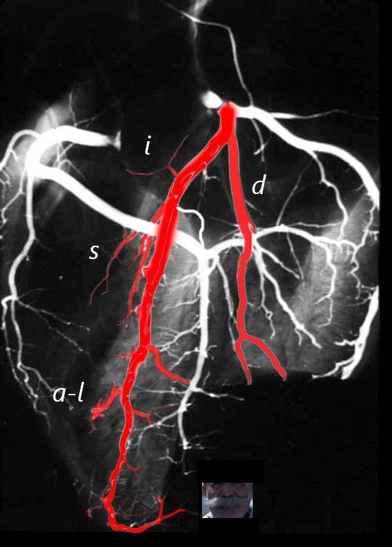
The anatomical specimen has been injected with barium and shows LAD arising from the left main coronary artery. The diagonal arteries (d) course toward the patients left to supply the anterior and anterolateral part of the LV. The infundibular artery (I, aka arc of Vieussen?s) courses over the RVOT to join its partner arising from the RCA. The septal arteries have a vertical course toward the diaphragm, and the branch to the anterolateral papillary muscle of the RV (a-l) courses toward the right of the patient. The LAD often terminates with a bifurcation reminiscent of a moustache.
Ashley Davidoff MD
CIRCUMFLEX CORONARY ARTERY ?
Note The distal circumflex is usually very small and sometimes not even visible but is recognised by its relationship to the A_V groove and coronary sinus
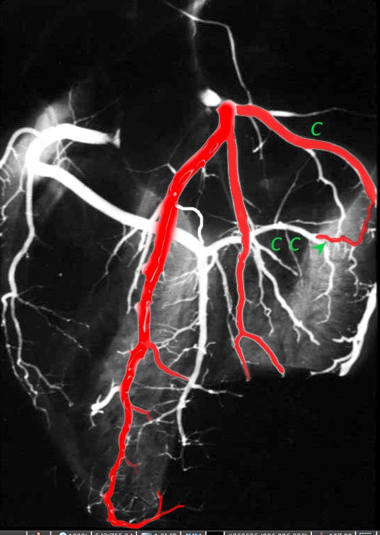
The anatomical specimen has been injected with barium and shows the circumflex (c) arising from the left main coronary artery. The obtuse marginal arteries are large vessels so the distal circumflex (c c green arrow) is a small vessel that runs in the posterior A-V groove with the coronary sinus
Ashley Davidoff MD
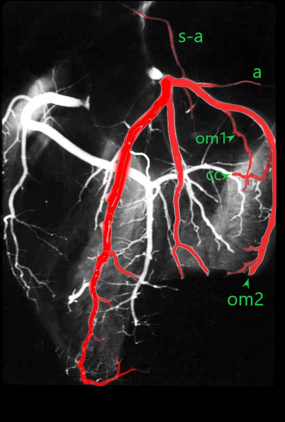
The anatomical specimen has been injected with barium and shows the circumflex (c) arising from the left main coronary artery. The obtuse marginal arteries are large vessels so the distal circumflex continuation (c c green arrow) is a small vessel that runs in the posterior A-V groove with the coronary sinus
The S-A nodal artery (s-a) sometimes arises from the circumflex as the first branch. Atrial branches (a) also arise from the proximal circumflex artery.
A number of obtuse marginal arteries (OM1 and OM2 ) supply the posterior and posterolateral parts of the LV and are almost always larger than the distal circumflex itself (c c).
Ashley Davidoff MD
RCA
Note the septal perforators supply about 1/3 of the septum and are important collateral pathway when the LAD is occluded
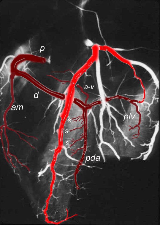
The coronary arteries have been injected with barium and shows left (bright red overlay) and right (maroon) coronary circulation The proximal RCA (p) courses anteriorly in the A-V groove arising from the right coronary ostium. The acute marginal (am) courses along the right margin of the heart, and the RCA continues posteriorly (p) to the crux of the heart where it gives off 3 branches. The posterior descending artery (PDA), A-V nodal artery (a-v) and posterior left ventricular artery (plv). The PDA supplies the inferior 1/3 of the interventricular septum with perforating septal arteries (s)
This is a dominant RCA
Ashley Davidoff MD
